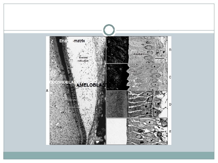
ENAMEL Physical characteristics Enamel forms a protective covering


 The inorganic material of the enamel is hydroxyapatite. Its")


- Slides: 44
ENAMEL
� Physical characteristics: � Enamel forms a protective covering of variable thickness over the entire surface of the crown. � On the cusps of human molars and premolars the enamel attains a maximum thickness of about 2 to 2. 5 mm, thinning down to almost a knife edge at the neck of the tooth. The enamel was found to be thicker in the lingual surfaces of maxillary molars and in the buccal surfaces of mandibular molars. As these are supporting cusps adaptation to functional demands. � Because of its high content of mineral salts and their crystalline arrangement, enamel is the hardest calcified tissue in the human body. The function of the enamel is to form a resistant covering of the teeth, rendering them suitable for mastication.
� The structure and hardness of the enamel render it brittle, which is particularly apparent when the enamel loses its foundation of sound dentin. The complex microstructure of enamel leads to large variations in mechanical behavior. At the surface the modulus of elasticity is higher and the hardness is more than on dentinoenamel junction. � The specific gravity of enamel is 2. 8. The density decreases from the surface to the deeper regions and from cuspal to incisal region. � Enamel has always been observed as a non electrical conductive material: it is in fact an insulator at room temperature. � Enamel is its permeable permitting complete or partial passage of certain molecules.
�The color of the enamel-covered crown ranges from yellowish white to grayish white is determined by differences in the translucency of enamel, yellowish teeth having a thin, translucent enamel through which the yellow color of the dentin is visible and grayish teeth having a more opaque enamel. This due to variations in the degree of calcification and homogeneity of the enamel. Grayish teeth frequently show a slightly yellowish color at the cervical areas. Incisal areas may have a bluish tinge where thin edge consists only of a double layer of enamel. Dehydration decreased the translucency but it was reversed on rehydration.
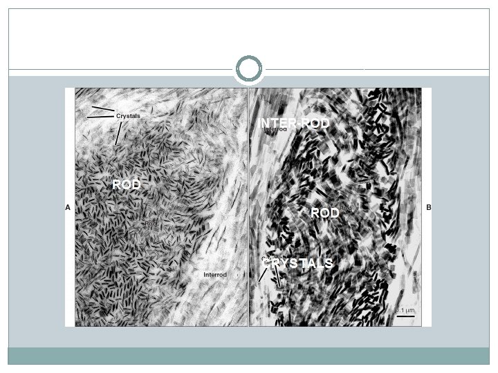
Chemical properties: �Inorganic material (96%) The inorganic material of the enamel is hydroxyapatite. Its chemical formula is Ca 10 (PO 4)6(OH 4)2. They are hexagonal in cross-section. The crystals are arranged to form enamel rods or enamel prisms. The hydroxyapatite crystal has a central core or C axis of hydroxyl ion around which calcium and phosphorus ions are arranged in the form of triangles. Carbonate rich crystals are preferentially attacked by acids in caries �Organic substance and water (4%). Water is present as a part of the crystal (hydroxyapatite), between crystals and between rods and surrounding the rods. Pores are present between the crystals, especially at the boundaries of the rods and these are filled with water.
�The organic material consists of some unique proteins, found exclusively in the enamel and lipids. Enamel proteins do not contribute to structuring of enamel. This is in contrast to collagen, which is the principal protein of dentin or bone, having a structuring function. � � The proteins found in the enamel are of two main groups—the amelogenins and the nonamelogenins.
� 1 -Amelogenins, are a heterogenous group of low molecular weight proteins, accounting for about 90% of the enamel proteins. They are hydrophobic � 2 -Nonamelogenins constitute about 10% of enamel matrix proteins. Enamelin, ameloblastin and tuftelin are the important proteins of this group. Nonamelogenins are high molecular weight proteins .
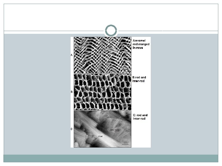
Enamel Structure: �The enamel is composed of 1 -enamel rods or prisms, 2 -rod sheaths, and in some regions a 3 - cementing interprismatic substance �Rods: �The enamel prisms are cylindrical, in longitudinal section, therefore the term rods. � The number of enamel rods has been estimated as ranging from 5 million in the lower lateral incisors to 12 million in the upper first molars. � From the dentino- enamel junction the rods run tortuous courses outward to the surface of the tooth.
�The length of most rods is greater than the thickness of the enamel because of the oblique direction and the wavy course of the rods. � The rods located in the cusps, the thickest part of the enamel, are longer than those at the cervical areas of the teeth. �The diameter of the rods averages 4 μm, but this measurement necessarily varies, since the outer surface of the enamel is greater than the dentinal surface where the rods originate. It is claimed that the diameter of the rods increases from the dentinoenamel junction toward the surface of the enamel at a ratio of about 1: 2.
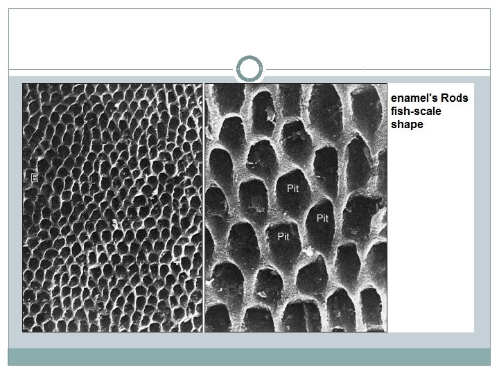
�In cross-sections of human enamel, many rods resemble fish scales. keyhole- or paddle- shaped prism in human enamel When cut longitudinally sections pass through the “heads” or “bodies” of one row of rods and the “tails” of an adjacent row. The “bodies” of the rods are nearer occlusal and incisal surfaces, whereas the “tails” point cervically.
�Direction of rods �Generally the rods are oriented at right angles to the dentin surface. In the cervical and central parts of the crown of a deciduous tooth they are approximately horizontal. Near the incisal edge or tip of the cusps they change gradually to an increasingly oblique direction until they are almost vertical in the region of the edge or tip of the cusps.
�Gnarled enamel: �Optical appearance of enamel Presents at the tip of the cusps due to twisted E. rods around each other in complex arrangement. They arranged in radial manner in horizontal plane.
Enamel Striations: � 1 -Cross striations : Each enamel rod is built up of segments separated by dark lines that give it a striated appearance. These cross striations demarcate rod segments and become more visible by the action of mild acids. The rods are segmented because the enamel matrix is formed in a rhythmic manner. �
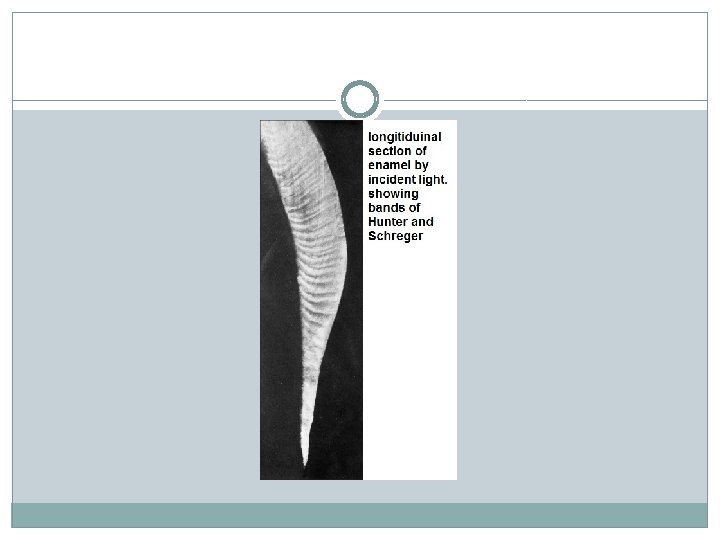
2 -Hunter–Schreger bands: �The more or less regular change in the direction of rods may be regarded as a functional adaptation, minimizing the risk of cleavage in the axial direction under the influence of occlusal masticatory forces. The change in the direction of rods is responsible for the appearance of an optical phenomenon the Hunter–Schreger bands. These are alternating dark and light strips of varying widths that can best be seen in a longitudinal ground section under oblique reflected light. They originate at the dentinoenamel border and pass outward, ending at some distance from the outer enamel surface.
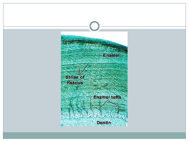
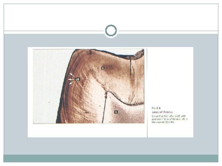
� 3 -Incremental lines of Retzius �The incremental lines of Retzius appear as brownish bands in ground sections of the enamel. They illustrate the incremental pattern of the enamel, that is, the successive apposition of layers of enamel during formation of the crown. In longitudinal sections they surround the tip of the dentin. In the cervical parts of the crown they run obliquely. In transverse sections of a tooth the incremental lines of Retzius appear as concentric circles. They may be compared to the growth rings in the cross-section of a tree.
�The term “incremental lines” reflect variations in structure and mineralization, either hypomineralization or hypermineralization, that occur during growth of the enamel. The mean daily rate of enamel formation of about 3. 5 microns increases from inner to outer enamel and decreases from incisal/cuspal region to cervical enamel. The evenly spaced striae of Retzius was shown to represent a 6– 11 day rhythm in enamel formation while other Retzius lines are suggested to be due to stress.
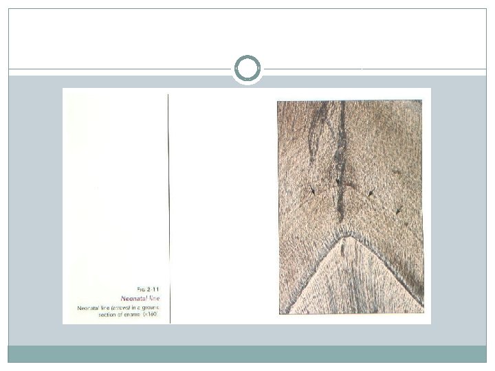
� 4 -The neonatal line or neonatal ring: The boundary between the two portions of enamel in the deciduous teeth is marked by an accentuated incremental line of Retzius. It appears to be the result of the abrupt change in the environment and nutrition of the newborn infant. The enamel of the deciduous teeth develops partly before and partly after birth. The prenatal enamel usually is better developed than the postnatal enamel. This is explained by the fact that the fetus develops in a wellprotected environment with an adequate supply of all the essential materials, even at the expense of the mother. Because of the undisturbed and even development of the enamel prior to birth.
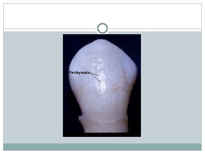
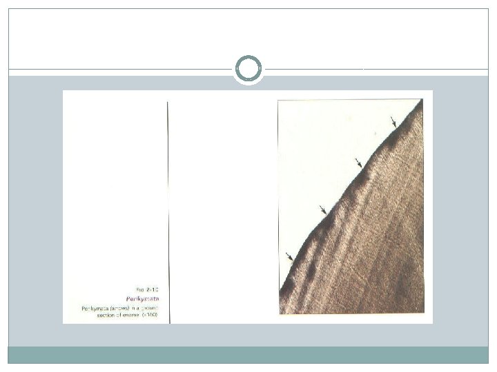
Surface structures: � 1 - Prismless enamel: A relatively structureless layer of enamel, approximately has been described in 70% of permanent teeth and all deciduous teeth. It is found least often over the cusp tips and most commonly toward the cervical areas of the enamel surface. � 2 -Perikymata is transverse, wave-like grooves, believed to be the external manifestations of the striae of Retzius. They are continuous around a tooth and usually lie parallel to each other and to the cementoenamel junction. The enamel rod ends are concave and vary in depth and shape. They are shallowest in the cervical regions of surfaces and deepest near the incisal or occlusal edges
3 - Cracks : �the narrow, fissure-like structures that are seen on almost all surfaces they are actually the outer edges of lamellae. They extend for varying distances along the surface, at right angles to the dentinoenamel junction, from which they originate. Most of them are less than a millimeter in length, but some are longer, and a few reach the occlusal or incisal edge of a surface. They are fairly evenly spaced, but long lamellae appear thicker than short ones. �
5 -Enamel cuticle: �A delicate membrane called Nasmyth’s membrane, after its first investigator, or the primary enamel cuticle covers the entire crown of the newly erupted tooth but is probably soon removed by mastication. Electron microscopic studies have indicated that this membrane is a typical basal lamina found beneath most epithelia secreted by the ameloblasts when enamel formation is completed. The function of enamel cuticle is to protect the surface of enamel from the resorptive activity of the adjacent vascular tissue prior to the eruption of the teeth
� 6 - Pellicle: erupted enamel is normally covered by a precipitate of salivary proteins. This pellicle reforms within hours after an enamel surface is mechanically cleaned. Within a day or two after the pellicle has formed, it becomes colonized by microorganisms to form a bacterial plaque
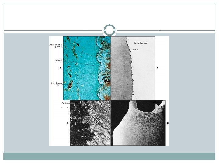
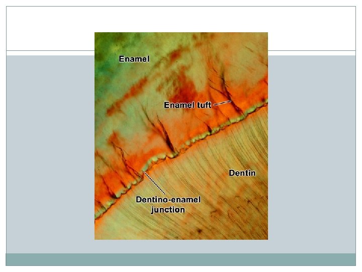
�Dentinoenamel junction �The surface of the dentin at the dentinoenamel junctions is pitted. Into the shallow depressions of the dentin fit rounded projections of the enamel. This relation assures the firm hold of the enamel cap on the dentin therefore, the DEJ appears not as a straight but as a scalloped line. The DEJ, which is a series of ridges is more pronounced in the occlusal area, where masticatory stresses are greater.
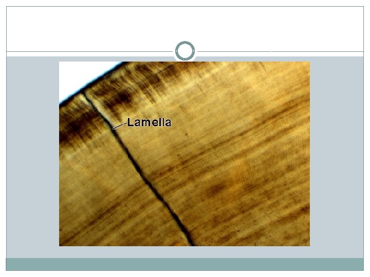
STRUCTURES RELATED TO DEJ: � 1 -Enamel lamellae: �Enamel lamellae are thin, leaf-like structures that extend from the enamel surface toward the DEJ. They may extend to, and sometimes penetrate into, the dentin. They consist of organic material, with but little mineral content makes possible the distinction between cracks and enamel lamellae.
Three types of lamellae can thus be differentiated: � 1 -type A, lamellae composed of poorly calcified rod segments restricted to the enamel � 2 -type B, lamellae consisting of degenerated cells; � 3 - type C more common. � types B and C may reach into the dentin. Lamellae arising in erupted teeth where the cracks are filled with organic matter, presumably originating from saliva. It has been suggested that enamel lamellae may be a site of weakness in a tooth and may form a road of entry for bacteria that initiate caries may act as pathways for caries producing bacteria.
2 -Enamel tufts �Enamel tufts arise at the DEJ and reach into the enamel to about one 13 -15 of its thickness. They resemble tufts of grass when viewed in ground sections. Tufts consist of hypocalcified enamel rods and interprismatic substance. Like the lamellae, they extend in the direction of the long axis of the crown. Therefore they are seen abundantly in horizontal section.
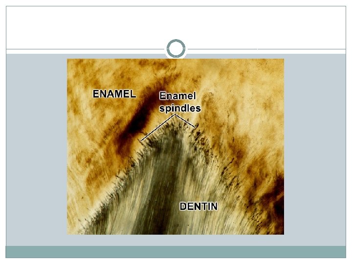
3 - Odontoblast processes and enamel spindles �: �Occasionally odontoblast processes pass across the dentinoenamel junction into the enamel. Since many are thickened at their end they have been termed enamel spindles. They seem to originate from processes of odontoblasts that extended into the enamel epithelium before hard substances were formed at right angles to the surface of the dentin. The structure of the spindles were similar to enamel tufts and that both of them were hypomineralized or partially mineralized structures.
Age changes: �Attrition or wear of the occlusal surfaces and proximal contact points as a result of mastication. This is evidenced by a loss of vertical dimension of the crown and by a flattening of the proximal contour. Facial and lingual surfaces lose their structure much more rapidly than do proximal surfaces, and anterior teeth lose their structure more rapidly than do posterior teeth. �Localized increases of certain elements such as nitrogen and fluorine, however, have been found in the superficial enamel layers of older teeth. This suggests a continuous uptake, probably from the oral environment, during aging.
�The teeth may become darker, and their resistance to decay may be increased. �Reduced permeability of older teeth to fluids. The decrease in permeability of enamel due to age is due to increase in the size of the crystal. The crystal size increases due to ions acquired by it from the oral fluids. The increase in size of the crystal decreases the pores between them causing a reduction in permeability.
CLINICAL CONSIDERATIONS �FLUORIDTION: �Is incorporated of fluoride ions into the hydroxyapatite crystal; the crystal becomes more resistant to acid dissolution. This reaction partly affects the reduction of dental caries which is an acid dissolution produced by certain bacteria. �Mottled teeth: are darkly stained teeth due to excessive fluoridation during tooth formation. Ameloblasts are sensitive to the fluorideion so the amount of fluoride should be controlled during tooth development.
ACID ETCHING: �is an important dental technique used for conditioning the enamel to adhere of fissure sealant, bonding of composite, cementing of orthodontic brackets to the tooth surface. �The material used mainly is 37% orthophosphoric acid that acts into 2 steps: � 1 -removing dental plaque and other debris from enamel surface. � 2 -exposing thin layer of enamel with increased porosity through selective dissolution of crystals; which provides a better bonding surface for the restorative and adhesive materials.