EMPYEMA LUNG ABSCESS SURGICAL LUNG INFECTIONS Dr Mahmoud
 Dr. Mahmoud ABU-ABEELEH The University of JORDAN. Faculty")
EMPYEMA LUNG ABSCESS (SURGICAL LUNG INFECTIONS) Dr. Mahmoud ABU-ABEELEH The University of JORDAN. Faculty of Medicine 4 -10 -2016

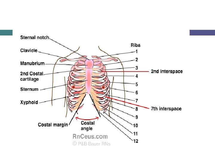
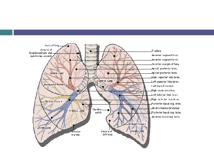
ANATOMY. Empyema Definition classification causes diagnosis management indications for surgery Lung abscesses definition causes clinical presentations diagnosis management

EMPYEMA Definition: Invading of the pleural space with bacteria which result in accumulation of pus. Classification : (American Thoracic Society) Stage 1 : Exudative , with swelling of the pleural membranes as a result of ↑ permeability of swollen membranes(Uncomplicated Acute stage) Stage 2: Fibropurulent(Transitional)with heavy fibrin deposits. Stage 3: Organizing or Chronic phase. With ingrowth of fiboblast and deposition of
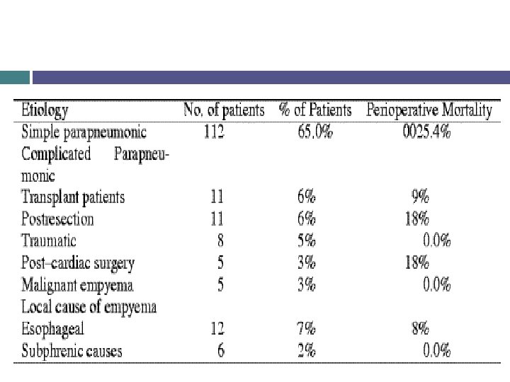
the most common Post trauma. Post surgery(esophageal or pulmonary)")
ETIOLOGY: PARAPNEUMONIC(secodary to a pneumonia)the most common Post trauma. Post surgery(esophageal or pulmonary) Subphrenic Abscess

Bacteriology : Before ABO 10% of Pts survived pneumonia developed EMPYEMA(Streptococci & Pneumococci are the most freuquent) After ABO the incidence as well as the mortality↓. Staph become more prevelent , 90% of empyema in children.

Incidence of Empyema according to Bacteria causing pneumonia EFFUSION EMPYEMA 50% <5% 70% 80% 40% 20% G –VE 50% 90% Anaerobes 35% 90% Aerobic G +VE Strep pneumonia Stap Aureus CHILDREN ADULT

Clinical presentation Pleuritic chest pain , fever, S. O. B , Tacycardia AS Pneumonia. If prolonged symptoms SUSPECT EMPYEMA. Anaerobic : indolent § P/E: Toxic anxious § pt, tacycardia, tacypnea, restricted chest wall excursion, ↓ air entry, dullness on percussion. Chronic pt Clubbing, Anaemia, wt loss.

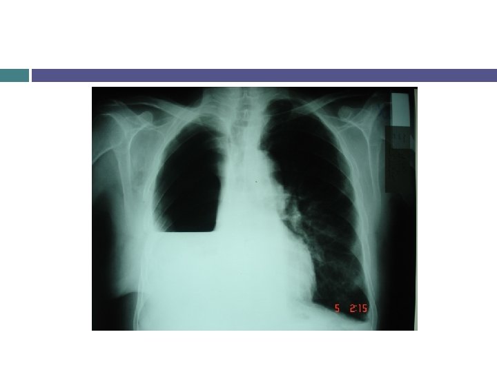
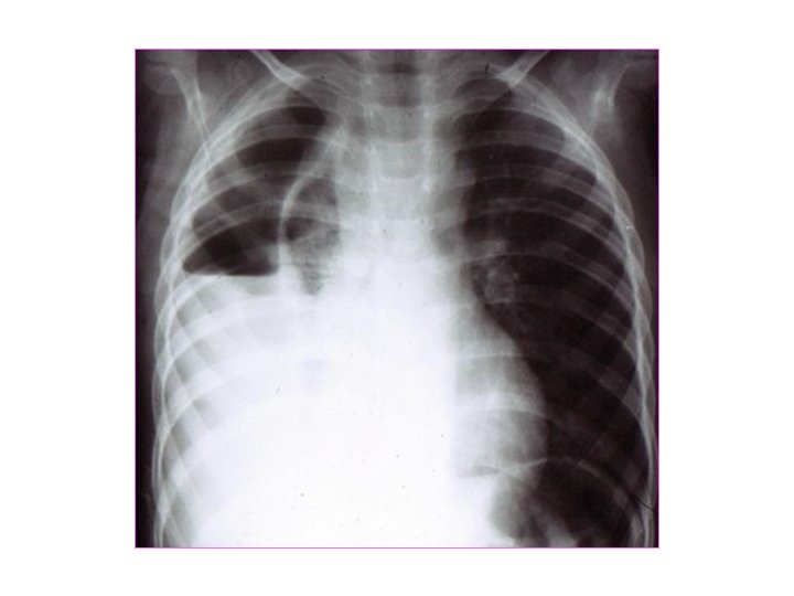
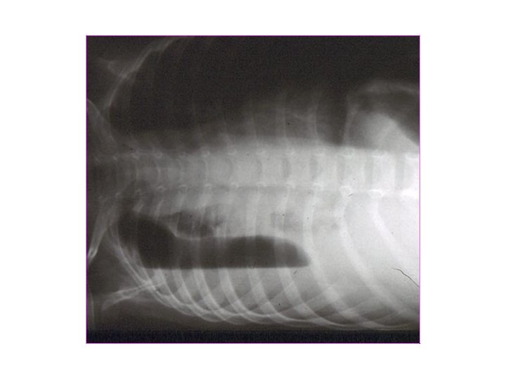
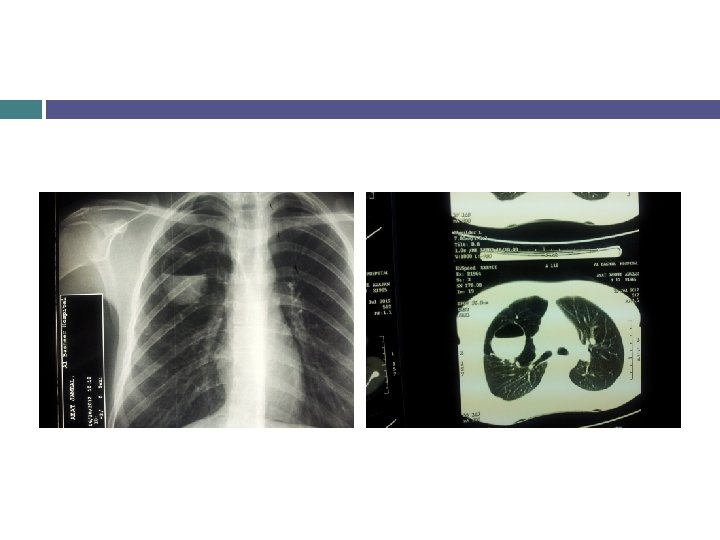
DIAGNOSIS: CBC: ↑ WBCwith shift to left, ↑ CRP ESR. CXR: Effusion, ↑thickness of the pleura, Air fluid level. THORACOCENTESIS: Empyema fluid PH <7. 0 Glucose < 40 mg/d. L LDH > 1000 IU/d. L Positive Gram stain Positive culture (50%) Specific gravity > 1. 018 WBC > 500 cells/mm 3 Protein > 2. 5 g/d. L
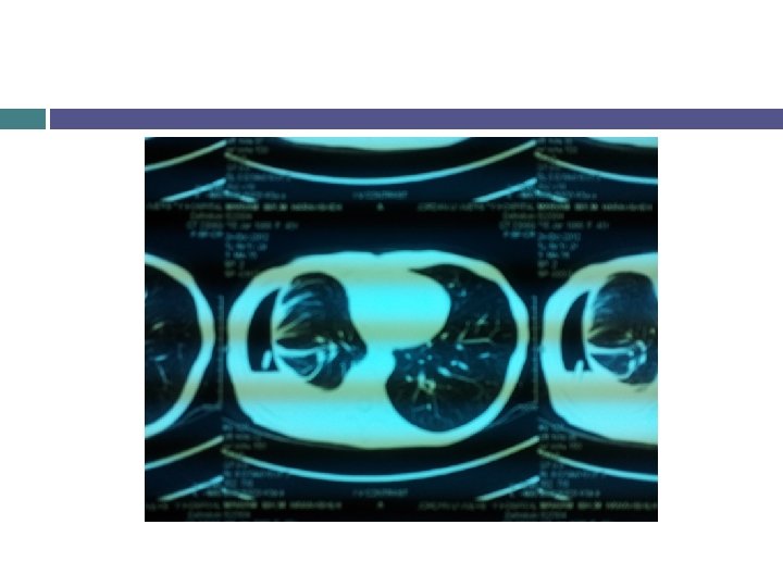
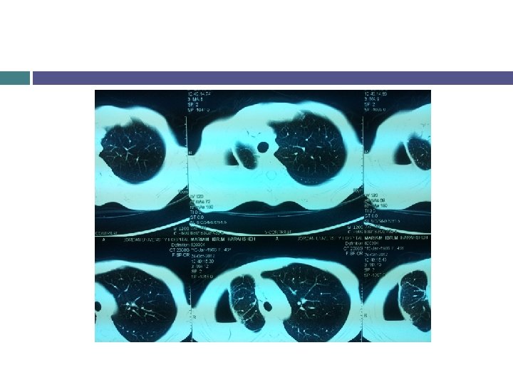

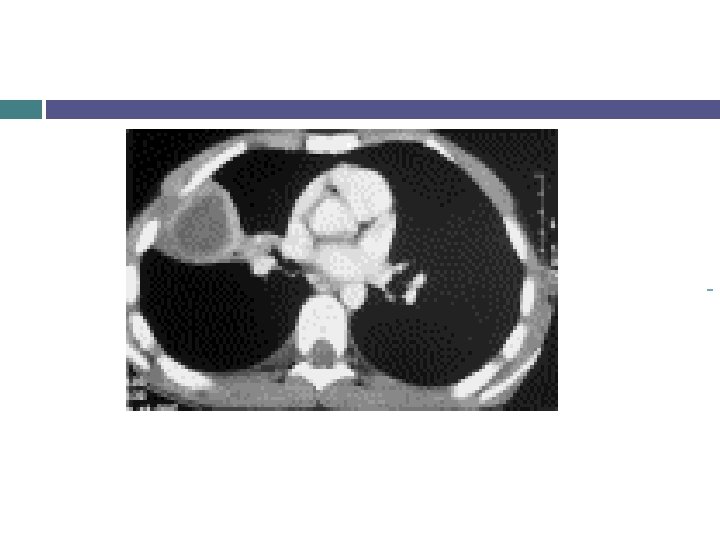
ü ü ü CT Scan: Localize collection. Identify the underlying parenchymal disease, . Distinguish it from lung abscess. Fluid density, loculations. Therapeutic: CT-guided aspiration.

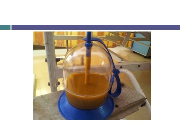
Managment 1. 2. 3. Antibiotics. 3 rd generation cephalosporine, clindamycin till the result of G stain , C&S. Evacuation of pus from the pleural space. In stage 1 thoracocentesis, other wise Chest tube insertion Obliteration of the empyema cavity.

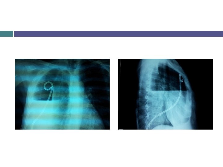
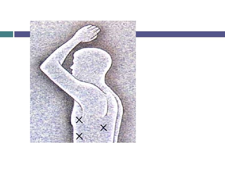
Chest Tube Insertion Procedure local anaesthesia Scrubing & draping An incision is made along the upper border of the rib By a curved clamp the track is developed by blunt dissection splitting the fibres. A track developed with the operator's finger The clamp is angled over the rib & dissection continued until pleura is entered.
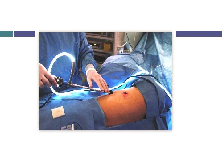
 chest tube is passed")
Chest Tube Insertion Procedure A large-bore (32 or 36 F) chest tube is passed into the pleural cavity. The tube is connected to an underwater seal and sutured / secured in place. a U-stitch A chest X-ray is taken to confirm placement & position.

Clinical improvement within 48 hrs. ≥ 80% of stage 1 managed conservatively. Stage 3 80% require thoracotomy. Intrapleural Fibrinolytic therapy; STK or Urokinase to break loculations produced by membranes composed of fibrin. V. A. T. S. THORACOTOMY: decortication.

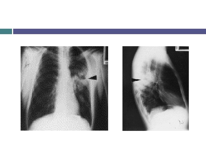
LUNG ABSCESS Definition: Sub acute pulmonary infection in which the chest X ray shows cavity within the lung parenchyma. ü ü ü § Before ABO era , high mortality, . ACUTE &CHRONIC: if duration< 6 weeks. PRIMARY &SECONDARY PRIMARY: 1. 2. Aspiration: The most frequent. Post-Pneumonic

Secondary: 1. 2. 3. 4. 5. § § Obstructing carcinoma. COPD Metastatic from extrathoracic source septisemia. F. B aspiration. Pulmonary infarctions. The individuals with high risk: ALCOHOL ABUSE, hx of Aspiration, Old TB, Epilepsy, drug abuse, COPD. In endemic areas TB: 20% of lung

BACTERIOLOGY: ANAEROBES: 75 -80% § § • Bacteroid fragilis. Fusibacterium bacilli. Peptostreptococci. Provetella. AEROBIC: § § Kleibsiella &Pseudomonus: IN obstructive infections &Nasocomial. Staph. Auereus. S. pneumonia H. influenza.

DIAGNOSIS: A. B. C. D. Symptoms: Fever intermittent &night sweats chills. Purulent Foul-smelling sputum is highly suggestive. Hx of Aspiration, . Sepsis→Respiratory failure. Signs: Tachpnea, consolidation, local chest wall tenderness. CXR: Pneumonitis pattern early→Air-fluid level.

SPUTUM analysis&culture Aerobic, anaerobic, fungal &TB. CT-scan. Fibrooptic Bronchoscopy: is mandatory Take samples for culture. Ø R/O endobronchial tumour or obstruction. Ø To assess if can be drained internally. Ø

SITES: § § • • Superior segment of Rt lower lobe. Lat. Part of Post. Segment of R. U. L. Superior segment of L. L. L. D. Dx of cavitary lung lesion: 1. 2. 3. 4. Cavitary carcinoma. T. B or fungal abscess. Pyogenic lung abscess. Empyema with bronchopleural fistula.

Managment: Principles of therapy: Identify the organism→proper ABO therapy for 6 -8 wks. Drainage: I. III. Chest physiotherapy. Bronchoscopy=internal darainaige or indwelling transbronchial catheter drainage. Percutaneous cath. Drainage. SURGERY. 80 -90%of Lung abscess respond to medical tt. Flagyl or Clindamycinfor anaerobes. Gentamicin or 3 rd generation cephalosporines for aerobes.
 2) 3) If remain septic. Failure to")
External drainage: I. III. IV. V. 1) 2) 3) If remain septic. Failure to wean from mechanical ventilation. Soiling of the contralateral lung. Abscess cavity >4 cm& under tension on CXR. ↑ size while on ABO. Chest tube thoracostom. CT-guided catheter. Open pneumonostomy =MONALDI procedure. 30% of Pt will need definitive surgery. Clinical improvement within 48 hrs.

INDICATIONS FOR SURGERY: 1. Acute : for complications 2. Bronchopleural fistula. Empyema. Hemoptysis. (Massive) Chronic =Definitive. Persistant symtoms despite long term ABO therapy. Suspecius of carcinoma. Complications: Empyema, bronchopleural fistula. Persistant cavity >6 cm after ABO therapy.

Lobectomy is the standard procedure. Mortality: 2. 5% after community acquired pneumonia. 66% with Nasocomial infections. Underlying diseases. Size of the abscess >6 cm. Organism: Pseudomonus & G –ve the highest.

QUESTIONS SUMMARY
- Slides: 39