Emergancy Pediatric and adolescent Gynecology Revital Arbel ShaareZedek

Emergancy Pediatric and adolescent Gynecology Revital Arbel Shaare-Zedek MC Jerusalem

Minor labia fusion

Minor labia fusion

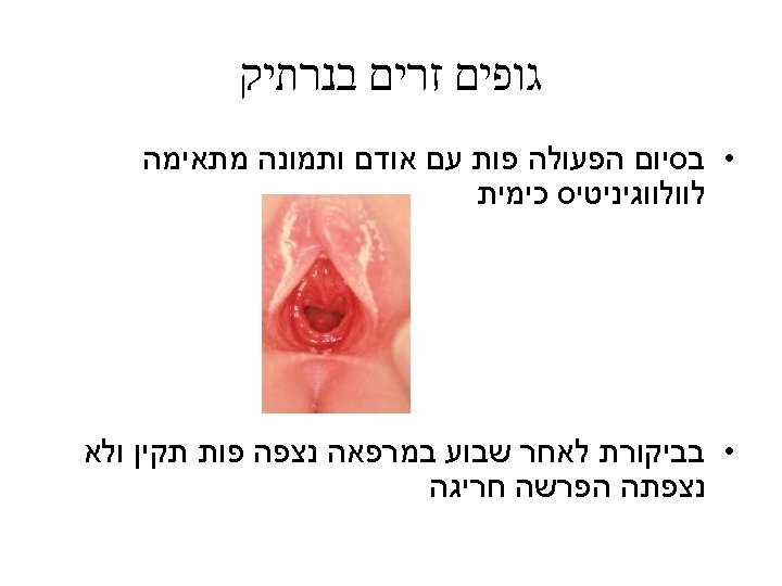
Minor labia fusion • Most girls with labial fusion are asymptomatic. The condition is usually discovered incidentally by a physician during a routine examination or by the mother while she is bathing the child. • Labial fusion may predispose the child to asymptomatic bacteriuria or to urinary tract infection. • Occasionally, the labial fusion causes urinary outflow obstruction with resultant bladder distention and hydronephrosis.

Approach • Treatment consists of precise application of a topical estrogen cream to the fused area twice a day until lysis of the adhesion is complete. Most cases resolve within 2 to 3 months. • As the labia minora separate, petroleum jelly should be applied to the edges to prevent readhesion • Manual seperation of labial fusion is an option

Vaginal discharge

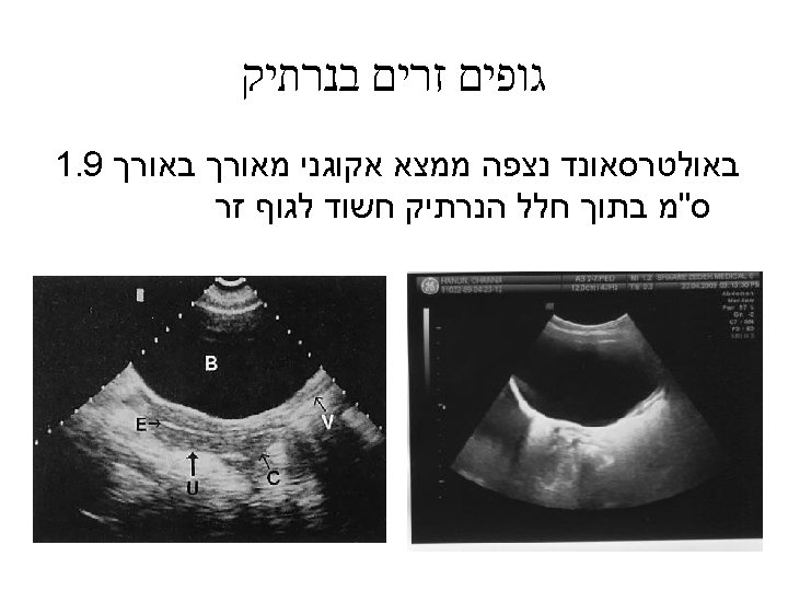
Vaginal bleeding in a pre-pubertal girl

Genital trauma
 • Sexual")
Mechanism of injury • Straddle injury • Accidental penetration (including foreign body) • Sexual abuse • Sudden abduction of lower extremities • Pelvic fractures

Straddle injury • Generally minor injuries • Involves mons, clitoris, urethra, Minor and Major labia • Hymeneal and vaginal injuries extremely rare

Evaluation • External inspection • Vaginal lavage to detect blood • Examination under anesthesia: bimanual and endoscopic

Penetration-I

Penetration-II • • Dimension of object Patient weight Force and direction Final resting place • Complex injuries

Abduction Injuries • • • Vaginal orifice tear Posterior vaginal tear posterior fornix tear Recto-vaginal septum involvement R/O Peritoneal involvement • Examination under anesthesia is required to fully evaluate and treat the tear

Fracture/Perineal injuries • Less common in girls • Associated Urethral injury • Vaginal tear can be a cause for severe Hemorrhage

AAST organ injury scaling of vulva and vagina Vulva Injury I Contusion/hematoma II Superficial laceration (skin only) III Deep laceration (fat/muscle) IV Avulsion (skin/fat/muscle) V Injury into adjacent organs (Anus/Rectum/Urethra/Bladder) Vagina injury I Contusion/hematoma II Superficial laceration (skin only) III Deep laceration (adjacent fat/muscle) IV Laceration-complex into cervix V Injury into adjacent organs (Anus/Rectum/Urethra/Bladder)

Perineal Injury If Hemodynamically Stable Bimanual and Endoscopic Exam Under Anesthesia Urethra Anorectal injury Hematuria Microscopic CT Upper. UT Grade I Urethral Catheter Vagina and perineal surface Macroscopic Urethrocysto gram Grade IIIV Urethral Catheter Suprapubic Catheter Primary Repair Hematoma Non Expanding Laceration Grade IIIIV Grade I-II Grade IIIIV Local closure Explore and ligate vessels Observe Preoperative ABX Closure of laceration Graft for avulsions Preoperative antibiotics Peroximal diverting colostomy Presacral drainage Washout of defunctionalized bowel Closure of laceration

Foreign body in the vagina

Etiology • Self administrated • Accidental • Abuse


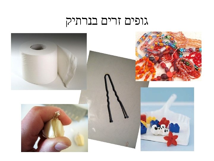

Suspicion of foreign body in vagina • Purulent or sanguineous discharge • Vulvar irritation • Persistent or recurrent vulvitis • Frequently the child denies the possibility of foreign body insersion

Evaluation • Direct visualization • Baby vaginal speculum • Rectal examination Lubrication Hard object felt anterior to the vagina

Ultrasound

X ray

Vaginal lavage • Usually Rectal examination, Ultrasonogrhphy and Radiographic studies fail detect most foreign bodies • Most common foreign objects recovered in pre-pubertal girls is wads of toilet paper • Initial efforts of removal include vaginal lavage using a 10 -12 F catheter

Vaginoscopy
")
Complications • • Genital puritus Low grade fever Abdominal pain Peritonitis (retrograde)

Long term complications • Foreign body embedded in vaginal wall • Inflammation produces a papillary growth of mucosa • Foreign body migration

Primary Amenorrhea emergancy Evaluation and Treatment
 Chemical control Norepiniphrine (+) Endorphines (-) Hypothalamus")
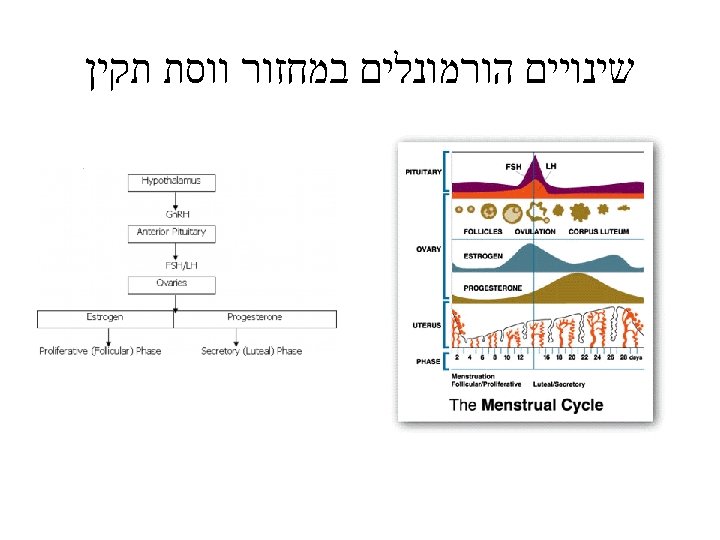
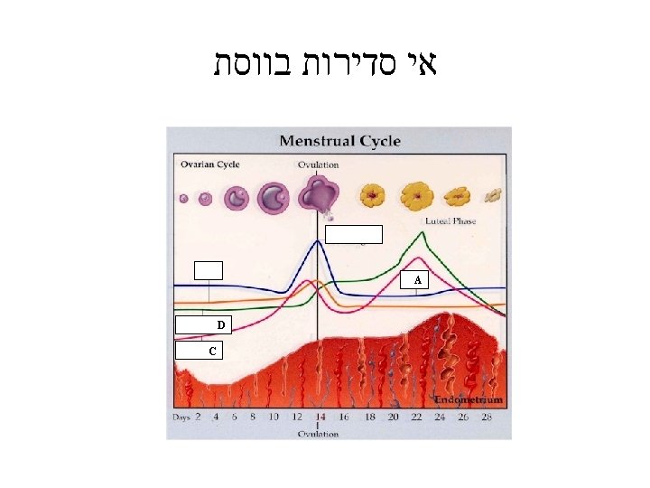
CNS-Hypothalamus-Pituitary Ovary-uterus Interaction Neural control Dopamine (-) Chemical control Norepiniphrine (+) Endorphines (-) Hypothalamus ± Gn-RH Ant. pituitary ? – FSH, LH Estrogen Ovaries Uterus Menses Progesterone

AMENORRHOEA AN APPROACH FOR DIAGNOSIS • HISTORY • PHYSICAL EXAMINATION • ULTRASOUND EXAMINATION Exclude Pregnancy Exclude Cryptomenorrhea

Cryptomenorrhea Outflow obstruction to menstrual blood - Imperforate hymen - Transverse Vaginal septum with functioning uterus - Isolated Vaginal agenesis with functioning uterus - Isolated Cervical agenesis with functioning uterus - Intermittent abdominal pain - Possible difficulty with micturition - Possible lower abdominal swelling Bulging bluish membrane at the introitus or absentvagina (only dimple)

Embryology

Imperforate hymen

Transverse vaginal septum presents as a complete membrane across the vagina usually beyond the hymenal ring

Transverse vaginal septa • Transverse vaginal septa occur at several locations and may be complete or incomplete. • Usually about 2 cm thick and located near the junction of the upper third and lower two thirds of the vagina; however, septa may be present in the middle or lower third of the vagina • Vaginal septa presumably result from failure of urogenital sinus derivatives and the müllerian duct derivatives to fuse or canalize.

Transverse vaginal septum

Resection of a transverse septum

Vaginal Agenesis • Absence of a portion of the vagina but presence of a normal uterus (vaginal atresia). • Absence of most of the vagina and all or almost all of the uterus (as part of müllerian aplasia) • The two conditions are embryologically, anatomically and clinically distinct

vaginal Agenesis • In vaginal atresia the urogenital sinus fails to contribute the caudal portion of the vagina. The lower fifth to third of the vagina is replaced by 2 to 3 cm of fibrous tissue, above which lie a well-differentiated upper vagina, cervix, uterine corpus, and fallopian tubes

Distal vaginal Agenesis with hematocolpus and hemaometria

Distal vaginal agenesis misdiagnosed as Imperforated Hymen • Presented as recurrent hematocolpus

Distal vaginoplasty

Absence or Atresia of the Uterine Cervix • Isolated congenital cervical atresia with normal vaginal development. • Congenital cervical atresia with complete vaginal agenesis.

Uterus didelphis with longitudinal oblique septum • Menstruation present with hematocolpus


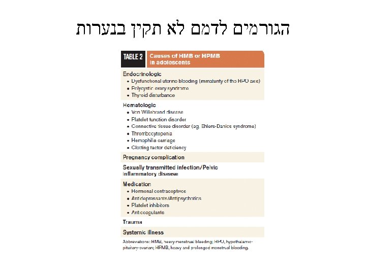
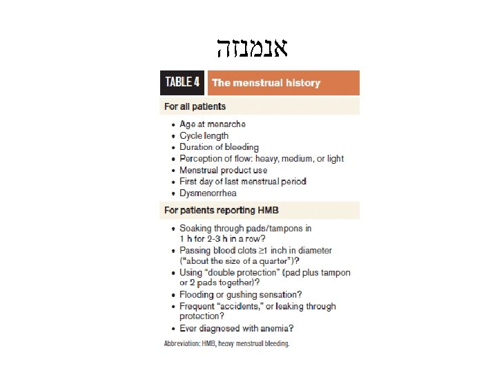
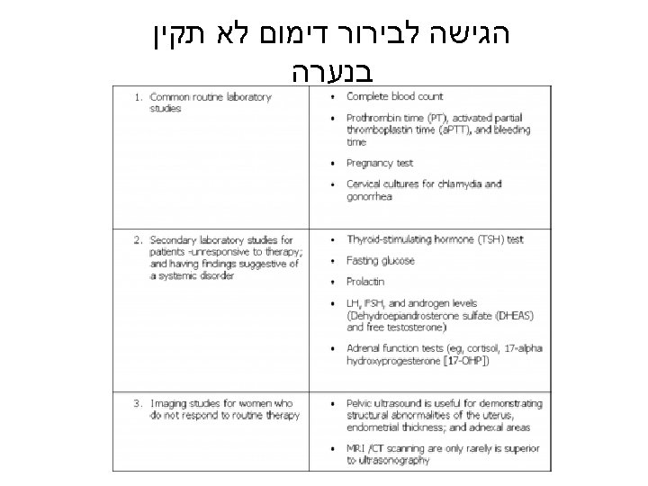
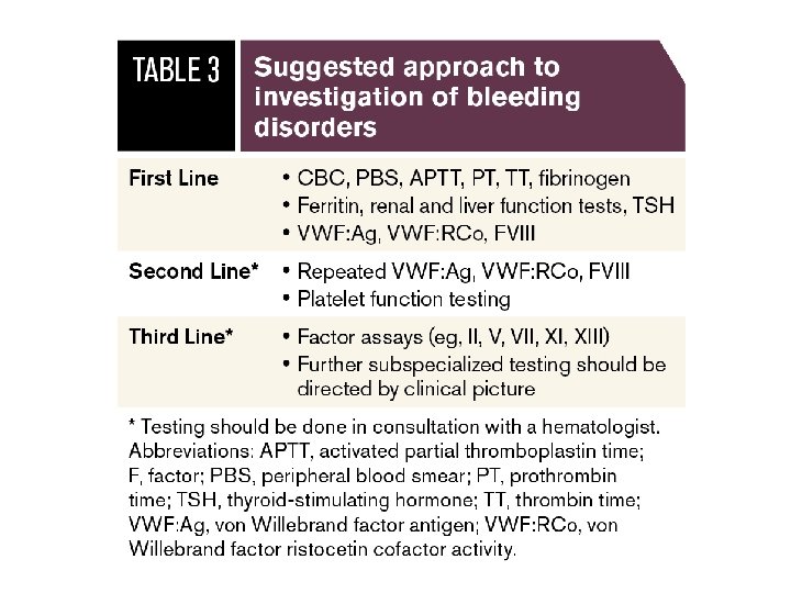
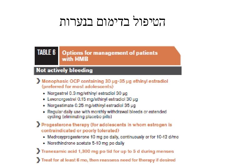
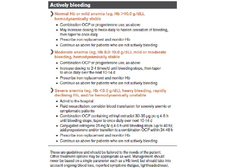
Vaginal bleeding in a pre-pubertal girl


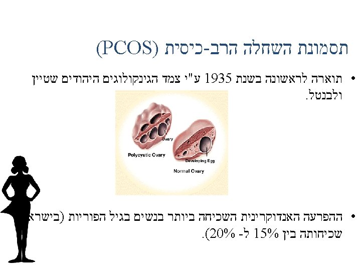
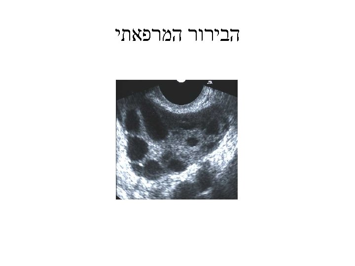
Polycystic ovary syndrome


- Slides: 88