
EMC I A STUDY UNIT 2 3 Basic
EMC I A STUDY UNIT 2 & 3 Basic Airway Management, Oxygenation, Ventilation & Pulse Oximetry
LEARNING OBJECTIVES 1. 2. 3. 4. 5. 6. 7. 8. Understand the physiology of oxygen transport State the normal values for Pa. O 2 & Pa. CO 2. State the equations to calculate Tidal Volume (TV) & Minute Volume (MV). Discuss the different types of Hypoxia Describe the principles of operation of a pulse oximeter & identify physiological & technical factors that can influence the accuracy of readings Explain the concepts of oxygenation & ventilation. Establish & maintain a patient’s airway using appropriate equipment & basic airway techniques. Effectively oxygenate & ventilate a patient using appropriate equipment & BLS techniques.
Our Two Main Goals? 1. 2. Blood goes round & round Air goes in & out Any deviation is………. .
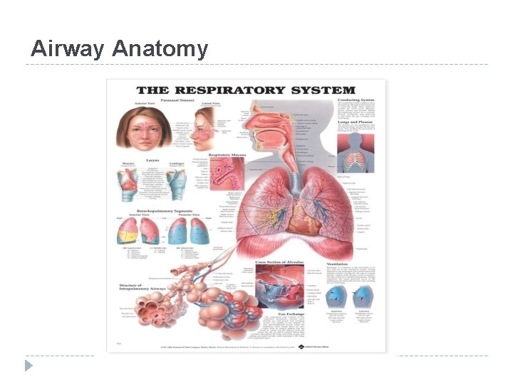
Airway Anatomy
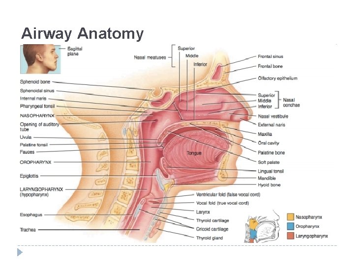
Airway Anatomy
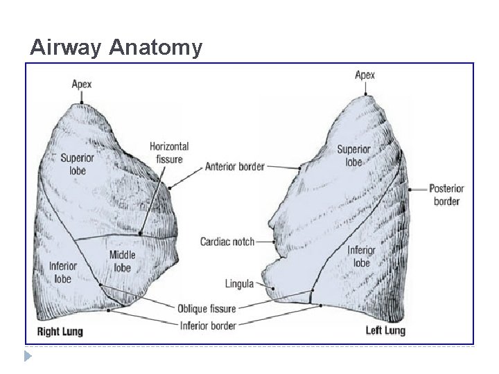
Airway Anatomy
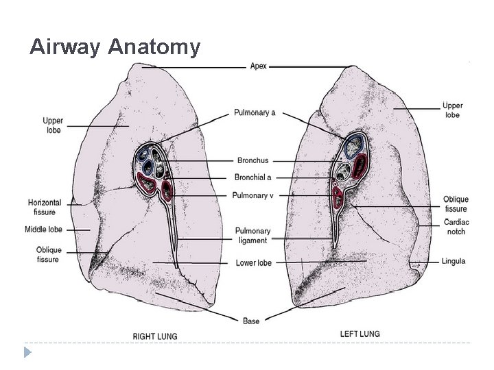
Airway Anatomy
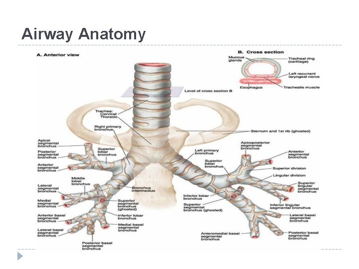
Airway Anatomy
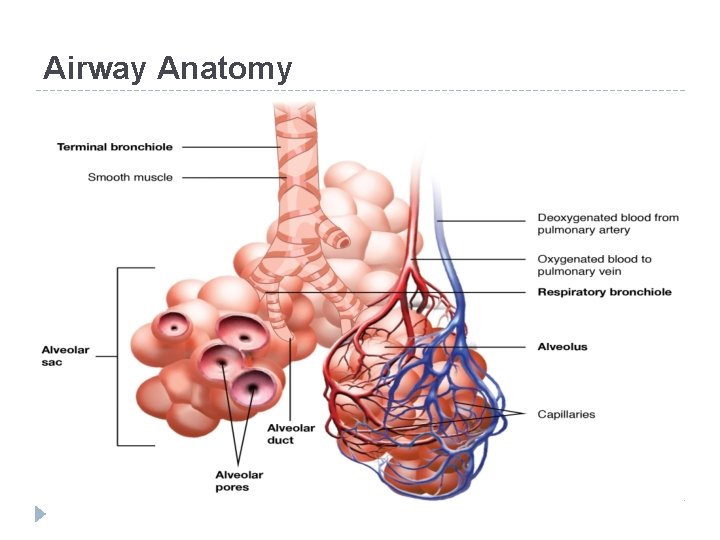
Airway Anatomy
Airway Assessment � Inspection – DCAP BTLS � Palpitation � Auscultation � Percussion – All lung fields, note sounds (or lack there of)
Airway Assessment Assessing Airway & Breathing: O - Onset (Sudden or gradual? ) P - Provokes (What was the trigger? ) Q - Quality (What type of dyspnoea? ) R - Recurrence (Has it happened before, when, compare) S - Severity (How bad is it ? Scale 1 – 10) T - Timing (Continuous or intermittent? ) A - Associated symptoms and signs
Physiology Of Oxygen Transport Air = 21% Oxygen 78% Nitrogen 1% Other At sea level partial pressure of? 760 mm. Hg Therefor the partial pressure of oxygen when inspired is? 159 mm. Hg The addition of humidification causes? Drops the partial pressure of each of the inspired gases by 47 mm. Hg. Therefore the partial pressure of inspired O 2 is? 150 mm. Hg This gives you PAO 2 – partial pressure of oxygen in the alveoli.
Understand The Physiology Of Oxygen Transport The PAO 2 then drops to 100 mm. Hg. Why? Oxygen uptake by the pulmonary capillaries. Partial pressure of deoxygenated blood? 40 mm. Hg This allows for? Diffusion!!!!!
Physiology Of Oxygen Transport Oxygen is now transported in two forms: 1. Hemoglobin: - Chemically combines with 1. 31 ml of Oxygen(i. e. Oxygen Capacity) - Blood hemoglobin concentration = 15 g/d. L (150 g/L) - Meaning each litre of blood can carry 197 ml of O 2 150 g/L x 1. 31 ml/g = 197 m. L
Physiology Of Oxygen Transport 2. Dissolved in Plasma: - At pressure 0. 3 ml/d. L or 3 ml/L can dissolve in plasma. - Increases to 20 ml/L with 100% O 2. - Increased further under hyperbaric conditions. All this O 2 dissolved in the arterial blood (i. e. arterial partial pressure) is call? Pa. O 2
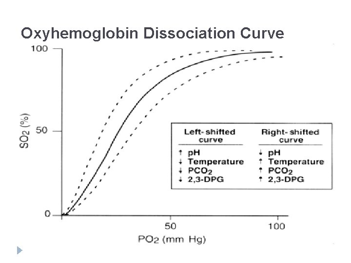
Oxyhemoglobin Dissociation Curve
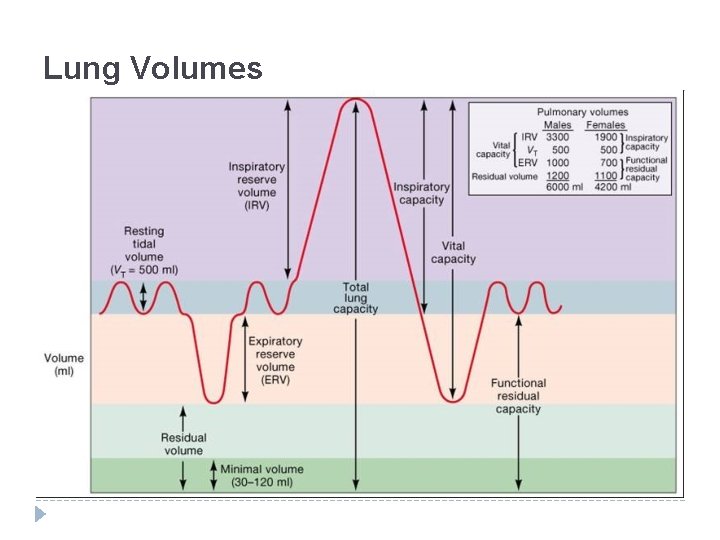
Lung Volumes
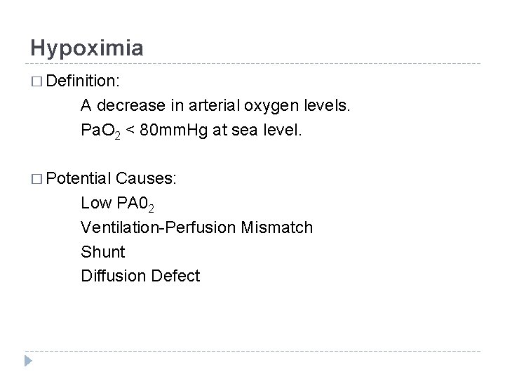
Hypoximia � Definition: A decrease in arterial oxygen levels. Pa. O 2 < 80 mm. Hg at sea level. � Potential Causes: Low PA 02 Ventilation-Perfusion Mismatch Shunt Diffusion Defect
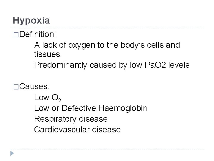
Hypoxia �Definition: A lack of oxygen to the body’s cells and tissues. Predominantly caused by low Pa. O 2 levels �Causes: Low O 2 Low or Defective Haemoglobin Respiratory disease Cardiovascular disease
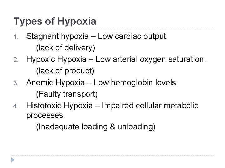
Types of Hypoxia 1. 2. 3. 4. Stagnant hypoxia – Low cardiac output. (lack of delivery) Hypoxic Hypoxia – Low arterial oxygen saturation. (lack of product) Anemic Hypoxia – Low hemoglobin levels (Faulty transport) Histotoxic Hypoxia – Impaired cellular metabolic processes. (Inadequate loading & unloading)
Pulse Oximetry What do they do? Noninvasively measure the percentage of Hb that is saturated with oxygen. Indirect means of detecting atrial oxygen saturations. Aim is to detect hypoxia.
Pulse Oximetry � So how does it work? Two principal assumptions: 1) There is a pulsatile signal generated by arterial blood flow. (allows monitor to differentiate between arterial and venous blood flow) 2) The fact that Hb and O 2 Hb absorb two different wave lengths. (and this will give us a ratio)
Pulse Oximetry The two LED lights on a pulse oximeter emit different wave lengths for light: 660 nm (red) 940 nm (infrared) O 2 Hb will have a greater affinity to infrared light Hb will have a greater affinity to red light
Pulse Oximetry Limitations: 1. Does not measure amount of Hb 2. Does not measure amount of blood flow 3. Oxyhaemoglobin dissociation curve 4. Only detects O 2 Hb and Hb
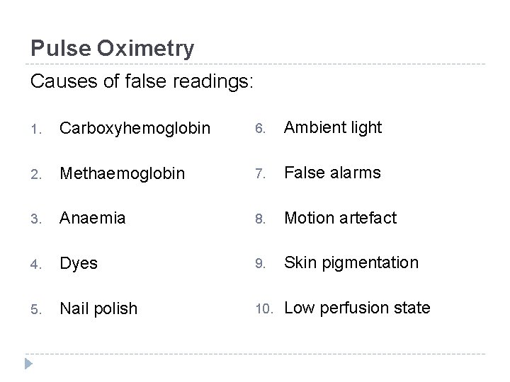
Pulse Oximetry Causes of false readings: 1. Carboxyhemoglobin 6. Ambient light 2. Methaemoglobin 7. False alarms 3. Anaemia 8. Motion artefact 4. Dyes 9. Skin pigmentation 5. Nail polish 10. Low perfusion state
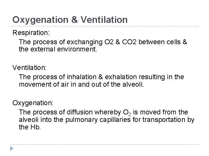
Oxygenation & Ventilation Respiration: The process of exchanging O 2 & CO 2 between cells & the external environment. Ventilation: The process of inhalation & exhalation resulting in the movement of air in and out of the alveoli. Oxygenation: The process of diffusion whereby O 2 is moved from the alveoli into the pulmonary capillaries for transportation by the Hb.
Supplemental Oxygen An effort to increase the patients Fi. O 2 and thereby increase their PAO 2. Who do we provide this higher Fi. O 2 to? Hypoxic patients
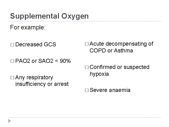
Supplemental Oxygen For example: � Decreased � PAO 2 � Any GCS or SAO 2 < 90% respiratory insufficiency or arrest � Acute decompensating of COPD or Asthma � Confirmed or suspected hypoxia � Severe anaemia
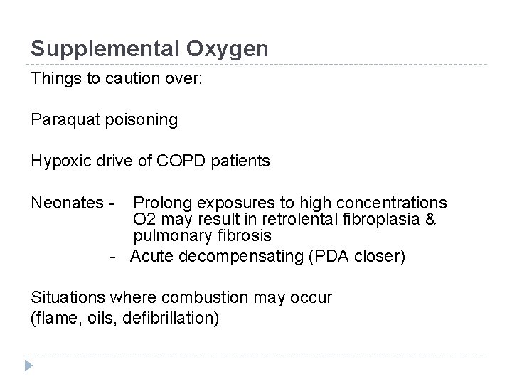
Supplemental Oxygen Things to caution over: Paraquat poisoning Hypoxic drive of COPD patients Neonates - Prolong exposures to high concentrations O 2 may result in retrolental fibroplasia & pulmonary fibrosis - Acute decompensating (PDA closer) Situations where combustion may occur (flame, oils, defibrillation)
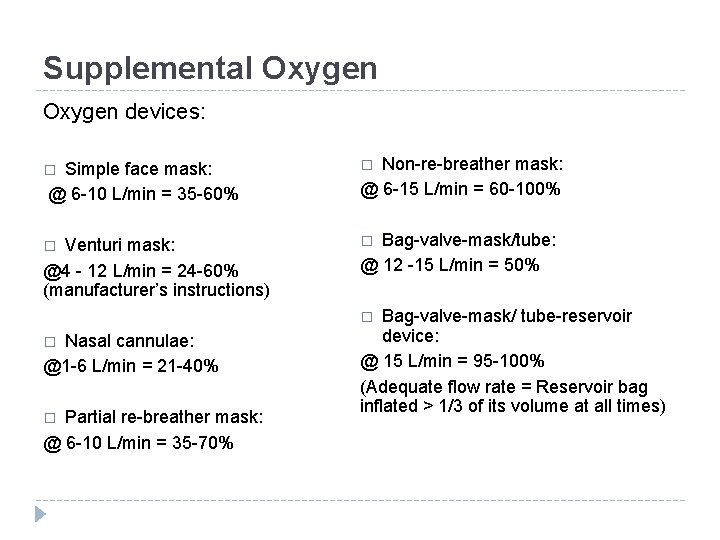
Supplemental Oxygen devices: Non-re-breather mask: @ 6 -15 L/min = 60 -100% � � Venturi mask: @4 - 12 L/min = 24 -60% (manufacturer’s instructions) � Simple face mask: @ 6 -10 L/min = 35 -60% � Bag-valve-mask/tube: @ 12 -15 L/min = 50% Bag-valve-mask/ tube-reservoir device: @ 15 L/min = 95 -100% (Adequate flow rate = Reservoir bag inflated > 1/3 of its volume at all times) � Nasal cannulae: @1 -6 L/min = 21 -40% � Partial re-breather mask: @ 6 -10 L/min = 35 -70% �
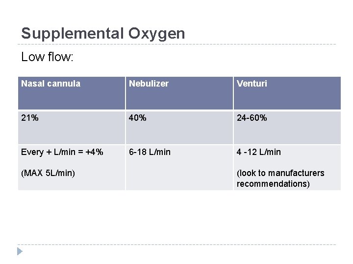
Supplemental Oxygen Low flow: Nasal cannula Nebulizer Venturi 21% 40% 24 -60% Every + L/min = +4% 6 -18 L/min 4 -12 L/min (MAX 5 L/min) (look to manufacturers recommendations)
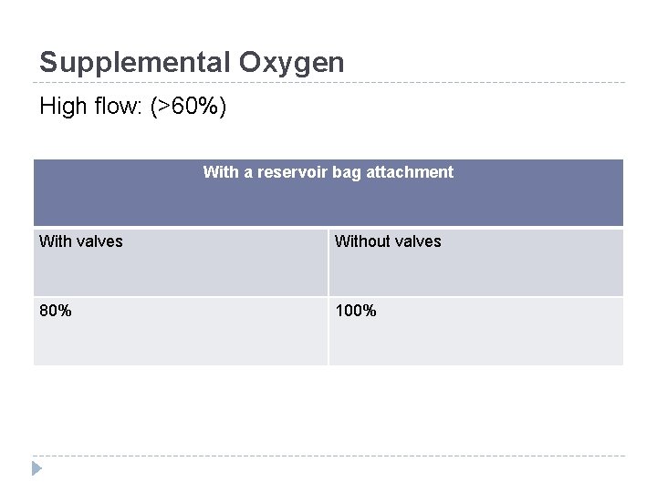
Supplemental Oxygen High flow: (>60%) With a reservoir bag attachment With valves Without valves 80% 100%
Ventilation � Two 1. 2. goals: Get O 2 in Get CO 2 out Can be achieved by patients internal drive or by our external efforts (PPV). PPV provided to?
Ventilation � Increased intrathoracic pressure � Increased ICP � Decreased venous return � Decreased cardiac output � Barotrauma � Over-ventilation causing Respiratory alkalosis CO 2 + H 2 O H 2 CO 3 H + HCO 3
Ventilation & Oxygenation �Problems 1) Ventilation-Perfusion Mismatch: Ratio of ventilation to blood flow (normal 0. 8) Decreased ventilation with constant blood flow = ↓ V/Q (↓PAO 2 → ↓Pa. O 2) Decreased blood flow with constant ventilation = ↑ V/Q (↑ PAO 2 But ↓Pa. O 2)
Ventilation & Oxygenation 2) Shunt: Occurs when alveoli are perfused but receive on ventilation. (extreme form of a V/Q mismatch) Causes: � Atelectasis � Lung consolidation � Fluid filled alveoli � Small airway closure
Ventilation & Oxygenation 3) Diffusion Defect: � Physical impairment of oxygen diffusion across the alveolar membrane. � Often due to a thickening of the alveolar membrane. � May also be due to the decreasing of global surface area of alveolar. � Commonly caused by pulmonary edema & pulmonary emphysema.
Predicting Difficult Bag-Mask Ventilation B - Beard & other mask seal issues O - Obesity O - Older (age >55 years) T - Toothless S - Sounds, Stridor, Snoring, Stiff lungs (wheezing, Rales)
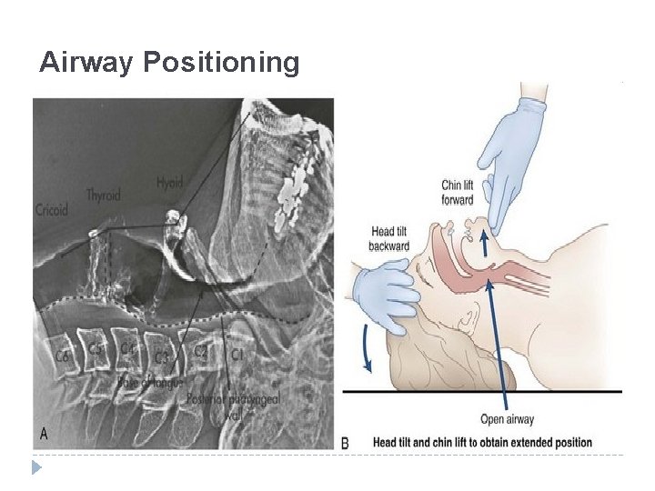
Airway Positioning
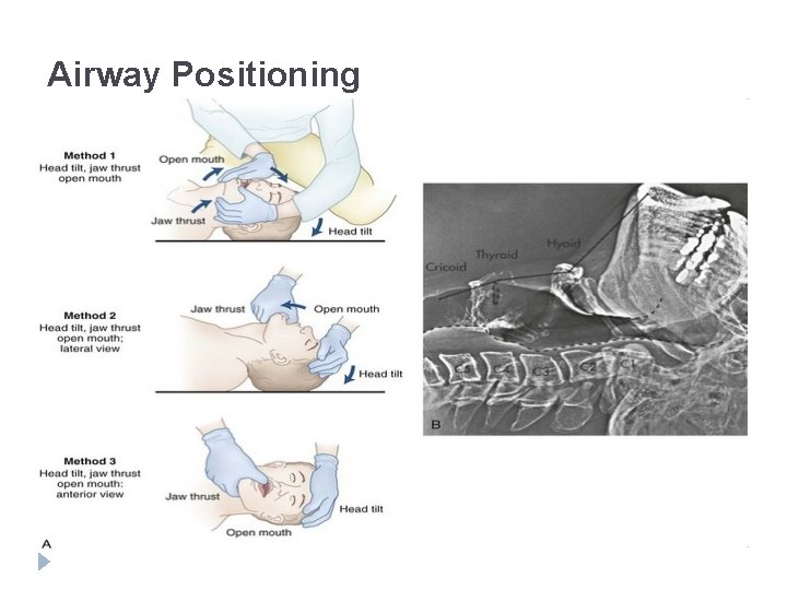
Airway Positioning
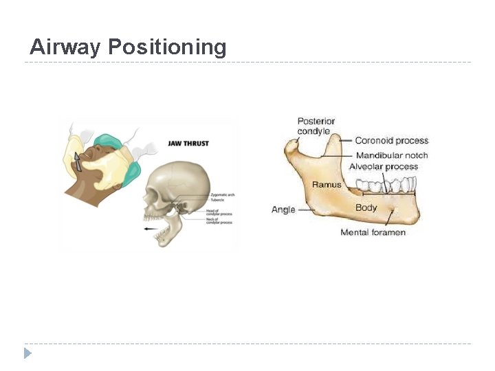
Airway Positioning
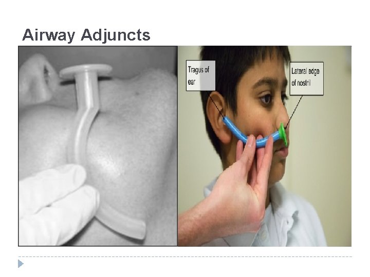
Airway Adjuncts
References 1. 2. 3. Martini F and Nath JL. Fundamentals of anatomy and physiology. 8 th edition. 2009. Pearson Benjamin Cummings. Brickley LS. Bates guide to physical examination and history taking. 10 th edition. 2009. Lippincott Williams & Wilkins. Kovacs G and Law AJ. Airway management in emergencies. 2008. Mc. Graw-Hill.
- Slides: 43