embryonic development in the fruit fly John Noto

embryonic development in the fruit fly John Noto BIO 441 Lecture 24 April 2017

Development refers to interaction of the genome with the cytoplasm and external environment to produce a programmed sequence of typically irreversible events. Differentiation refers to the formation of cell types, tissues, and organs through specific gene regulation. A single cell with one genotype produces a variety of specialized tissues and organs. Development and differentiation can be studied at many levels: 1. Morphology 2. Biochemistry 3. Genetics

Development refers to interaction of the genome with the cytoplasm and external environment to produce a programmed sequence of typically irreversible events. Differentiation refers to the formation of cell types, tissues, and organs through specific gene regulation. A single cell with one genotype produces a variety of specialized tissues and organs. Development and differentiation can be studied at many levels: 1. Morphology 2. Biochemistry 3. Genetics

Development refers to interaction of the genome with the cytoplasm and external environment to produce a programmed sequence of typically irreversible events. Differentiation refers to the formation of cell types, tissues, and organs through specific gene regulation. A single cell with one genotype produces a variety of specialized tissues and organs. Development and differentiation can be studied at many levels: 1. Morphology 2. Biochemistry 3. Genetics

Flies are a great model organism

Flies are a great model organism

Flies are a great model organism
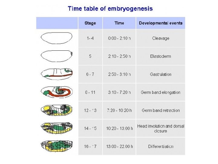
 Egg Larva (3 instars) Pupa Adult")
Developmental stages of Drosophila (10 -12 days) Egg Larva (3 instars) Pupa Adult

EMS Mutation screening Wieschaus, Lewis and Nusslein-Volhard Nobel Prize, genetics basis of development

EMS Mutation

Recombination


Fertilization and subsequent development
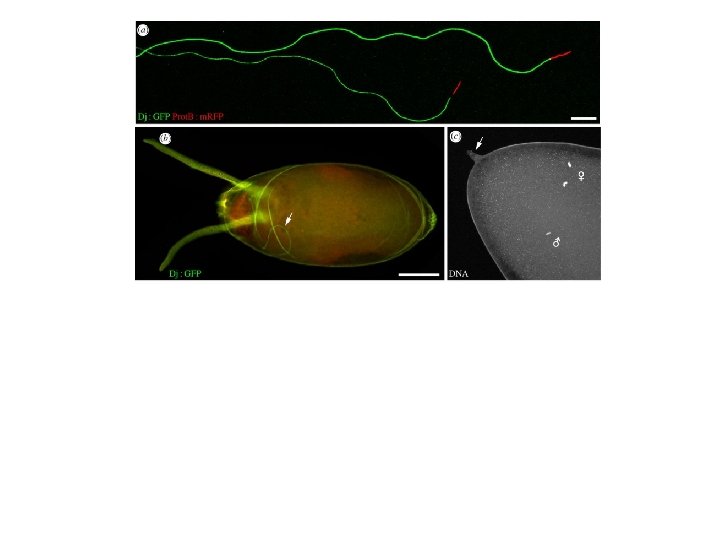
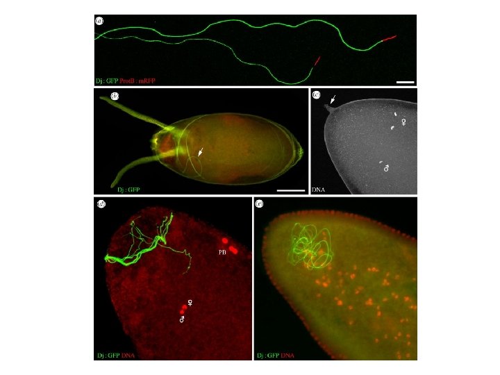

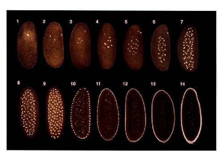
Embryonic development in Drosophila: • Development begins with fertilization. • Prior to fertilization, molecular gradients exist within the eggs. Polar cytoplasm occurs at the posterior end---example of maternal effect. • 2 nuclei fuse after fertilization to form a zygote. • 9 mitotic divisions occur without cell division, and after 7 divisions, some nuclei migrate to the polar cytoplasm (posterior) creating germ-line precursors. • Other nuclei migrate to the cell surface and form blastoderm precursor. • 4 more mitotic divisions occur and all nuclei are separated by cell membranes.

Embryonic development in Drosophila: • Development begins with fertilization. • Prior to fertilization, molecular gradients exist within the eggs. Polar cytoplasm occurs at the posterior end---example of maternal effect. • 2 nuclei fuse after fertilization to form a zygote. • 9 mitotic divisions occur without cell division, and after 7 divisions, some nuclei migrate to the polar cytoplasm (posterior) creating germ-line precursors. • Other nuclei migrate to the cell surface and form blastoderm precursor. • 4 more mitotic divisions occur and all nuclei are separated by cell membranes.

Embryonic development in Drosophila: • Development begins with fertilization. • Prior to fertilization, molecular gradients exist within the eggs. Polar cytoplasm occurs at the posterior end---example of maternal effect. • 2 nuclei fuse after fertilization to form a zygote. • 9 mitotic divisions occur without cell division, and after 7 divisions, some nuclei migrate to the polar cytoplasm (posterior) creating germ-line precursors. • Other nuclei migrate to the cell surface and form blastoderm precursor. • 4 more mitotic divisions occur and all nuclei are separated by cell membranes.

Embryonic development in Drosophila: • Development begins with fertilization. • Prior to fertilization, molecular gradients exist within the eggs. Polar cytoplasm occurs at the posterior end---example of maternal effect. • 2 nuclei fuse after fertilization to form a zygote. • 9 mitotic divisions occur without cell division, and after 7 divisions, some nuclei migrate to the polar cytoplasm (posterior) creating germ-line precursors. • Other nuclei migrate to the cell surface and form blastoderm precursor. • 4 more mitotic divisions occur and all nuclei are separated by cell membranes.

Embryonic development in Drosophila: • Development begins with fertilization. • Prior to fertilization, molecular gradients exist within the eggs. Polar cytoplasm occurs at the posterior end---example of maternal effect. • 2 nuclei fuse after fertilization to form a zygote. • 9 mitotic divisions occur without cell division, and after 7 divisions, some nuclei migrate to the polar cytoplasm (posterior) creating germ-line precursors. • Other nuclei migrate to the cell surface and form blastoderm precursor. • 4 more mitotic divisions occur and all nuclei are separated by cell membranes.

Embryonic development in Drosophila: • Development begins with fertilization. • Prior to fertilization, molecular gradients exist within the eggs. Polar cytoplasm occurs at the posterior end---example of maternal effect. • 2 nuclei fuse after fertilization to form a zygote. • 9 mitotic divisions occur without cell division, and after 7 divisions, some nuclei migrate to the polar cytoplasm (posterior) creating germ-line precursors. • Other nuclei migrate to the cell surface and form blastoderm precursor. • 4 more mitotic divisions occur and all nuclei are separated by cell membranes.

Embryonic development in Drosophila: • Development begins with fertilization. • Prior to fertilization, molecular gradients exist within the eggs. Polar cytoplasm occurs at the posterior end---example of maternal effect. • 2 nuclei fuse after fertilization to form a zygote. • 9 mitotic divisions occur without cell division, and after 7 divisions, some nuclei migrate to the polar cytoplasm (posterior) creating germ-line precursors. • Other nuclei migrate to the cell surface and form blastoderm precursor. • 4 more mitotic divisions occur and all nuclei are separated by cell membranes.

Embryonic development in Drosophila: • Development begins with fertilization. • Prior to fertilization, molecular gradients exist within the eggs. Polar cytoplasm occurs at the posterior end---example of maternal effect. • 2 nuclei fuse after fertilization to form a zygote. • 9 mitotic divisions occur without cell division, and after 7 divisions, some nuclei migrate to the polar cytoplasm (posterior) creating germ-line precursors. • Other nuclei migrate to the cell surface and form blastoderm precursor. • 4 more mitotic divisions occur and all nuclei are separated by cell membranes.

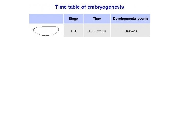
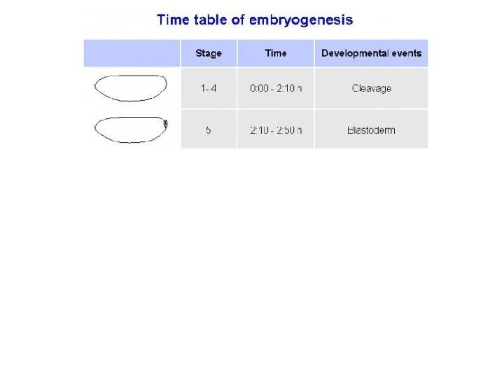
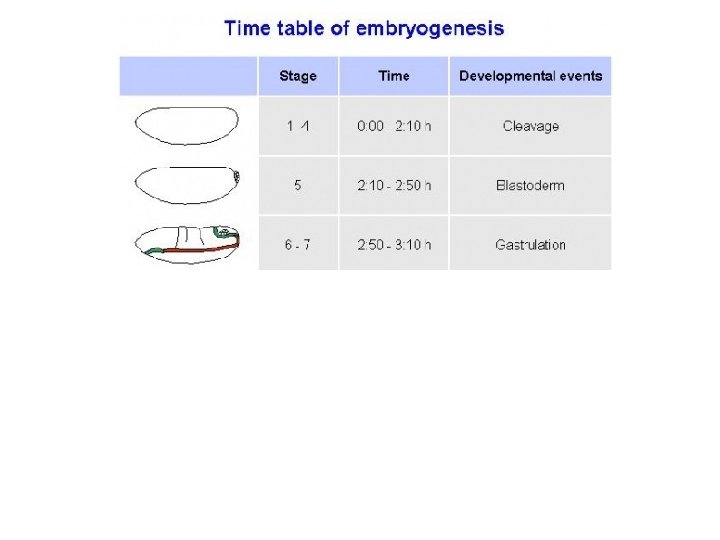
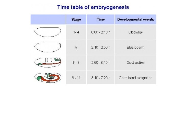
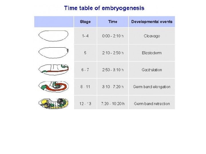
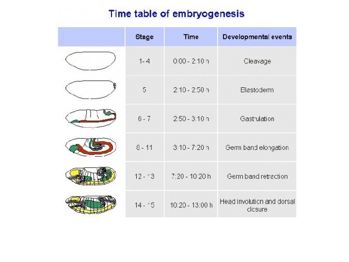
Embryonic development in Drosophila.

Embryonic development in Drosophila.

Embryonic development in Drosophila.

Embryonic development in Drosophila.

Embryonic development in Drosophila.

• ZEISS Lightsheet Z. 1 - Imaging of Drosophila embryo for cell tracking • ZEISS Lightsheet Z. 1 - Drosophila cell tracking using a color gradient • https: //www. youtube. com/watch? v=FCh. S 4 KU 5 j. DM

Cytoskeletal proteins create cytoplasmic islands around nuclei DNA Actin microfilaments microtubules

Subsequent development depends on two processes: 1. Anterior-posterior and dorsalventral molecular gradients exist in the egg---m. RNAs and proteins placed in egg by mother confer maternal effect. 2. Formation of (1) parasegments and (2)embryonic segments, which give rise to (3) adult segments. Adult segmentation reflect Embryo segmentation

Subsequent development depends on two processes: 1. Anterior-posterior and dorsalventral molecular gradients exist in the egg---m. RNAs and proteins placed in egg by mother confer maternal effect. 2. Formation of (1) parasegments and (2)embryonic segments, which give rise to (3) adult segments. Adult segmentation reflect Embryo segmentation

Three major classes of genes control development and differentation *Mutations identified by presence lethal or abnormal structures during development. 1. Maternal effect genes 2. Segmentation genes 3. Homeotic genes

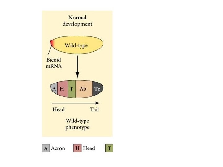
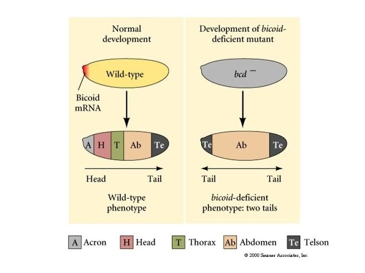
1. Maternal effect genes Expressed by the mother during egg production; they control polarity of the egg and the thus embryo. bicoid gene • Regulates formation of anterior structures (mutants possess posterior structures at each end). • Gene is transcribed during egg production, and expressed after fertilization. nanos gene • Regulates abdomen formation (m. RNAs collect in posterior of the egg). torso gene • Transcription and translation occur during egg production. • Occurs throughout the eggs, but is only active at the poles.

1. Maternal effect genes Expressed by the mother during egg production; they control polarity of the egg and the thus embryo. bicoid gene • Regulates formation of anterior structures (mutants possess posterior structures at each end). • Gene is transcribed during egg production, and expressed after fertilization. nanos gene • Regulates abdomen formation (m. RNAs collect in posterior of the egg). torso gene • Transcription and translation occur during egg production. • Occurs throughout the eggs, but is only active at the poles.

1. Maternal effect genes Expressed by the mother during egg production; they control polarity of the egg and the thus embryo. bicoid gene • Regulates formation of anterior structures (mutants possess posterior structures at each end). • Gene is transcribed during egg production, and expressed after fertilization. nanos gene • Regulates abdomen formation (m. RNAs collect in posterior of the egg). torso gene • Transcription and translation occur during egg production. • Occurs throughout the eggs, but is only active at the poles.

1. Maternal effect genes Expressed by the mother during egg production; they control polarity of the egg and the thus embryo. bicoid gene • Regulates formation of anterior structures (mutants possess posterior structures at each end). • Gene is transcribed during egg production, and expressed after fertilization. nanos gene • Regulates abdomen formation (m. RNAs collect in posterior of the egg). torso gene • Transcription and translation occur during egg production. • Occurs throughout the eggs, but is only active at the poles.

Distribution of bicoid m. RNA and protein in the egg A = Anterior P = Posterior

Distribution of bicoid m. RNA and protein in the egg A = Anterior P = Posterior

Distribution of bicoid m. RNA and protein in the egg A = Anterior P = Posterior

Bicoid protein

Bicoid protein m. RNA Protein

2. Segmentation genes: Determine the segments of the embryo and adult, and thus divide the embryo into regions that correspond to the adult segmentation patterns. 1. Gap genes v Subdivide the embryo along the anterior-posterior axis. v 2. Pair rule genes v Divide the embryo into regions, each containing parasegments. v 3. Mutation results in the deletion of several adjacent segments. Mutations cause deletions of the same part of a pattern in every other segment. Segment polarity genes v Determine regions that become segments of larvae and adults v Mutants possess parts of segments replaced by mirror images of adjacent half segments.

2. Segmentation genes: Determine the segments of the embryo and adult, and thus divide the embryo into regions that correspond to the adult segmentation patterns. 1. Gap genes v Subdivide the embryo along the anterior-posterior axis. v 2. Pair rule genes v Divide the embryo into regions, each containing parasegments. v 3. Mutation results in the deletion of several adjacent segments. Mutations cause deletions of the same part of a pattern in every other segment. Segment polarity genes v Determine regions that become segments of larvae and adults v Mutants possess parts of segments replaced by mirror images of adjacent half segments.

2. Segmentation genes: Determine the segments of the embryo and adult, and thus divide the embryo into regions that correspond to the adult segmentation patterns. 1. Gap genes v Subdivide the embryo along the anterior-posterior axis. v 2. Pair rule genes v Divide the embryo into regions, each containing parasegments. v 3. Mutation results in the deletion of several adjacent segments. Mutations cause deletions of the same part of a pattern in every other segment. Segment polarity genes v Determine regions that become segments of larvae and adults v Mutants possess parts of segments replaced by mirror images of adjacent half segments.

2. Segmentation genes: Determine the segments of the embryo and adult, and thus divide the embryo into regions that correspond to the adult segmentation patterns. 1. Gap genes v Subdivide the embryo along the anterior-posterior axis. v 2. Pair rule genes v Divide the embryo into regions, each containing parasegments. v 3. Mutation results in the deletion of several adjacent segments. Mutations cause deletions of the same part of a pattern in every other segment. Segment polarity genes v Determine regions that become segments of larvae and adults v Mutants possess parts of segments replaced by mirror images of adjacent half segments.

2. Segmentation genes: Determine the segments of the embryo and adult, and thus divide the embryo into regions that correspond to the adult segmentation patterns. 1. Gap genes v Subdivide the embryo along the anterior-posterior axis. v 2. Pair rule genes v Divide the embryo into regions, each containing parasegments. v 3. Mutation results in the deletion of several adjacent segments. Mutations cause deletions of the same part of a pattern in every other segment. Segment polarity genes v Determine regions that become segments of larvae and adults v Mutants possess parts of segments replaced by mirror images of adjacent half segments.
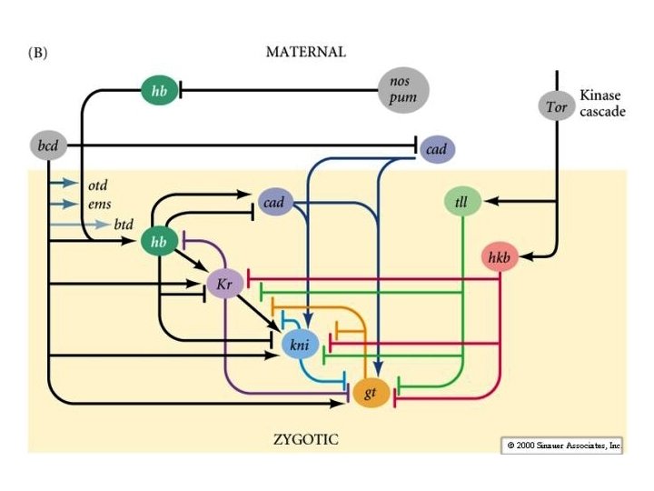

Functions for segmentation genes defined by mutations.

3. Homeotic genes: • Homeotic genes specify the body part to develop at each segment. • Adult body parts develop from undifferentiated larval tissues called imaginal discs. • Homeotic mutants develop a different body part at a particular segment (imaginal disc) than the usual body part. • Different homeotic gene groups share similar sequences of ~180 bp called homeoboxes that code proteins. • Homeoboxes regulate development and produce proteins that bind upstream of the gene units. • Homeotic gene complexes are abbreviated Hox. • Hox genes also specify body plans in vertebrates and plants.

Homeotic genes: HOX genes

Fig. 19. 21, Locations of homologous imaginal discs in larva and adult.

Fig. 19. 21, Locations of homologous imaginal discs in larva and adult.

Examples of homeotic Drosophila mutant with the bithorax mutation What is wrong with one of these flies?

Antennapedia and aristapedia mutants

antennapedia

antennapedia

Aristapedia

Ectopic expression: Dpp>eyeless

Fig. 19. 28, Organization of bithorax homeotic genes in a 300 kb region of the Drosophila genome. T = thoracic A = abdominal

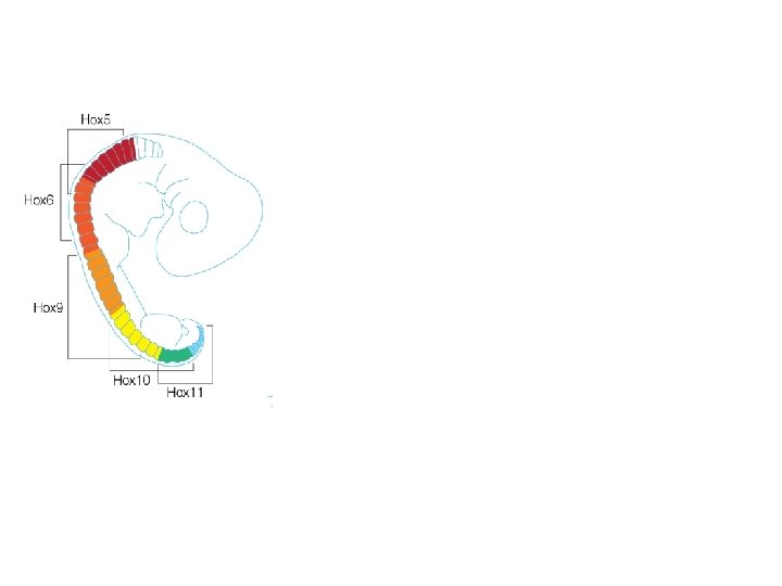
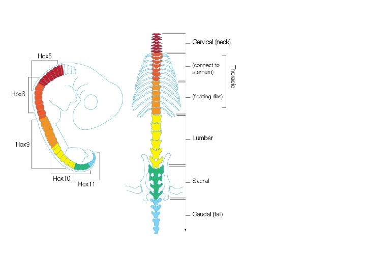
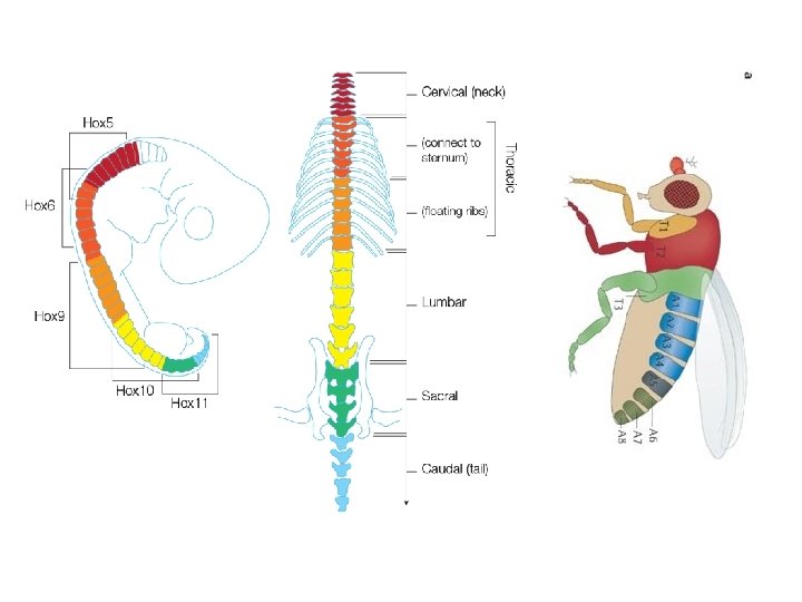
Fig. 19. 29 Homologous Hox gene clusters occur in Drosophila and the mouse.

How do development biologists study differential expression of genes during development and differentiation? Immunofluorescene assays that bind to specific m. RNAs and proteins.

How do development biologists study differential expression of genes during development and differentiation? Quantitative real-time RT-PCR of c. DNA from m. RNA transcripts.

How do development biologists study differential expression of genes during development and differentiation? Quantitative real-time RT-PCR of c. DNA from m. RNA transcripts.

RNA-Seq http: //www. nature. com/nbt/journal/v 28/n 5/full/nbt 0510 -421. html

Ribosome Profiling – sequencing of ribosome-bound m. RNAs

How do development biologists study differential expression of genes during development and differentiation? Gene knockout using transformation or transduction, or other gene silencing techniques like RNAi.

http: //ja. wikipedia. org/wiki/RNAi RISC = RNA-induced silencing complex

How do development biologists study differential expression of genes during development and differentiation? CRISPR/Cas 9 DNA editing tools---snip DNA and replace “clustered regularly interspaced short palindromic repeats” present in bacteria 1. CRISPR tell Cas 9 where to snip the DNA 2. Cas 9 recognizes sequences about 20 bp long 3. Guide RNA to match target sequence
- Slides: 83