Embryology organogenesis Week 4 Development and teratology of

Embryology /organogenesis/ Week 4 Development and teratology of reproductive system.

Male or female sex is determined by spermatozoon Y in the moment of fertilization ♂ ♀

SRY gene, on the short arm of the Y chromosome, initiates male sexual differentiation. • The SRY initiates transformation of indifferent gonads to form testes, which produce hormones supporting development of male reproductive organs. • Developed testes produce: - testosterone (T) - stimulates the Wolffian ducts development (epididymis and deferent ducts) and - anti-Müllerian hormone (AMH) - suppresses the Mullerian ducts development (fallopian tubes, uterus, and upper vagina).
")
• Indifferent stage – until the 7 th week • Differentiated stage 1) Development of gonads 2) Development of reproductive passages 3) Development of external genitalia 1, 2, 3 - is studied independently, although everything releates to everything
 Dorsal wall of body: urogenital ridge (medially), consisting")
Development of gonads mesonephric ridge (laterally) Dorsal wall of body: urogenital ridge (medially), consisting of mesenchyme and coelomic epithelium (Wolffian duct) gonad
 2")
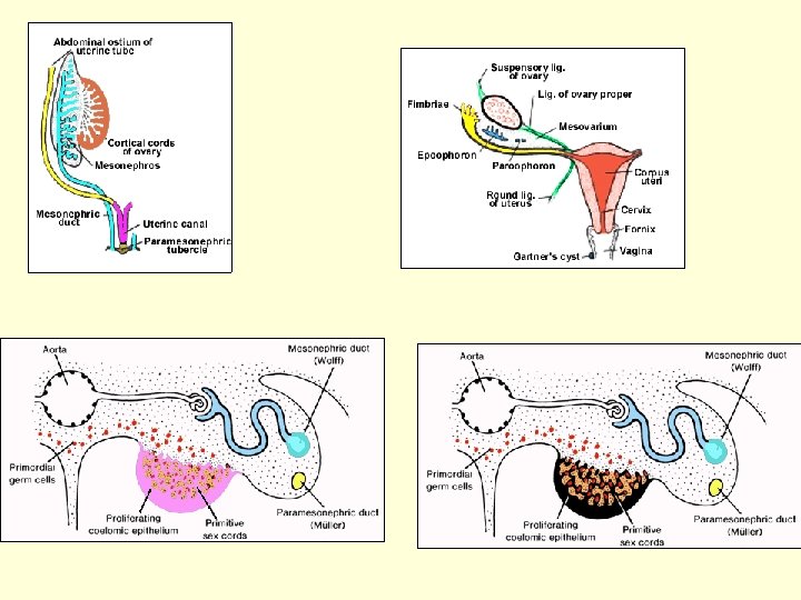
Three sources of gonad development: 1 – mesenchyme of gonadal ridges (plica genitalis) 2 – coelomic epithelium (mesodermal origin) 3 – gonocytes (primordial cells) gonocytes

Primordial germ cells – gonocytes – in endoderm of dorsal wall of yolk sac. Gonocytes migrate along dorsal mesentery of hindgut into the gonadal ridges and induce (!) gonad development.

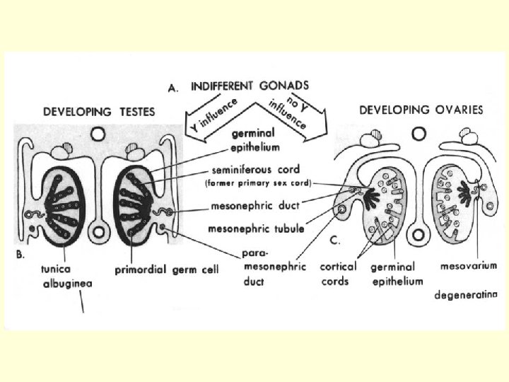
Indifferent gonad development • Gonocytes induce coelomic epithelium to proliferate Together with gonocytes, cells of coelomic epithelium in mesenchyme form - - primary sex cords in indifferent gonad

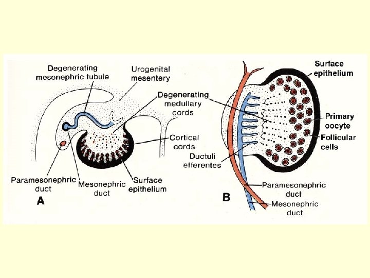
Primary proliferation Secondary proliferation TESTIS ♂ INDIFFERENT GONAD Tunica albuginea primary sex cords = medullary cords OVARY ♀ secondary sex cords = cortical cords ONLY in ovary

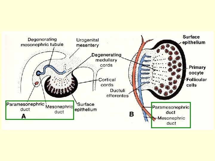
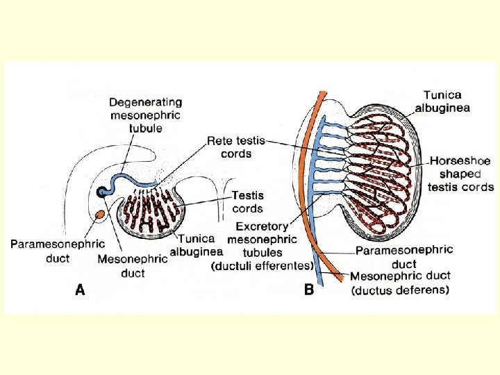
TESTIS: ♂ Primary sex cords tubuli semuniferi contorti Gonocytes spermatogonia Coelomic cells Sertoli cells Mesenchyme Leydig cells, interstitial connective tissue Tunica albuginea OVARY: Primary sex cords degenerate in ovarian medulla Secondary sex cords disintegrate into the follicles: Gonocytes oogonia Coelomic cells follicular cells Mesenchyme ovarian stroma ♀
 • In mesonephric ridge) – 2")
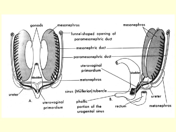
Development of reproductive passages (indifferent – differentiated stage) • In mesonephric ridge) – 2 ducts: Ductus mesonephricus (Wolffi) Ductus paramesonephricus (Mülleri)

Indifferent stage: Wolffian duct ♂ Müllerian duct ♀ genital ridge mesonephric ridge gubernaculum

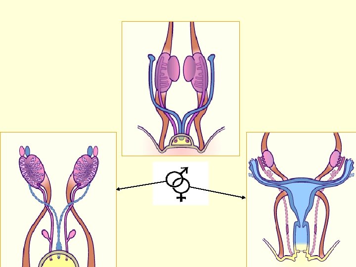
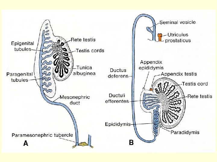
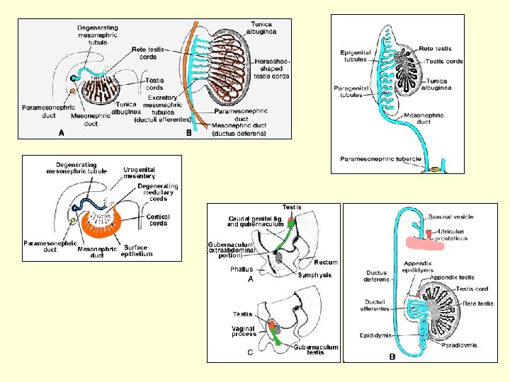
Differentiated stage of development: Müllerian duct: Wolffian duct: Oviduct Uterus Cranial part of vagina Ductus epididymidis Ductus deferens Ductus ejaculatorius Ductuli efferentes in epididymis and rete testis originate from mesonephric tubules (see mesonephros)

EPOOPHORON APPENDIX EPIDIDYMIDIS PAROOPHORON gubernaculum GARTNER‘S DUCT + RUDIMENTARY STRUCTURES

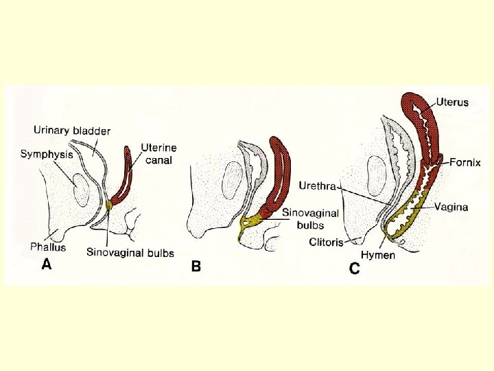
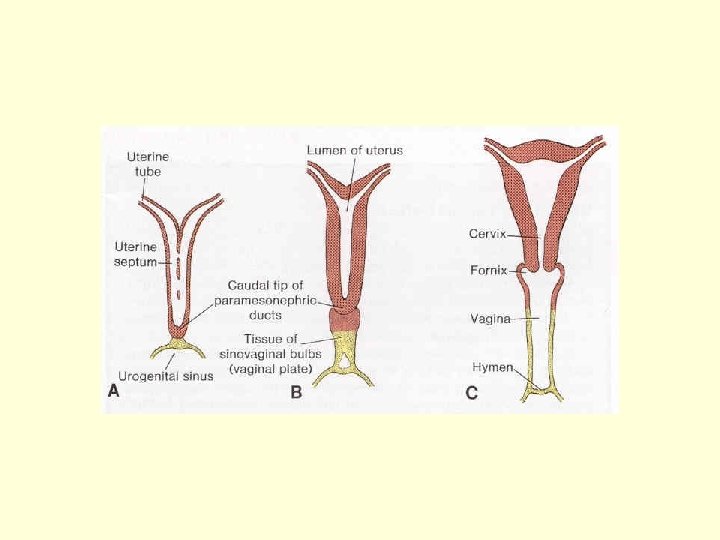
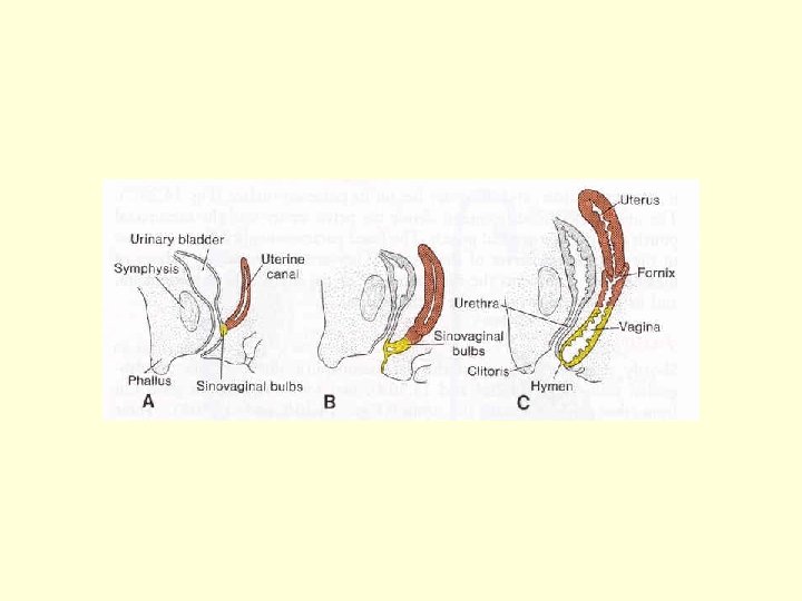
Oviduct Utero-vaginal canal

UTEROVAGINAL CANAL
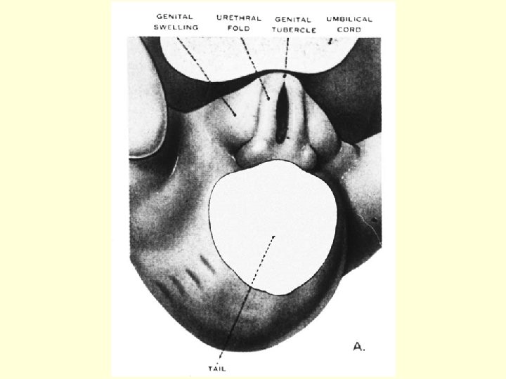
 Genital tubercle [tuberculum genitale Urethral (cloacal)")
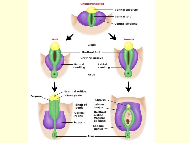
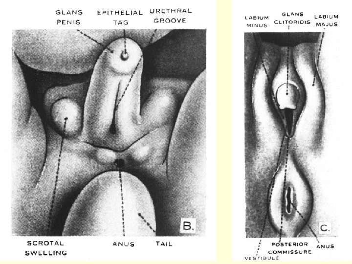
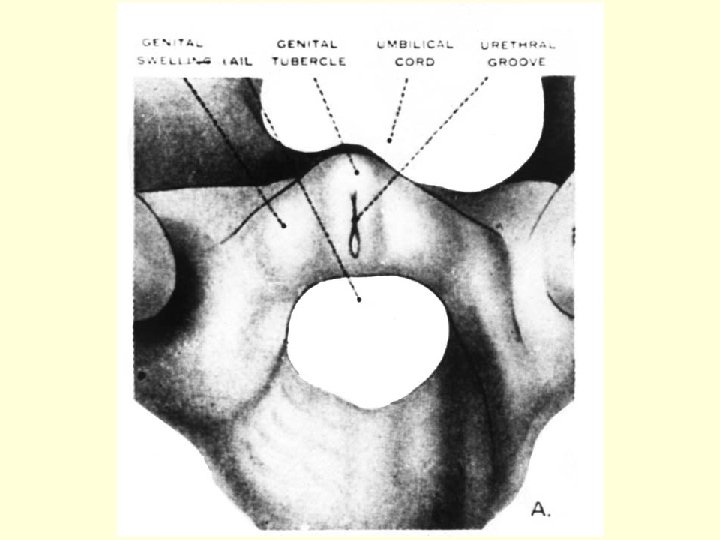
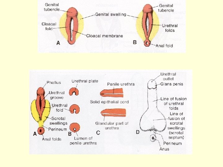
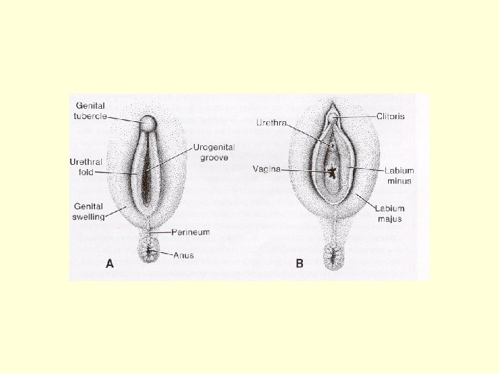
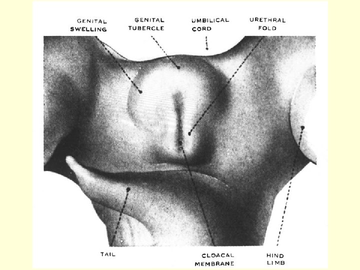
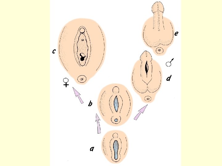
Development of external genatalia (indifferent – differentiated stage) Genital tubercle [tuberculum genitale Urethral (cloacal) folds [plicae genitales Labio-scrotal swellings [tori genitales

Labioscrotal folds

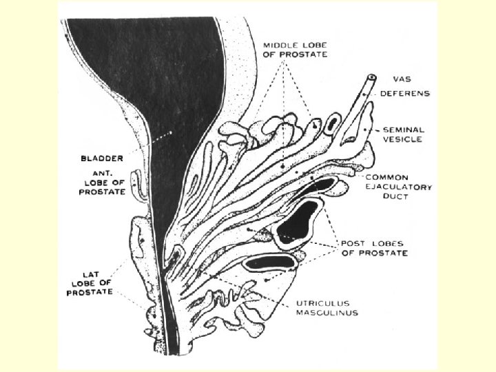
Accessory glands development Seminal vesicles – develop as diverticles of ductus deferens (from Wolffian duct) Prostate – develops around urethra as numerous diverticles (from pelvic part of sinus urogenitalis)

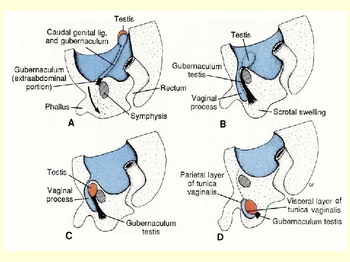
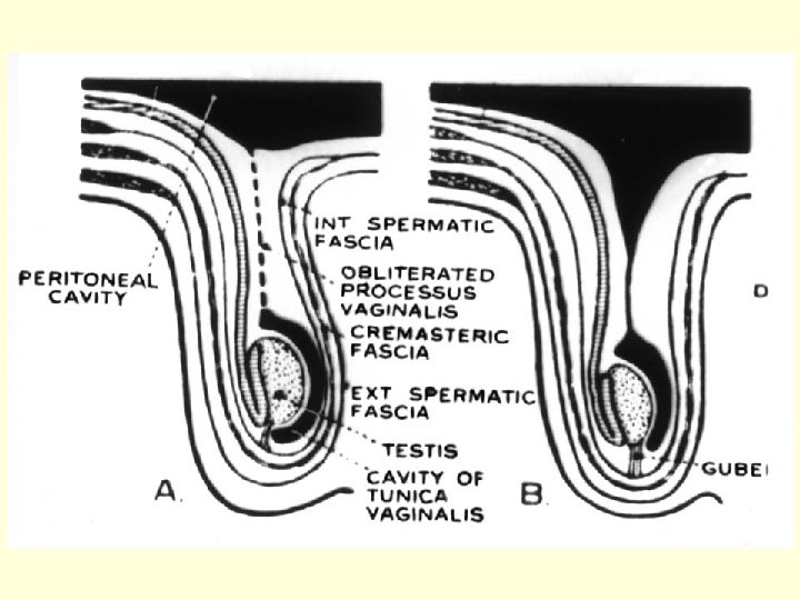
Position of gonads during development • Gonad develops in only short, lumbal part of genital (gonadal) ridge (Th 6 – S 2) • Cranial part - disappeares • Caudal part transforms into gubernaculum • Testes – descensus into the scrotum • Ovaries – change also their position due to fusion of Müllerian ducts and formation of broad ligament (lig. latum uteri)

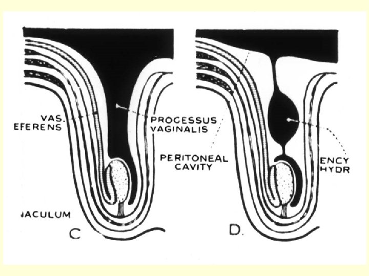
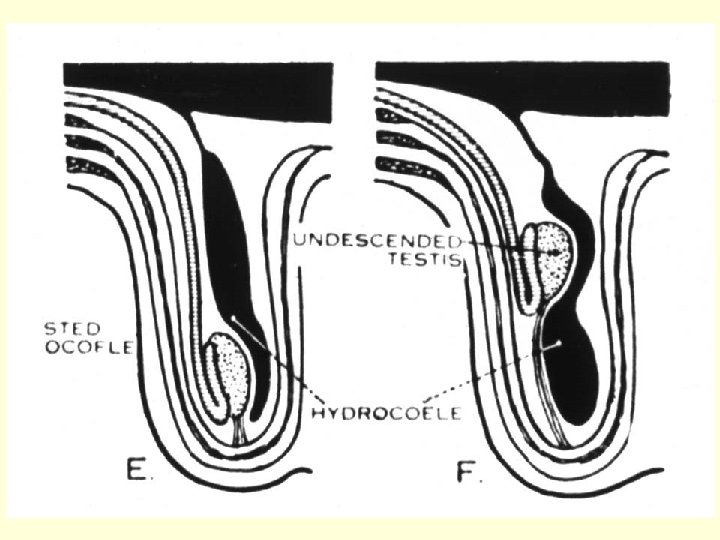
Testis – descens into the scrotum gubernaculum

Ovaries – change their position due to fusion of Müllerian ducts and formation of broad ligament
 agenesis • Hermafroditism (ovotestes, ovary+testis)")
Congenital malformations - 1 • Genetic anomalies: • Gonad(s) agenesis • Hermafroditism (ovotestes, ovary+testis) + chromosomal aberations (45 X/46 XX, 45 X/46 XY, 47 XXY/46 X, etc. ) • Pseudohermafroditism – karyotype and gonads do not correspond to external genitalia • Gonadal hypolasia – Turner sy. (45 X 0), Klinefelter sy. (47 XXY)

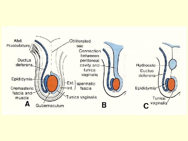
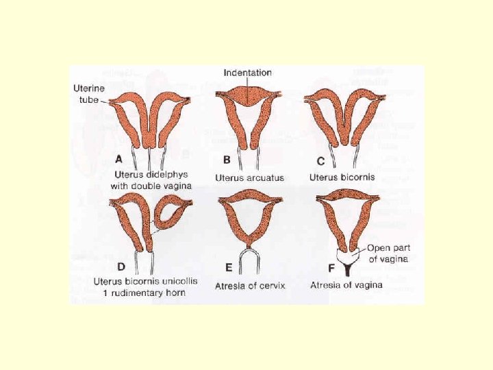
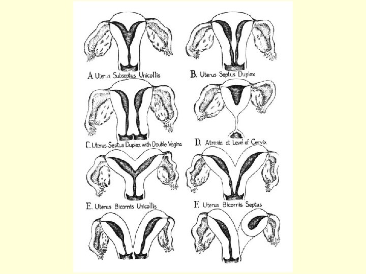
Congenital malformations - 2 • • • Kryptorchism Hydrocele testis Hypospadias, epispadias _________________ Developmental defect of uterus (and vagina) uterus et vagina separatus, uterus bicornis, uterus septus or subseptus, uterus unicornis etc.


19. Indifferent stage in development of reproductive system. 20. Development of male and female gonad. 21. An overview of development of male and female genital duct. 22. Development of external genital organs. 23. Developmental malformations of urogenital system.

Repetition of blood • • Composition of the blood Hematocrit Hemoglobin Erythrocytes – shape, size, density per 1 l Reticulocytes Anisocytosis Poikilocytosis Polycythemia (= polyglobulia)

• • Granulocytes Agranulocytes Number of leukocytes per 1 l Anemia Leukocytopenia Thrombocyte Number of thrombocytes per 1 l Hyalomere, granulomere

• • • Bone marrow structure Erythropoiesis Granulocytopoiesis Megakaryocyte Endomitosis • Differential white cell count (DWCC) !!! • Shift to the left or to the right

Neutrophilic granulocytes: 10 -12 m in „band“ 4 % in DWCC „segment“ 67 % in DWCC

Basophilic granulocyte: 8 m in , only 1 % in DWCC

Eosinophilic granulocyte: up to 14 m in , 3 % in DWCC

Lymfocyte Monocyte 20 % in DWCC 5 % in DWCC

- Slides: 63