Embryology of the nervous system Major divisions of

Embryology of the nervous system

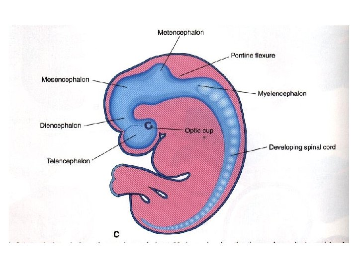
Major divisions of the developing brain: 3 parts, then 5 parts • 3 parts: Prosen-, Mesen-, and Rhomencephalon • 5 parts: Prosencephalon divides into telen- and diencephalon, Rhombencephalon divides into met- and myelencephalon

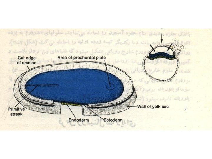
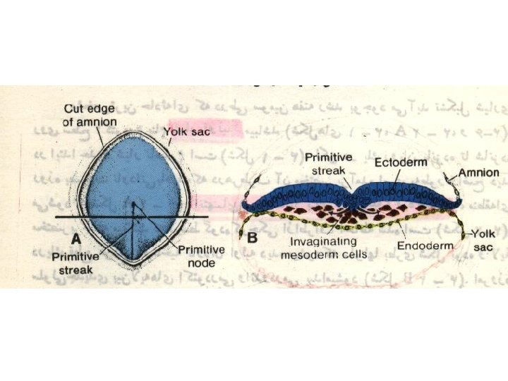
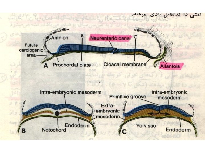
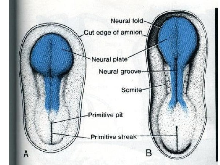
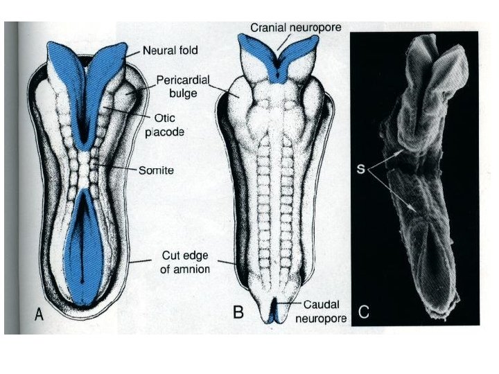
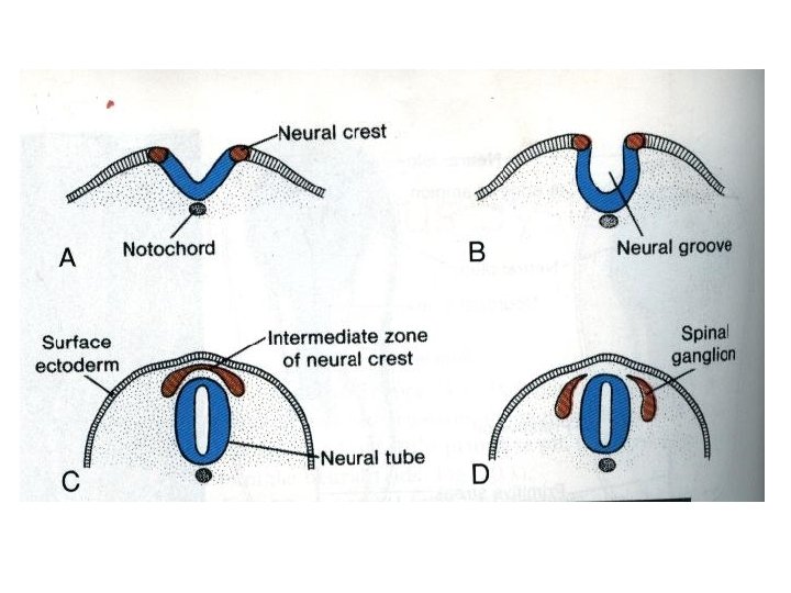
THIRD WEEK • Neural Plate : • It is a dorsal midline thickening of the ectoderm overlying the notochord (Neuroectoderm). • Neural Folds : • They are the elevated lateral margins of the neural plate. • They are on each side of the longtudinal midline (Neural Groove).

NEURAL TUBE • It is formed from the apposition and fusion of the neural folds which seal the neural groove and create the tube.

FOURTH WEEK • The neural tube is completed and transformed into the adult CNS. • This growth is maximal at the rostral part which becomes the brain. • The caudal portion becomes the spinal cord. • The axis of the neural tube (neuroaxis) is straight.
 prosencephalon")
Formation of brain and spinal cord • 1 - primary brain vesicles: 1) prosencephalon (forebrain) 2) mesencephalon (midbrain) 3) rhombencephalon (hindbrain) • 2 - Flexures
 flexure")
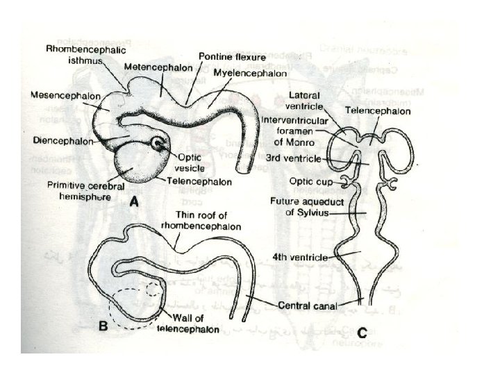
FLEXURES The neuraxis is bent by two flexures : Cephalic ( Mid brain) flexure at the junction of the fore and mid brains. Cervical flexure : Between the brain and spinal cord. Anterior (rostral) Posterior (caudal) Metencephalon Mesencephalon Diencephalon Midbrain Cervical Telencephalon Flexures Spinal cord Myelencephalon (a) Week 5 Figure 12. 3 a

DIFFERENTIATION OF BRAIN • 5 th week: • Three primary brain vesicles appear : • Fore brain (Prosencephalon). • Mid brain (Mesencephalon). • Hind brain (Rombencephalon).

1. PROSENCEPHALON • Is divided into : A. Telencephalon. B. Diencephalon.

A. TELENCEPHALON Telencephalon consists of : A median part and Two lateral pouches It is the largest brain vesicle. • It has the greatest degree of development. • It forms the two Cerebral Hemispheres. • •

B. Diencephalon • Thalamus • Epithalamus: - pineal - habenular nuclei • Hypothalamus • Subthalamus - subthalamic nuclei

3. ROMBENCEPHALON • Is divided into : A. Metencephalon. B. Myelencephalon.

A. METENCEPHALON • It is differentiated into: • Pons. • Cerebellum.

B. MYELENCEPHALON • It will form : • The Medulla Oblongata.

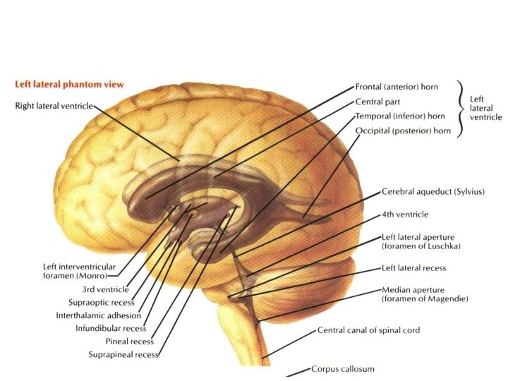
Ventricular spaces and central canal • • 1 -Telencephalon------ lateral ventricles 1&2 2 -Diencephalon-------third ventricle 3 -Mesencephalon----cerebral aqueduct 4 - Rhombencephalon--- forth ventricle 5 -Lumen of Spinal cord----- central canal 6 - in the end of spinal cord----- terminal ventricle Monro Foramen (interventricular Foramina)

DIFFERENTIATION OF SPINAL CORD • The Grey matter is located centrally around the central canal. • The White matter forms the outer coat.

Spinal Cord • The wall of neural tube consists of neuroepithelial cells. • These cells form a thick pseudostratified epithelium. • They divide rapidly, producing more and more neuroepithelial cells. they differentiate into 3 layers: • 1 -neuroepithelial (inner or ependymal) layer or neuroepithelium.

Spinal Cord • Neuroepithelial cells give rise to another cell type, these are the primitive nerve cells, or neuroblasts. • They form the 2 - mantle layer, a zone around the neuroepithelial layer. q The mantle layer later forms the grey matter of the spinal cord. q The outermost layer of the spinal cord, the 3 -marginal layer, contains nerve fibers emerging from neuroblasts in the mantle layer. q As a result of myelination of nerve fibers, this layer takes on a white appearance and therefore is called the white matter of the spinal cord.

Ventricular layer = ependymal layer = proliferative zone birthplace of CNS cells Mantle layer = newly born, post-proliferative cells Marginal zone = axonal layer

The developing neural tube is divided into 4 plates • • Roof plate. Alar plate: sensory Basal plate: motor Floor plate.

DIFFERENTIATION OF SPINAL CORD • Sulcus Limitans : • It is a longtudinal groove along the inner surface of the lateral walls of the developing spinal cord. • It differentiates the grouping of cells (grey matter) into dorsal (Alar) plate and a ventral (Basal) plate.

DIFFERENTIATION OF SPINAL CORD • The Alar plate is predominantly sensory in function (posterior horns). • The Basal plate is predominantly motor in function (anterior horns).

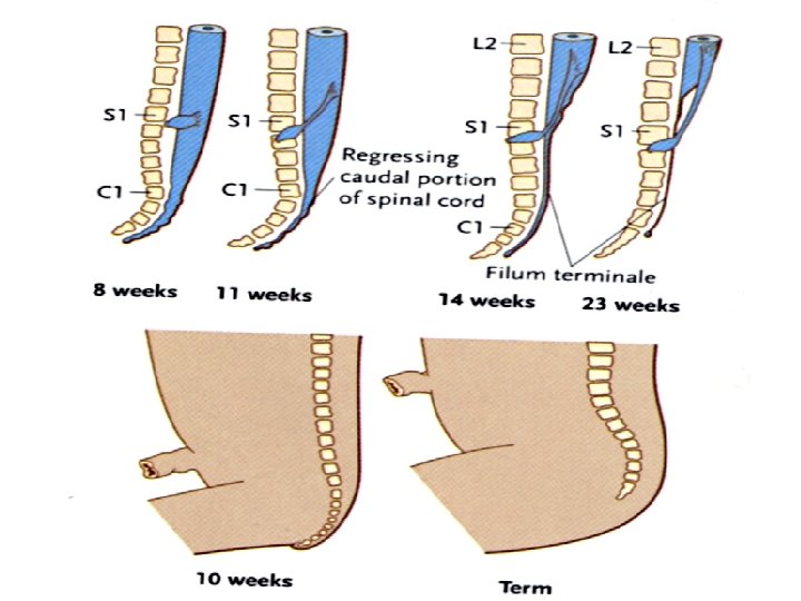
POSITIONAL CHANGES OF THE CORD • In the 3 rd month of development the spinal cord extends the entire length of the embryo, and spinal nerves pass through the intervertebral foramina at their level of origin. • With increasing age, the vertebral column and dura lengthen more rapidly than the neural tube, and the terminal end of the spinal cord gradually shifts to a higher level. • At birth, this end is at the level of the third lumbar vertebra. • As a result of this disproportionate growth, spinal nerves run obliquely from their segment of origin in the spinal cord to the corresponding level of the vertebral column. • The dura remains attached to the vertebral column at the coccygeal level. • In the adult, the spinal cord terminates at the level of L 1 to L 2, • The dural sac and subarachnoid space extend to S 2. • Below L 1 to L 2, a threadlike extension of the pia mater forms the filum terminale, which is attached to the periosteum of the first coccygeal vertebra and which marks the tract of regression of the spinal cord. • Nerve fibers below the terminal end of the cord collectively constitute the cauda equina.

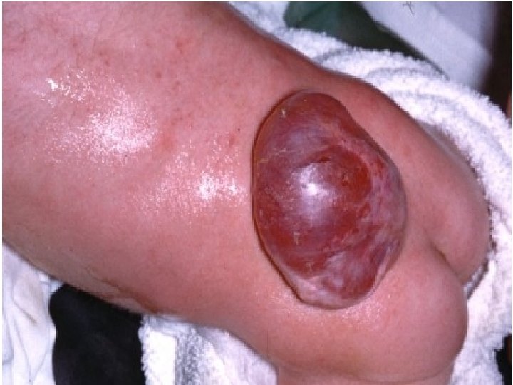
Congenital anomalies of spinal cord Spina bifida and Meningomyelocele • Spina bifida is a defect in the spine resulting from failure of the two halves of the vertebral arch to fuse. 34

• Types: – Spina bifida occulta: The bone only is affected, while the spinal cord and the membranes are intact. There may be a patch of hairy skin or a dimple over the affected area. It has a good prognosis. No treatment is required. – Spina bifida cystica ‘ overta’ which includes: » Meningocele. It is protrusion and herniation of the meninges, through a bony deficit to the skin. » Meningomyelocele: It is a protrusion of heterotopic neural tissue with the meninges. The defect is in the midline and affects the skin of the back, muscles, bones of the vertebral arches and neural tube. The membrane is easily ruptured » Myelocele: No skin or meninges to cover the lesion. It is usually incompatible with life. 35

occulta meningomyelocele meningocele myeloschesis

Thank You
- Slides: 38