Embryology nd 2 lecture Gastrulation n Gastrulation major

Embryology nd 2 lecture
 layers: n Ectoderm: skin,")
Gastrulation n Gastrulation: major cellular reorganization into 3 tissue (germ) layers: n Ectoderm: skin, nervous system n Endoderm: lining of gut and internal organs n Mesoderm: muscles, bones, heart During this stage cell movements result in a massive reorganization of the embryo from a simple spherical ball of cells, the blastula, into a multi-layered organism.


n n n n n The first days and weeks after conception: Day 1: first cleavage - 1 cell becomes 2 Day 2: second cleavage - 4 -cell stage Day 3: 6 -12 cell stage - can test at this stage for genetic diseases if done by IVF Day 4: 16 -32 cell stage - solid ball of cells - morula Day 5: Solid morula develops into hollow, fluid-filled blastula The embryo will develop from the inner cell mass, or embryonic disc Day 6 -7: Blastocyst attaches to the endometrium and burrows in: implantation The blastocyst starts to secrete HCG - human chorionic gonatotropin Stimulates estrogen and progesterone to prevent menstrual flow Causes "morning sickness" in some women. . . Pregnancy test measures the amount of this hormone n n n

n Fluid filled amniotic cavity starts to form n. Yolk sac starts to form (will make blood cells, germ cells) n. Embryo starts to form from embryonic disc n. Chorion (placenta) starts to form

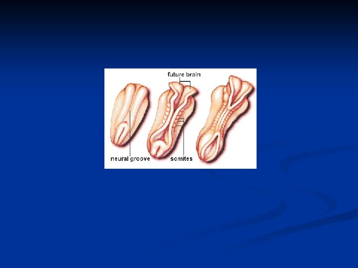
n Days 15 - 21: Emergence of body plan n Primitive streak starts to form - this is the site of gastrulation (formation of the 3 tissue layers - ecto, endo, and mesoderm) n Neural groove (future spinal cord and brain) begins to form n Somites (bands of tissue that will become muscles and bones) begin to form n Pharangeal arches (future face, neck, mouth, nose) begin to form

n Primitive streak starts to form - this is the site of gastrulation (formation of the 3 tissue layers - recto, endo, and mesoderm) n Neural groove (future spinal cord and brain) begins to form n Somites (bands of tissue that will become muscles and bones) begin to form n Pharyngeal arches (future face, neck, mouth, nose) begin to form
 - Development of all organ systems n")
n Week 3 - Week 8 (Embryo) - Development of all organ systems n Day 22: Cardiac cells (early heart) begins to beat

n Week 4 -8 is when all the major organ systems of the body are formed and when most teratogens have their greatest effect

n After 12 weeks or so, the baby's development is largely "finished" except brain and lung development n The fetus just spends much of the 2 nd and 3 rd trimesters just growing (and doing various flip-turns and kicks inside the amniotic fluid)
 Endoderm Brain")
Zygote Blastula Gastrula Ectoderm Epidermis & associated structures (skin, hair, nails etc) Endoderm Brain & NS Embryonic gut Inner lining of digestive tract Inner lining of respiratory tract Glands including liver & pancreas

Mesoderm Notochord Somites Muscle Excretory organs Outer covering of internal organs Gonads Mesenchyme (loose migratory cells) Bones & cartilage Dermis (inner skin layer) Circulatory system (heart, blood vessels)

craniofacial development

Patterning of the Craniofacial Region n n Morphogenesis: the cells undergo cell divisions, cell migration, or programmed cell death (PCD), for example, to allow fingers to form 2 factors determine morphogenesis: - Genetic - Environment Differential growth controlled by differences in cell behaviour: - cell death - cell migration - change in shape - change in size etc If any of these are perturbed, likely outcome is some type of birth defect Of the approximately 5, 000 known human inherited conditions, over 700 are craniofacial abnormalities.

At the end of Week 3, ectoderm differentiates into neuroectoderm and epidermis. The latter covers the outside of the body. n Signals from the underlying mesoderm cause the new neural precursor cells called the neural plate to invaginate and form a neural tube n Neuroectoderm forms neural tube and neural crest. n Hollow neural tube eventually becomes the central nervous system (top of tube becoming the brain and farther down, the spinal cord). n When the neural tube forms, the intermediate cells between the tube and the ectoderm become neural crest cells. Neural crest cells are migratory and begin leaving the neural crest at about Week 5 to reach various target areas where further specialization occurs. These cells migrate out and become cells of the peripheral nervous system n

n Just beneath the neural tube lies another structure, the notochord. n This notochord functions as the midline segmental organiser for all of embryologic development. n As adults, only vestigial remnants of this once dynamic notochord remain. n However a line of function, a midline that organises our physiology, persists to guide us throughout life. As the development of the head progresses, neural crest cells (and lateral plate mesoderm) both migrate into rapidly forming pharyngeal arches, a series of bumplike structures on both sides of the embryonic head n

n Neural crest cells, in addition to forming nerve tissue, produce the bones of the cranium. n Within the pharyngeal arches, neural crest cells and lateral plate mesoderm give rise to bones of the jaw and lower face, the viscerocranium n Lateral plate mesoderm also contributes to the formation of the cartilages of the larynx
")
n Early in the fourth week of human development the cranial and cervical (neck) regions make up approximately 1/2 of the embryo's length

Pharyngeal Apparatus
")
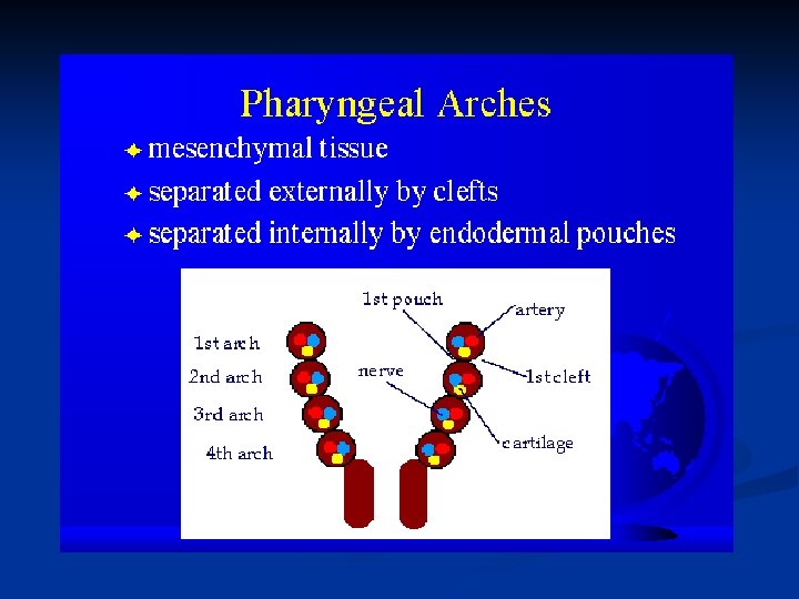
Pharyngeal Apparatus n The key to understanding craniofacial development are the Pharyngeal Apparatus (PA) n Also known as Branchial Apparatus n Pharyngeal Apparatus include: - Pharyngeal arches - Pharyngeal clefts - Pharyngeal pouches - Pharyngeal membranes (mesoderm) (ectoderm) (endoderm)

Organisation of the Pharyngeal Apparatus Pharyngeal Arches: - Out pocketings of surface ectoderm that are lined on the inside by endoderm. - Contain mesoderm in between. Ectoderm Mesoderm Endoderm

n n Arches consist of somitometric mesoderm & neural crest cells Each pharyngeal arch has cranial nerve associated with it n n A/ somitometric mesoderm differentiate in to an artery (aortic arches 1 -6)& muscle tissue B/ neural crest differentiate in to bone or cartilage & c. t neural crest cells are unique in that they can differentiate in to non neural components

n The PA consists of 4 pharyngeal arches, pharyngeal pouches and pharyngeal grooves. Neural crest cells in the cranial region migrate to the pharyngeal apparatus They cause enlargement of the arches and these cells contribute to the development of bones and connective tissues of the head and neck. Pharyngeal arches develop into components of the face. Arch mesoderm forms muscles of face

The developing face is represented by the frontonasal region, and the first pharyngeal (branchial, visceral) arch Removal of the surface ectoderm (some of which remains here) in the cranial and cervical region reveals the underlying mesenchyme (loosely organized cell populations. ) The majority of the mesenchyme immediately subjacent to the surface ectoderm is derived from the neural crest. Cells of mesodermal origin also contribute to the mesenchyme n

Components of the Pharyngeal Arches n 6 arches develop. We study 4 as the 5 th arch degenerates and the 4 th and 6 th arches fuse to form one n Each pharyngeal arch has its own vascular supply, cranial nerve innervation, muscular components and skeletal components (cartilage). n Grooves separate arches externally. Pouches separate arches internally.

Pharyngeal Arch 1: n First arch splits giving rise to 2 regions: - Rostral part called Maxillary Process - Caudal region called Mandibular Process n Maxillary process gives rise to upper jaw (Maxilla) n Mandibular process gives rise to lower jaw (Mandible) n The first pharyngeal arch is often called the mandibular arch. It is from this arch that the jaws develop.

SOThe first pharyngeal arch has both a maxillary and a mandibular prominence. Dorsal to the first arch is an elevation formed by the underlying trigeminal ganglion, the sensory ganglion for the nerve that supplies tissues derived from the first arch

n Union of the medial nasal prominence with the lateral nasal prominence and maxillary prominence is required for normal development of the upper lip

n the floor of the oral cavity and pharynx is formed by the arches.

n The first arch contributes to the surface of the anterior two-thirds of the tongue, while third arch contributes the posterior onethird

n The pharyngeal arches contribute to the developing tongue and epiglottis as shown

n The junction of the anterior two-thirds and posterior third of the tongue is at the terminal sulcus. The foramen cecum is the site at which the thyroid gland forms and invar

Pharyngeal Arch 2: n Often called the hyoid arch as part of the hyoid bone develops here. Where is the hyoid bone located and how is it important to communication? n Hyoid bone acts as a movable base for tongue. It is an attachment point for neck muscles that raise and lower larynx during swallowing and speech

Components of the Pharyngeal Arches II n The remaining parts of the hyoid bone develop in the third pharyngeal arch and the fourth pharyngeal arch and contributes to development of laryngeal cartilages. n It can be seen that each pharyngeal arch contributes to the development of structures that will play a role in communication. n In first arch syndromes (e. g. Treacher collins syndrome), there may be underdevelopment of first arch structures. Identify ways in which this may affect communication I

Pharyngeal Arch Cranial Nerve Muscle derivative Skeletal Derivative 1 Mandibular Trigeminal nerve Muscle of mastication Tensor tympani Incus, ant. Lig of malleus 2 Hyoid 3 Facial nerve Muscles of facial Stapes, expression Hyoid bone Glossopharyng eal nerve stylopharyngeas 4 Vagus nerve Striated muscles of oesophagus 5 None none 6 Merges with 4 Horns of hyoid none

n The fourth arch contributes to the epiglottis.

Each of the pharyngeal arches is supplied by a specific cranial nerve. n The cells that contribute to the sensory ganglia are derived from neural crest cells and from epibranchial placodes n

Epibranchial placodes are specialized regions of surface ectoderm, the cells of which invaginate to contribute to the formation of the sensory ganglia of cranial nerves V, VII, IX, and X. n The first, second, third and fourth arches are visible externally. The sixth arch does not form an external elevation n

The frontonasal prominence is composed of the tissue that surrounds the forebrain n Following closure of the anterior neuropore, the ectoderm that will line the nasal cavities (olfactory placodes) is located on the lateral aspects of the frontonasal prominence n

In the fifth week of human gestation, the olfactory placodes line the nasal pits. Medial and lateral nasal prominences form around the nasal pits n the mesenchyme of the medial and lateral nasal prominences and the thick olfactory placode epithelium. Axons extend from this epithelium, forming the olfactory nerves. n

1. pharyngeal arch 1 = mandibular arch ass with CN V – TRIG. N 2. pharyngeal arch 2=hyoid arch ass with CN VII- FACIAL. N 3. pharyngeal arch 3 ass with CN IX – GLASSOPHARYNGEAL. N 4. pharyngeal arch 4 ass with CN X= superior laryngeal branch of VAGUS. N 5. pharyngeal arch 6 ass with CN X = RECCURRENT LARYNGEAL BRANCH OF THE VAGUS. N

By the time that the anterior neuropore closes, the first and second pharyngeal arches are evident n The regions between the pharyngeal arches are termed pharyngeal clefts. The indentation just dorsal to the second pharyngeal cleft is the developing inner ear, the otic pit n

The Pharyngeal Pouches: n A human embryo has five pairs of pharyngeal pouches although only 4 develop. n Composed of Endoderm n Only the first pharyngeal pouch develops into structures that will have an impact on communication. n The first pharyngeal pouch develops into a tubotympanic recess that subsequently develops into the Middle ear cavity, the Tympanic membrane and the Eustachian tube

Pharyngeal Derivative Pouch 1 Epithelium of tympanic cavity and auditory tube 2 Epithelium of tonsil 3 4 Inferior parathyroid glands and epithelium of thymus Superior parathyroid glands.

Schematic sagittal section of a head, neck and upper thoracic regions of a 20 -week fetus, showing the adult derivatives of the pharyngeal pouches
: - located between arches - these are spaces, thus contain no")
Pharyngeal Clefts (grooves): - located between arches - these are spaces, thus contain no germ layer components - initially 4 clefts of which only one develops as 2 nd arch grows over all other clefts filling them in - 1 st cleft forms the External Auditory Meatus

Pharyngeal Membranes: - sites on bottom of arches - where ectoderm is joined to endoderm - 4 membranes initially - as most clefts are filled in, only first membrane develops. - this lies close to external auditory meatus and develops into the Tympanic membrane

n Neural crest cells form the majority of the facial and cranial skeleton. However, mesodermal cells also contribute to the cranium

n The pharyngeal arches are organized around blood vessels that extend dorsally from the developing heart

Five pairs of aortic arch vessels form temporally in a cranial to caudal sequence. The cranial-most vessels regress as the caudal ones develop. n LATER ON OTHER LECTURE CVS n

Thank you for your attention
- Slides: 53