Embryology Development of digestive system Embryo folding incorporation

Embryology: Development of digestive system

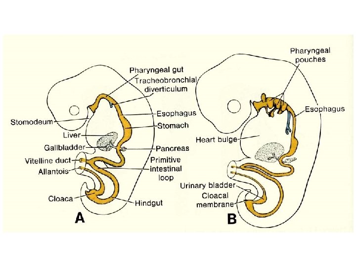
Embryo folding – incorporation of endoderm to form primitive gut. Outside of embryo – yolk sac and allantois. Vitelline duct
 the oral cavity + the salivary glands Proctodeum primitive anal pit")
Stomodeum (primitive mouth) the oral cavity + the salivary glands Proctodeum primitive anal pit Primitive gut whole digestive tube + accessory glands

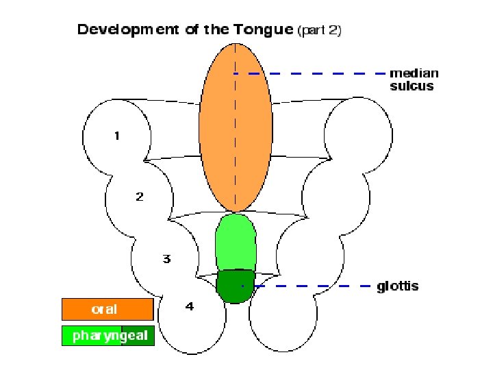
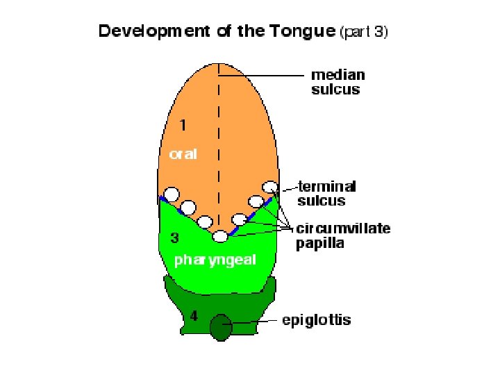
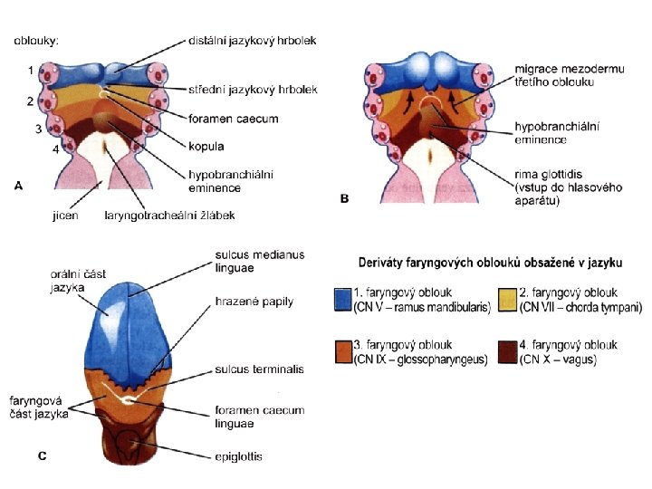
pharynx forgut midgut hindgut

• The epithelium and glandular cells of associated glands of the gastrointestinal tract develop from endoderm • The connective tissue, muscle tissue and mesothelium are derived from splanchnic mesoderm • The enteric nervous system develops from neural crest

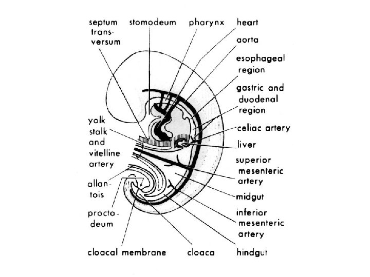
primitive gut foregut midgut pharyngeal membrane above ductus omphalomesentericus and yolk sack hindgut cloacal membrane
, stomach, cranial part of duodenum")
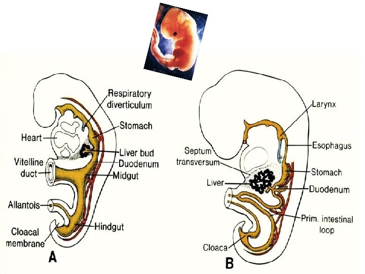
Derivatives of forgut – pharynx, esophagus (+ respiratory diverticul), stomach, cranial part of duodenum midgut – caudal part of duodenum (+ liver, gall bladder, pancreas), small intestine and part of large intestine (to the flexura coli sin. ) hindgut – large intestine (from flexura coli sin. ), rectum, upper part of anal canal

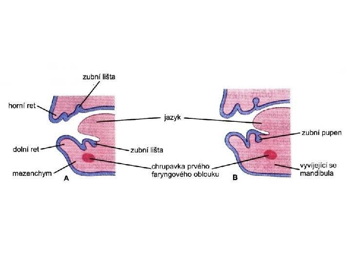
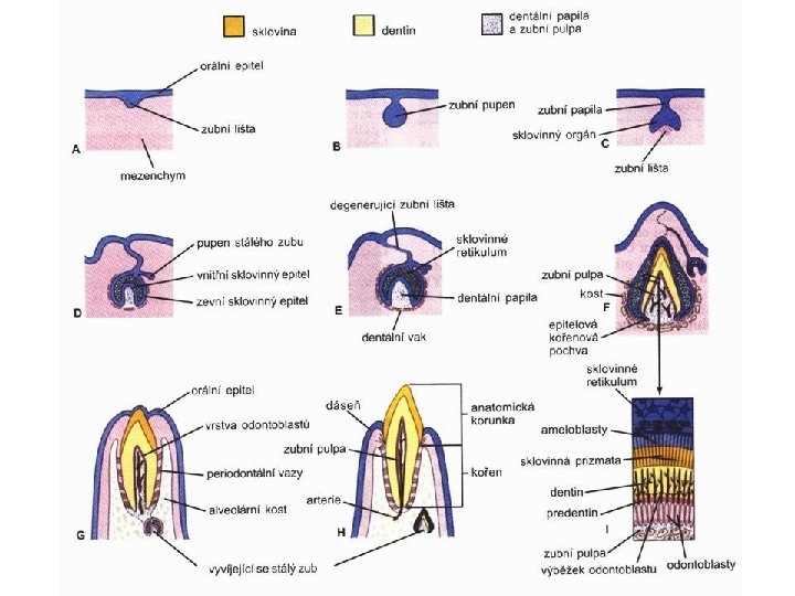
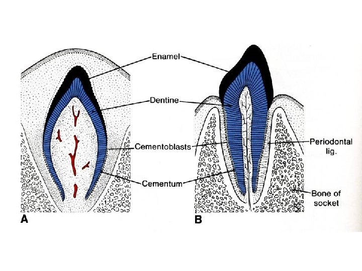
Oral cavity • primitive mouth pit – stomodeum • lined with ectoderm • surrounded by: - processus frontalis (single) - proc. maxillares (paired) - proc. mandibulares (paired) • pharyngeal membrane (it ruptures during the 4 th week, primitive gut communicates with amnionic cavity
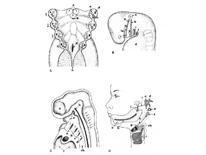
 apparatus Pharyngeal arches • appear in weeks 4 - 5 • on")
Pharyngeal (branchial) apparatus Pharyngeal arches • appear in weeks 4 - 5 • on the ventral side of the pharyngeal gut. • each arch has cartilage, cranial nerve, aortic arch artery and muscle • pharyngeal clefts and pouches are located between the arches • membrana obturans
")
Endodermal pharyngeal pouches Ectodermal pharyngeal clefts (grooves)

Fate of pharyngeal pouches and clefts Tympanic nenbrane + tympanic cavity Sinus cervicalis early development later development

endoderm ectoderm membrana obturans
 2 (hyoid) Nerve trigeminal (V) facial")
Structures derived from Arches ARCH 1 (maxillary/mandib ular) 2 (hyoid) Nerve trigeminal (V) facial (VII) Muscles Skeletal Structures malleus, incus Ligaments ant lig of malleus, sphenomandibula r ligament stapes, styloid process, lesser stylohyoid cornu of hyoid, ligament upper part of body of hyoid bone 3 glossopharyngeal (IX) greater cornu of hyoid, lower part of body of hyoid bone 4&6 superior laryngeal and recurrent laryngeal branch of vagus (X) thyroid, cricoid, arytenoid, corniculate and cuneform cartilages

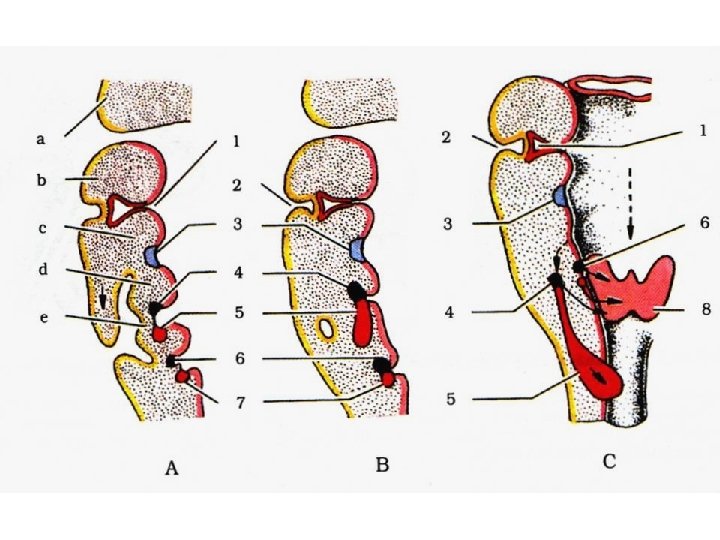
Structures derived from Pouches Each pouch is lined with endoderm and generates specific structures. POUCH Overall Structure Specific Structures 1 tubotympanic recess tympanic membrane, tympanic cavity, mastoid antrum, auditory tube 2 intratonsillar cleft crypts of palatine tonsil, lymphatic nodules of palatine tonsil 3 inferior parathyroid gland, thymus 4 superior parathyroid gland, ultimobranchial body

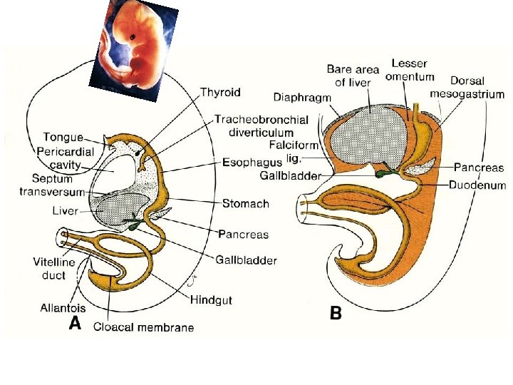
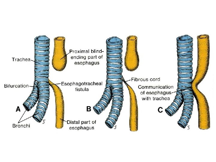
Esophagus development below respiratory diverticle, behind larynx and trachea primitive pharynx thyroid gl. laryngotracheal diverticle (respiratory divertcle) esophagus

Esophagus development • differentiation of epithelium from endoderm • during the 2 nd month endoderm proliferates and temporarily closes esophageal lumen • other tissues and structures in the wall arrise from splanchnic mesoderm

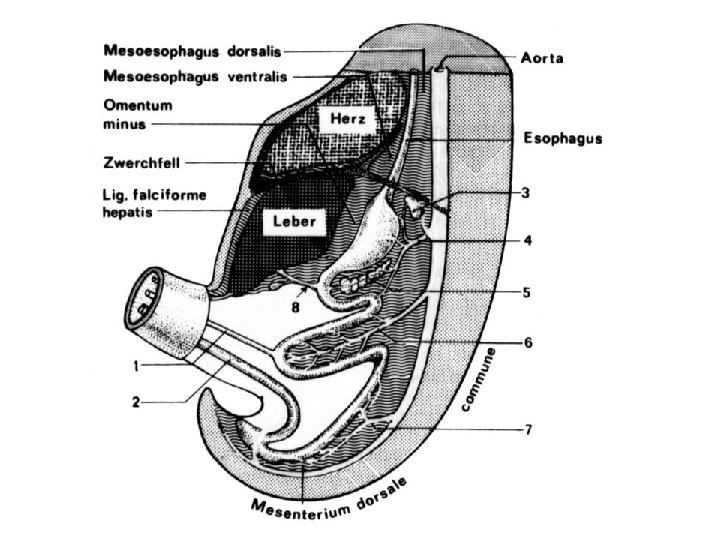
Mesenteries – suspensory duplicature derived from mesoderm and mesenchyme (a fold of tissue that attaches organs to the body wall) mesooesophageum esophagus mesoesophageum dorsale gives rise to dorsal mediastinum and mediastinal pleura mesoesophageum ventrale disappears

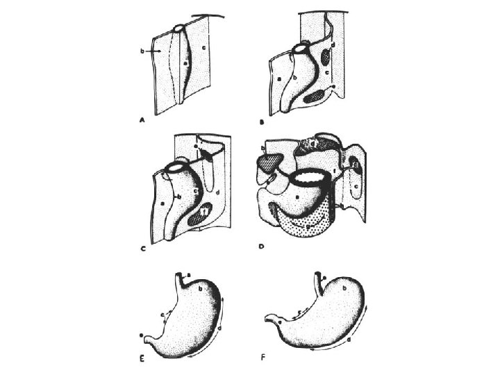
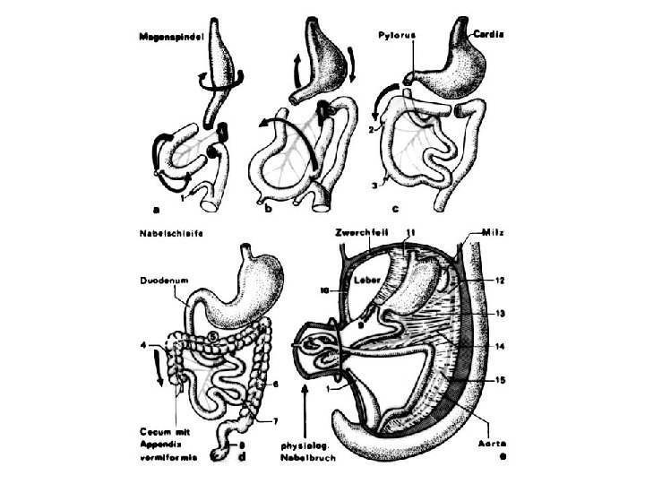
Stomach development in the 4 th week – spindle dilatation of distal forgut endoderm – epithelium and glandular cells splanchnic mesoderm – other tissues of stomach wall

Rotation around longitudinal axis: - left side → ventrally, - right side → dorsally. Uneven growth of ventral and dorsal wall: - curvatura minor (to the right), - curvatura major (to the left). Rotation around sagital axis : - curvatura minor (cranial position), - curvatura major (caudal position).

Sagital rotation axis
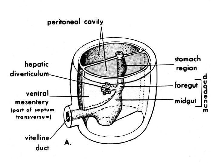
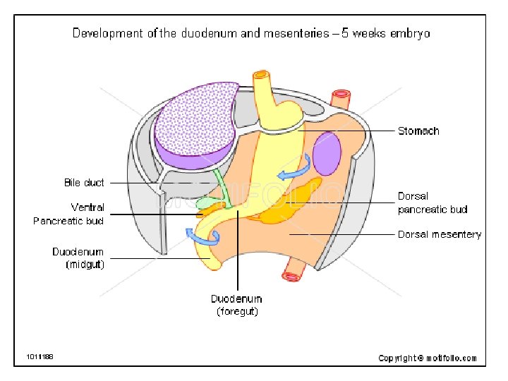
 appears at the distal end of the foregut (week")
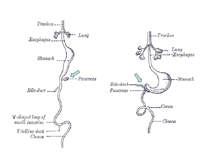
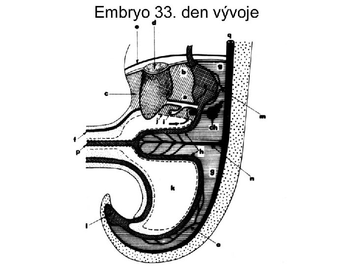
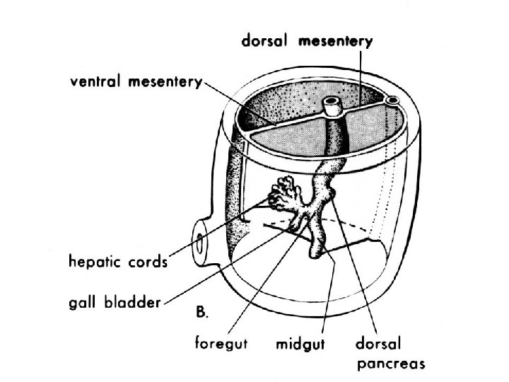
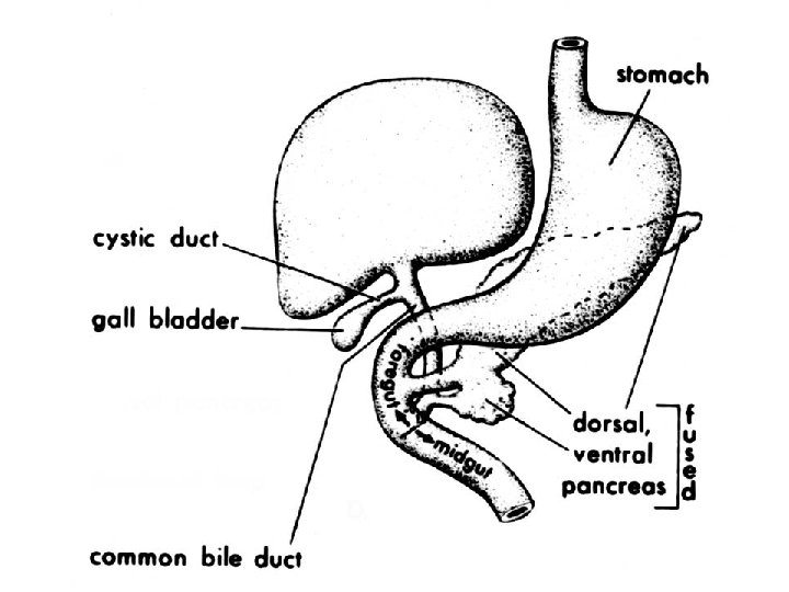
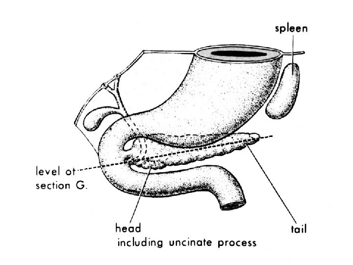
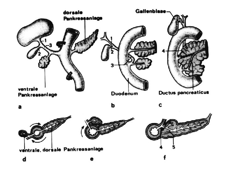
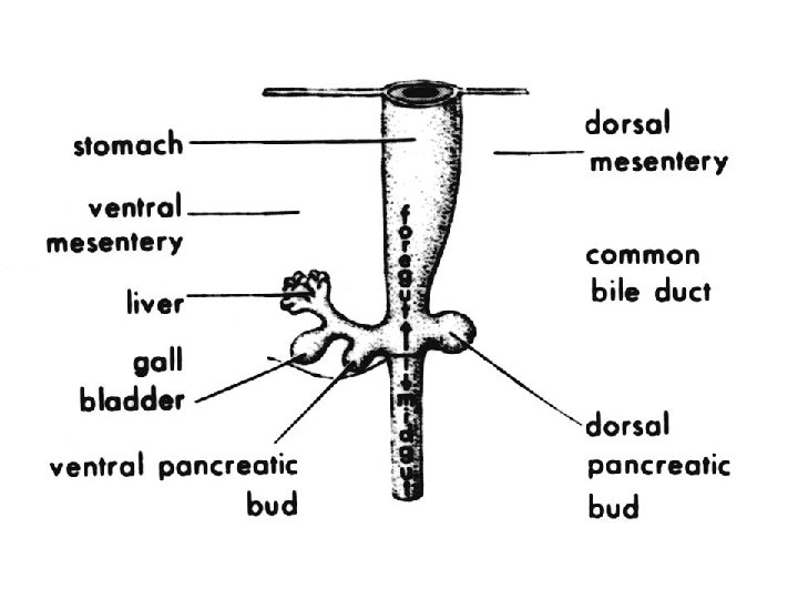
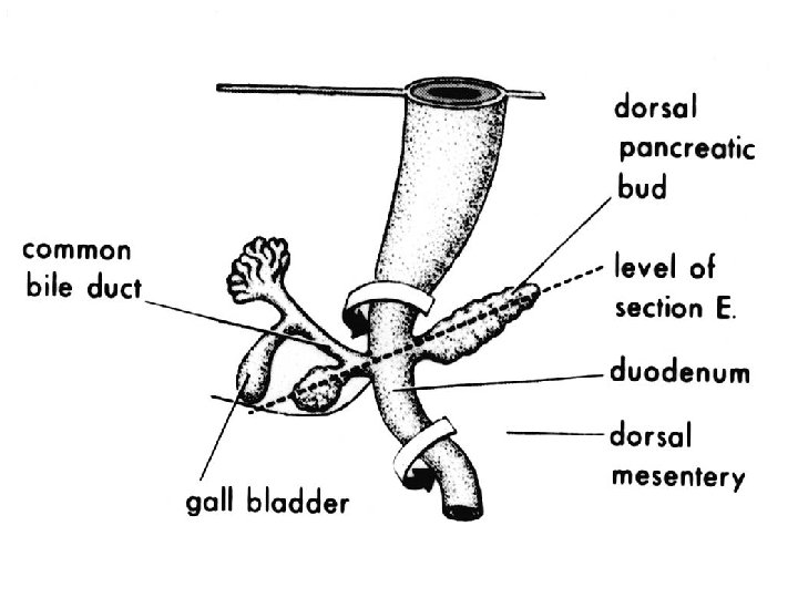
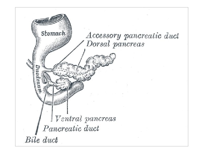
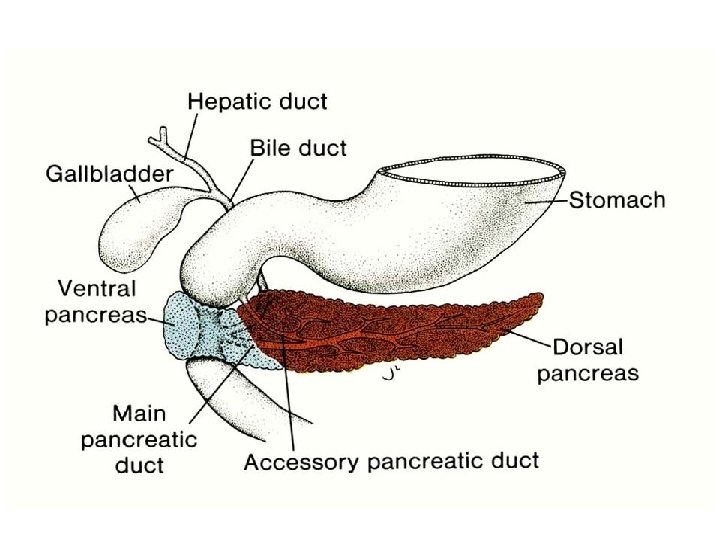
The liver bud (hepatocystic diverticcle) appears at the distal end of the foregut (week 4) and divides into hepatic and cystic diverticles, later ventral pancreatic bud and dorsal pancreatic bud (week 5). Both pancreatic buds meet and fuse (week 6).

liver

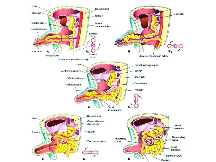
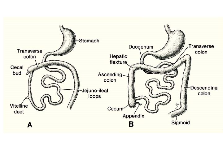
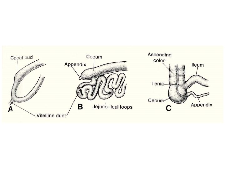
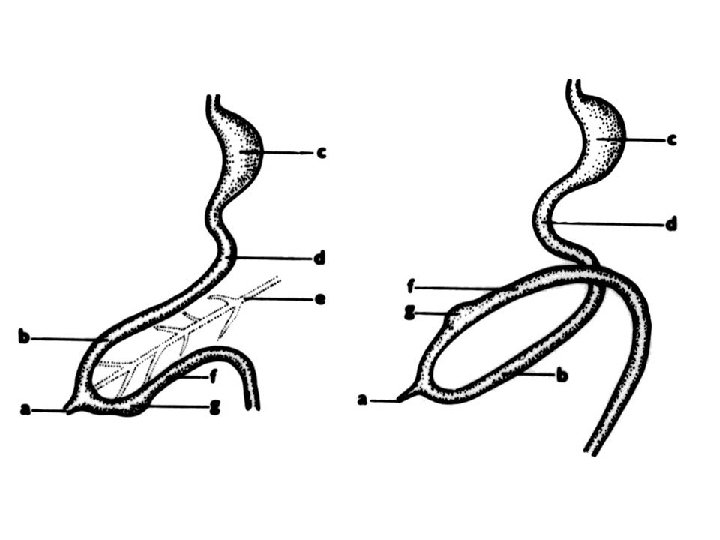
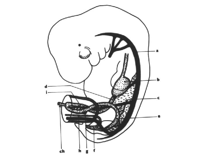
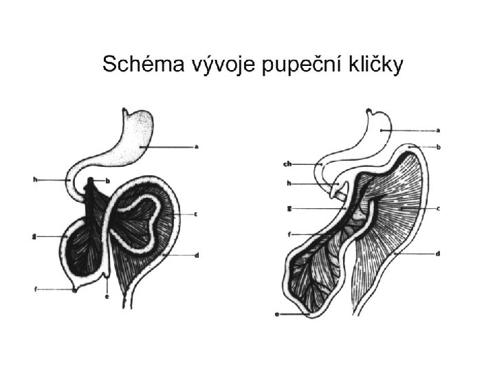
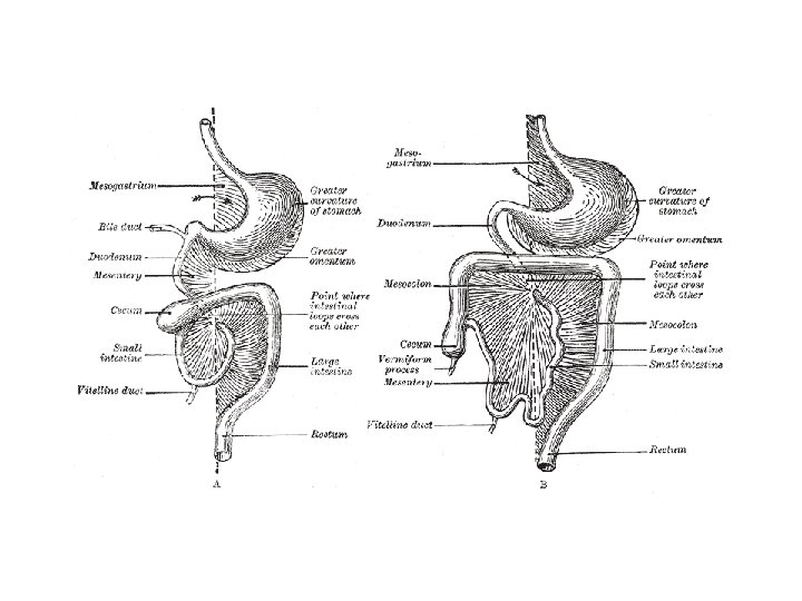
Midgut The midgut is divided into two regions at the viteline duct: the cranial and caudal limbs. The derivatives of the cranial limb - the distal duodenum, jejunum, and proximal ileum. The derivatives of the caudal limb - the distal ileum, cecum, appendix, ascending colon, and proximal 2/3 of transverse colon. the midgut grows faster than that of the embryo, creating: - duodenal loop - umbilical loop

Duodenal loop and umbilical loop Flexura duodenojejunalis forgut midgut Umbilical loop herniates into the umbilical cord (physiologic herniation, in week 6 -10)
 lower limb")
Duodenum development • Duodenal loop – 2 limbs: upper limb (from forgut) lower limb (from midgut) • On top of loop – diverticles (for liver, gallbladder, pancreas)

Due to rotation of umbilical loop, duodenal loop changes its position (from front to the right) and becomes retroperitoneal organ (together with pancreas)

Intestines development • Umbilical loop – 2 limbs: cranial – jejunoileal limb (jejunum, major part of ileum) caudal – ileocecal limb (rest of ileum, caecum + appendix, colon ascendens and 2/3 of colon transversum) • A. mesenterica sup. – axis of rotation • week 6 – physiologic herniation into the umbilical cord, week 10 – reposition into abdominal cavity

Umbilical loop rotation • • • The midgut loop rotates 90° counterclockwise in the umbilical cord around the axis of the superior mesenteric artery Upon returning, the gut undergoes another 180° counterclockwise rotation, placing the cecum and appendix near the right lobe of the liver. The total rotation of the gut is 270°.

90º º 180º after 270º rotation

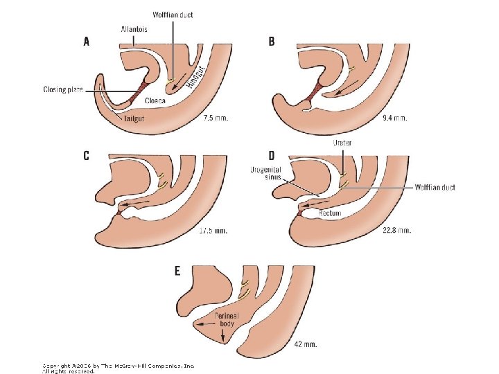
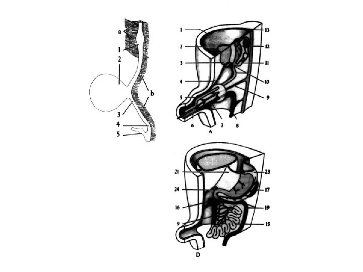
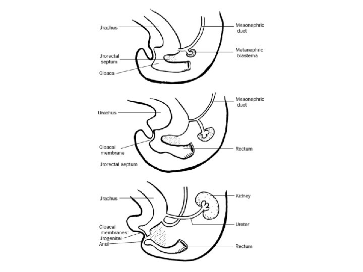
Hindgut The distal end of the hindgut – the cloaca. Derivatives of the hindgut: the distal 1/3 of the transverse colon, descending colon, sigmoid colon, rectum and upper part of anal canal (above the pectinate line).

Division of the cloaca - urorectal septum divides the cloaca into a ventral primitive urogenital sinus and a dorsal primitive anorectal canal. The cloacal membrane breaks down in the 7 th week. Distal to the pectinate line (site of the former cloacal membrane), the epithelium of the anal canal is derived from ectoderm of proctodeum (primitive anal pit)

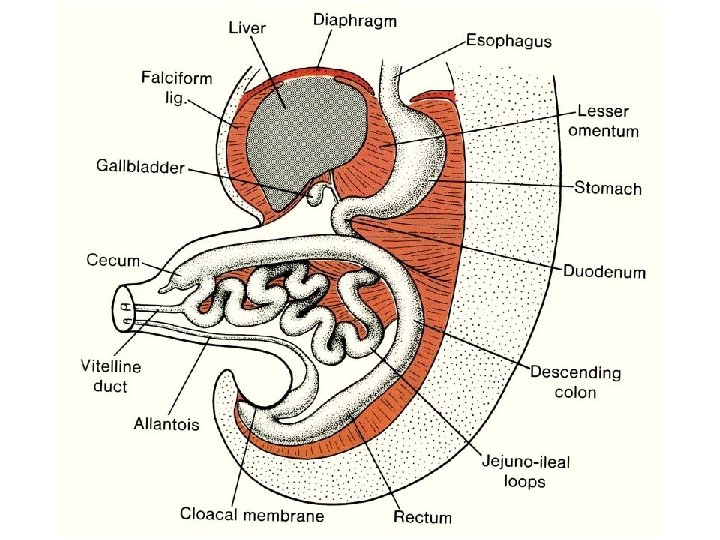
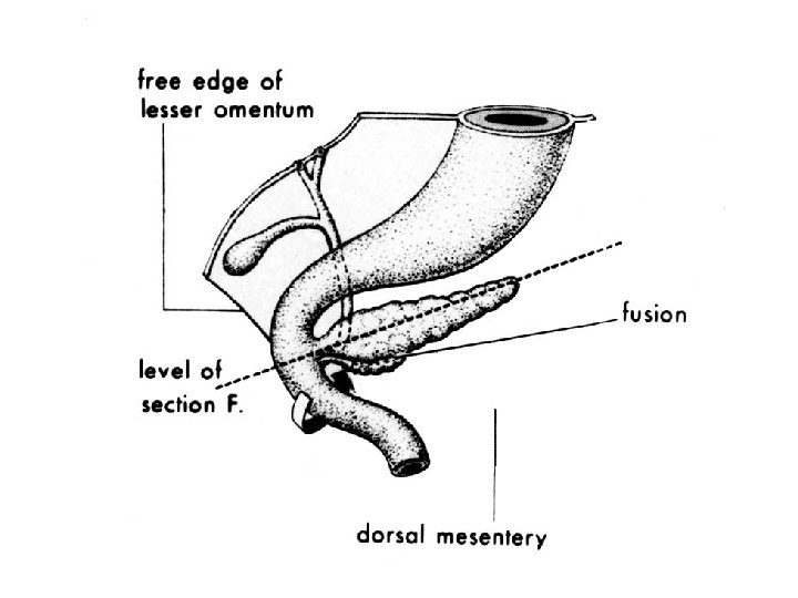
Mesenteries • double layer of peritoneum enclosing organs and connecting them to the body wall Ventral mesentery exists only in region of distal part of esophagus, stomach (lesser omentum) and upper part of duodenum Dorsal mesentery forms dorsal mesogastrium (greater omentum), dorsal mesoduodenum, mesentery proper (jejunum, ileum)

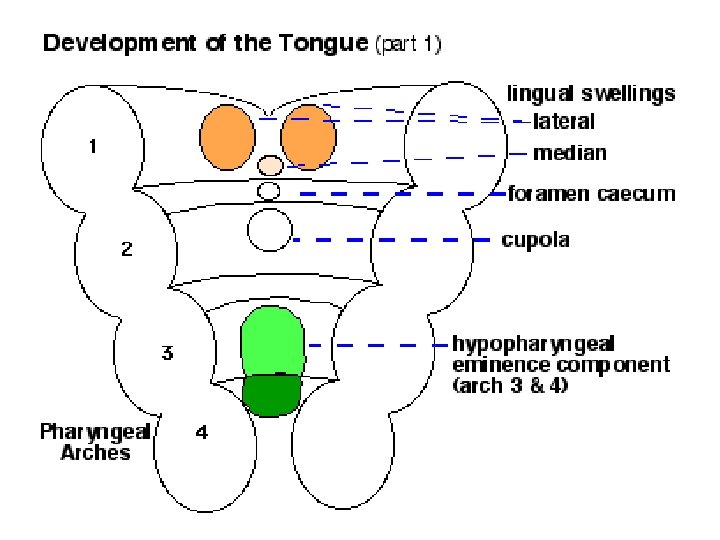
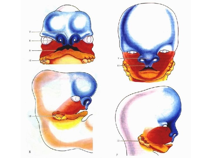
Face development • During 2 nd month i. u. • Stomodeum • Mesenchymal processes covered with ectoderm - processus frontonasalis - processus mandibulares - processus maxillares

Stomodeum The head and neck of a human embryo 32 days old, seen from the ventral surface. The floor of the mouth and pharynx have been removed.
 Nasal placode (plate)")
Frontonasal process Maxillar process Mandibular process Stomodeum Intermaxillary segment (intermaxillare) Nasal placode (plate)

Frontal view of an embryo at 4 to 5 weeks of age. Observe the branchial arch formation and the ruptured buccopharyngeal membrane.

Developing face week 4 4 -5 5 -6 6 -7
: human embryo stage 15 (8. 0 -mm), × 52. stage")
Scanning electron micrograph (SEM): human embryo stage 15 (8. 0 -mm), × 52. stage 17 (11. 7 -mm), 57 x stage 17 (11. 7 -mm), 14 x

Nasal placodes Nasal pits Nasal canals Proc. frontalis Proc. nasalis medialis et lateralis
 medial palatine plate (1) – from processus nasalis")
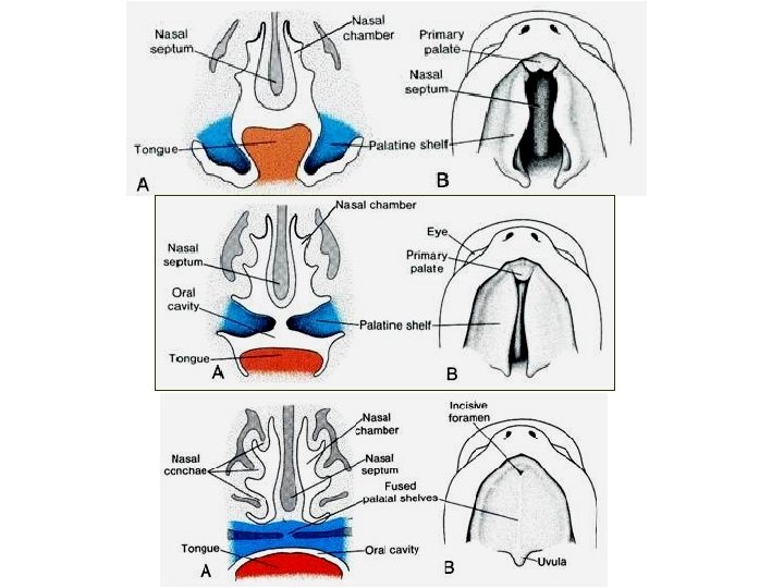
Palate development 3 ectoderm-mezenchymal plates: a) medial palatine plate (1) – from processus nasalis medialis (intermaxillare) primary palate b) lateral palatine plates (2) – from medial side of maxillary processes secondary palate Fusion of plates = raphe palati

Clefts of maxilla and palate Maxilla Cleft between lateral incisivus and caninus Uni- or bilateral (cheilognathoschisis unilateralis, cheilognathoschisis bilateralis)
 1: 2500")
Palate Uni- or bilateral Single or combined (cheilo – gnatho – palatoschisis) 1: 2500 heredity- autosomal dominant

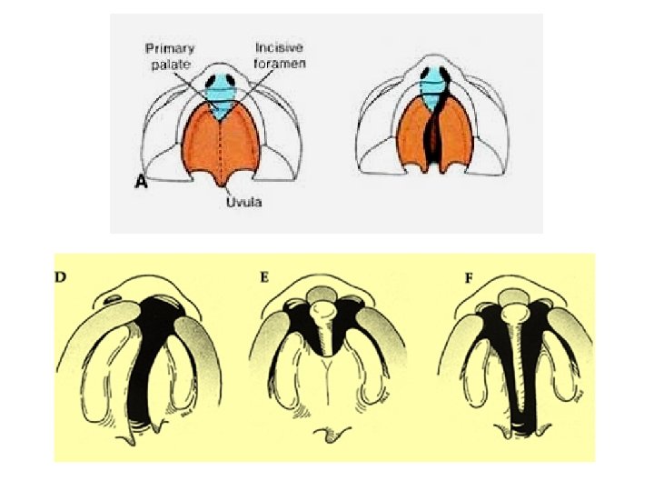
Clefts of primary palate Ventrally from foramen incisivum One or both lateral plates don‘t fuse with primary palate Clefts of secondary and primary palates Ventrally and dorsaly from foramen incisivum Lateral palatine plates are not fused with primary palate Nasal septum is free
 behind foramen incisivum Nonfused palatine plate in middle plane")
Clefts of secondary palate (palatoschisis) behind foramen incisivum Nonfused palatine plate in middle plane (completly – soft and hard palate and uvula) staphyloschisis (uvula bifida)

H – fissura orbitofacialis bilat. Transverse clefts J – macrostomia K - microstomia

Pharyngeal arches, pouches and clefts
")
Pancreas – ducts and parenchyma development (from endoderm)
 Endodermal pharyngeal pouches Thyroid gl. Laryngotracheal diverticle primitive pharynx")
Ectodermal pharyngeal clefts (grooves) Endodermal pharyngeal pouches Thyroid gl. Laryngotracheal diverticle primitive pharynx

Pharyngeal arches, pouches and clefts

At 6 weeks, the pancreatic buds meet and fuse.
- Slides: 97