Embryology 5 Mesoderm HOX genes and limb development

Embryology 5. Mesoderm, HOX genes and limb development. Dr. Nandor Nagy Semmelweis University
 Dev Cell 8: 401")
Shh expression in vertebrates Xenopus zebrafish Khokha et al (2005) Dev Cell 8: 401 -11 chick
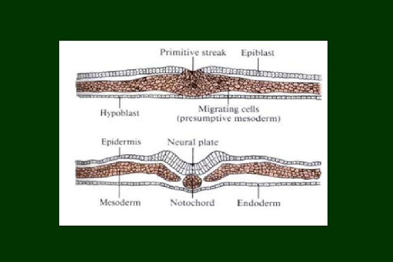

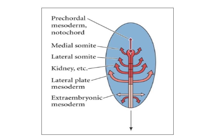
• -thick bands of mesodermal cells alongside neural tube • -unsegmented mesoderm separates into blocks of cells (somites) somite paraxial mesoderm Paraxial mesoderm and somite neural tube
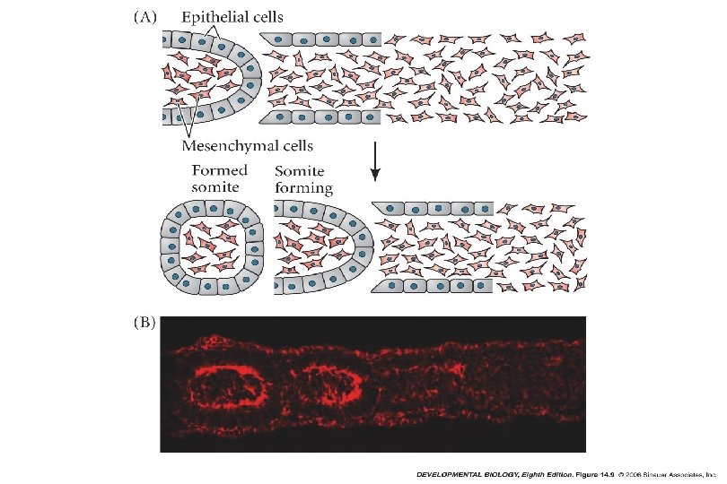

The somites are transient segmented structures derived from paraxial mesoderm. They contain the progenitors of the axial skeleton, trunk musculature and associated tendons, trunk dermis, endothelial cells, and meninges of the spinal cord.

• The Limb Field Morphogenetic field Limb. Includes Bud all cells with – Ectoderm the common fate of forming a limb – Mesoderm: • Paraxial (somite) Specified by Hox genes • Lateral plate (somatic) And retinoic acid Limb formation begins in the limb field

They were able to identify and classify a small number of genes that are of key importance in determining the body plan and the formation of body segments.

Hox genes • The Hox genes contain a 180 bp conservated region: the homeobox • The homeobox encodes a 60 AA length homeodomain = a DNAbinding helix turn helix motif • The homeodomain proteins are transcription factors • The Hox genes are selector genes (regulate the expression of other so-called realisator genes) • The expression of several Hox genes is region-specific in the embryo

Hox genes determine the number and types of vertebrae • Hoxc-6 determines that in the chicken the 7 vertebrae will develop into ribs • Snake: Hoxc-6 is expanded dramatically toward the head and toward the rear so all these vertebrae develop ribs.

Limb field specified by the HOX genes
; FGF-2 FGF induction")
Fibroblast growth factor-10 (FGF-10); FGF-2 FGF induction
 Ektodermakappe: Kölliker A. (1879) Mesoderm will induce ectoderm to")
AER: (Saunders J. W. 1948) Ektodermakappe: Kölliker A. (1879) Mesoderm will induce ectoderm to form the Apical Ectodermal Ridge (AER) AER develops along the distal margin of the limb bud

The vertebrate limb develops from a limb bud • Removal of the AER will result in loss of distal structures depending upon the time of surgery.
")
The outgrowth-promoting signal produced by the AER is FGF-8 (fibroblast growth factor-8)

Apical Ectodermal Ridge • The AER is needed for outgrowth and proximo-distal patterning of the limb : Proximal-distal axis Mesoderm determines type of limb and structure First to leave progress zone Proximal structures Last to leave progress zone Distal structures • Maintaining proliferation of the mesoderm (the AER produces FGF 8). The mesenchyme (progress zone) in contact with the AER proliferates instead of differentiating into cartilage and muscle.

Anterior-Posterior Axis of Limb The polarizing region specifies position along the A/P axis • The limb bud has a zone of polarizing activity (ZPA) – • group of mesenchymal cells at the caudal part of the limb bud. • involves production of sonic hedgehog (SHH) and interactions with AER

Shh mediates the signaling activity of the ZPA

Ectopic expression of mouse sonic hedgehog in the anterior limb causes extra digit formation

2012

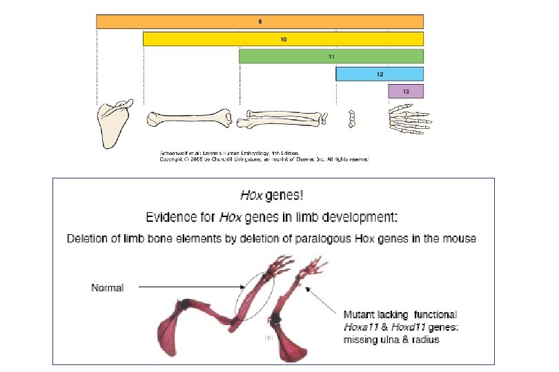
Members of Hox. D and Hox. A genes patterns the limb bud Hoxd 9 -13 seems to control A/P (i. e. finger) identity. Hoxa 9 -13 are expressed in a nested proximo-distal pattern. -i. e. Hoxa 9 only in the presumptive upper limb (humerus), -Hoxa 9 -11 in the lower limb region (radius, ulna). -Hoxa 9 -13 in the upper limb region to give rise to wrist and digits.
")
Loss of Hox. D 13 affects digit patterning (polysyndactyly)

Dorsal-Ventral Axis of Limb is controlled by the ectoderm • Probably induced by presence of a specific paracrine factor (Wnt 7 a) in the dorsal ectoderm of the limb bud Dorsal ventral

Molecular Interactions by which Limb Bud Formation and Growth are Initiated and Maintained proliferation Proximodistal Dorso-ventral Limb bud is mostly a regulative developmental field but two organizing centres exist. 1) apical ectodermal ridge (AER) at the limb bud tip and 2) zone of polarizing activity (ZPA) at the posterior mesenchyme.

Joint formation

Rotations of the human limb: The lower limb rotates medially so that the knee points cranially and the original ventral surface of the limb bud becomes the caudal surface of the limb.
- Slides: 29