EMBOLISM DR R S GOPIKA Prof Ho D

EMBOLISM DR. R. S. GOPIKA Prof. & Ho. D Dept. Of Pathology

Embolism: � Embolism is the occlusion or obstruction of a vessel by an abnormal mass transported from a different site by the circulation �An embolus is a detached intravascular solid, liquid, or gaseous mass that is carried by the blood to a site distant from its point of origin. � 99% of all emboli represent some part of a dislodged thrombus-thromboembolism.

TYPES �Arterial or venous Arterial embolism is a sudden interruption of blood flow to an organ or body part due to an embolus adhering to the wall of an artery blocking the flow of blood, the major type of embolus being a blood clot (thromboembolism) .
 and �the brain")
�The major sites for arteriolar embolization are �the lower extremities (75%) and �the brain (10%), �with the intestines, kidneys, and spleen affected to a lesser extent. �The consequences of embolization in a tissue depend on vulnerability to ischemia, caliber of the occluded vessel, and the collateral blood supply; �in general, arterial embolization causes infarction of the affected tissues.

Venous embolism � an embolus formed in a systemic vein will always impact in the lungs, after passing through the right side of the heart pulmonary embolism that will result in a blockage of the main artery of the lung � The most common sites of origin of pulmonary emboli are the femoral veins. � The deep veins of the calf are the most common sites of actual thrombi.
 �also known as crossed embolism �an embolus from the veins")
Paradoxical (venous to arterial) �also known as crossed embolism �an embolus from the veins crosses to the arterial blood system. �found only with heart problems such as septal defects between the atria or ventricles. �Eg: patent foramen ovale

The direction of the embolus �Anterograde �Retrograde �In anterograde embolism, the movement of emboli is in the direction of blood flow. �In retrograde embolism, the emboli move in opposition to the blood flow direction; this is usually significant only in blood vessels with low pressure (veins) or with emboli of high weight.

Types – depending on the material �Solid �Liquid �Gaseous

Fat Embolism �Microscopic fat globules can be found in the circulation after fractures of long bones or after soft-tissue trauma. �Fat enters the circulation by rupture of the marrow vascular sinusoids or rupture of venules in injured tissues.

The sequel: �depends on the size and the number of fat globules >20μm in diameter: occlusion of the branching arterioles of pulmonary artery < 20μm in diameter: pass through the pulmonary circulation and enter left heart, and consequently cause embolism of cerebral microvasculature

Fat embolism syndrome �is characterized by pulmonary insufficiency, neurologic symptoms, anemia, and thrombocytopenia � the symptoms appear 1 to 3 days after injury, with sudden onset of dyspnoea, and tachycardia. �Neurologic symptoms include irritability and restlessness, with progression to delirium or coma.

Bone marrow embolus in the pulmonary circulation

Amniotic Fluid Embolism �AFE is a rare obstetric emergency in which amniotic fluid, fetal cells, hair, or other debris enter the maternal circulation, causing cardio respiratory collapse

AFE �occurs in obstetric terms or during labor � multiparous woman with a large baby �use of uterine stimulants �occurred during abortion �amnioinfusion �Amniocentesis �caesarian section �placenta accreta �ruptured uterus

pathology �Amniotic fluid and fetal cells enter the maternal circulation, possibly triggering an anaphylactic reaction to fetal antigens. (1) Clinical symptoms result from mast cell degranulation with the release of histamine and tryptase, (2)activation of the complement pathway.

AFE phase I: � pulmonary artery vasospasm with pulmonary hypertension and elevated right ventricular pressure cause hypoxia. � Hypoxia causes myocardial capillary damage and pulmonary capillary damage, left heart failure, and acute respiratory distress syndrome.

AFE �Women who survive these events may enter phase II. �This is a haemorrhagic phase characterized by massive haemorrhage with uterine atony and DIC

Clinical presentation The classic clinical presentation of the syndrome has been described by five signs that often occur in the following sequence: (1) Respiratory distress (2) Cyanosis (3) Cardiovascular collapse cardiogenic shock (4) Haemorrhage (5) Coma.

Air Embolism �Gas bubbles within the circulation can obstruct vascular flow. � Air may enter the circulation during obstetric procedures or as a consequence of chest wall injury. �more than 100 m. L of air are required to produce a clinical effect � bubbles can coalesce to form frothy masses sufficiently large to occlude major vessels.

Decompression sickness. �occurs when individuals are exposed to sudden changes in atmospheric pressure. � Scuba and deep-sea divers, and underwater construction workers �When air is breathed at high pressure increased amounts of gas (particularly nitrogen) become dissolved in the blood and tissues.
 too rapidly, the nitrogen expands in the")
� If the diver then ascends (depressurizes) too rapidly, the nitrogen expands in the tissues and bubbles out of solution in the blood to form gas emboli that can induce focal ischemia in a number of tissues, including brain and heart. � The rapid formation of gas bubbles within skeletal muscles and supporting tissues in and about joints is responsible for the painful condition called- the bends.

Pathology �In the lungs, gas bubbles in the vasculature cause oedema, haemorrhages, or emphysema, leading to respiratory distress, called the chokes. �A more chronic form of decompression sickness is called caisson disease, where persistence of gas emboli in the bones leads to multiple foci of ischemic necrosis; the heads of the femurs, tibias, and humeri.
 Pulmonary embolism The origin of emboli: > 90% from deep leg")
Thrombembolism � 1) Pulmonary embolism The origin of emboli: > 90% from deep leg vein � The sequel: Small emboli may lodge in the branching arterioles of pulmonary artery, and usually be clinical silent Large emboli often occlude the main pulmonary artery and cause sudden death

Large embolus derived from a lower extremity deep venous thrombosis and now impacted in a pulmonary artery branch

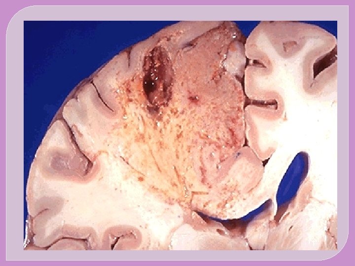
SYSTEMIC EMBOLISM The origin of emboli: Most arise from intracardiac mural thrombi or from ulcerated atherosclerotic plaques The sequel: Arteriolar embolization (e. g. the brain, kidneys , spleen and intesrines)

Arteriolar embolization of brain

Arteriolar embolization of spleen

Arteriolar embolization of intestine
- Slides: 29