Electron Microscope EM Electron microscopy EM is an
")
 is an electron beam which is focused into a small probe")


 are used to stain cells prior to examine via")


 * The mode of")


, a beam of highly focused")


 Laser scanning confocal microscopy is an invaluable tool")


- Slides: 19
Electron Microscope (EM)
Electron microscopy (EM) is an electron beam which is focused into a small probe across the surface of a specimen The first electromagnetic lens was developed in 1926 by Hans Busch.
* Electron microscope follows the same principle of compound microscope, but uses electrons beam as an illumination source instead of light.
Electron microscopes allow biologists to explore cells in more details. To observe the organelles such as: Mitochondria, Ribosomes, Endoplasmic reticulum (ER), Golgi apparatus and Lysosomes.
Heavy metals (such as lead) are used to stain cells prior to examine via EM. The stain is more visible in organelles than in the surrounding cytoplasm. Defects in a cell’s organelles are easily seen.
Electron microscopes are used in the scientific laboratories and many industries, such as : Nanotechnology Forensics Mining
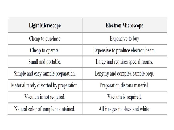
Disadvantages of electron microscopes 1 - It is a large machines 2 - Training is required 3 - It is very expensive 4 - Specimens are required a lot of preparation. 5 - The specimens are mounted in plastic, which means that only dead cells can be viewed.
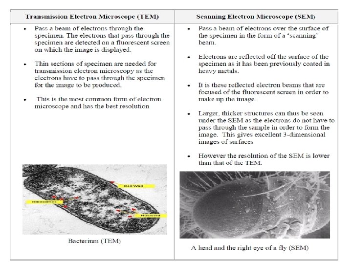
* Types of electron microscopes 1. Scanning electron microscope (SEM) * The mode of action for the SEM is similar to compound microscope however, an electron beams behave like waves which focus via using a magnetic field rather than uses of ordinary lenses.
• Metallic coating is required for the biological specimens. • The electron microscopes are used to achieve up to 100, 000 x magnification and more than 1000 x resolution than the light microscope.
Components of the SEM 1 - Lens: It is an electrical field and are not the optical materials (like glass Electron optics: a- Condenser lens: It is focusing the electron beam to the objective lens. b- Objective lens: It is responsible for size of electron beam that hit sample surface. 2 - Electron beam. 3 - Transducers (detectors).
2. Transmission Electron Microscope: In transmission electron microscopy (TEM), a beam of highly focused electrons are directed toward a thinned sample (<200 nm). Normally no scanning required helps the high resolution, compared to SEM.
* Advantages: 1 - TEMs offer the most powerful magnification, potentially over one million times or more 2 - Direct imaging of crystalline lattice. 3 - No metallic stain-coating is needed, thus convenient for structural imaging of organic materials. 4 - Images are high-quality and detailed. 5 - Electrons can only travel through a vacuum, so the specimen must be completely dehydrated. 6 - Electrons have poor penetrating ability which the image contrast results when electrons are scattered by the specimen. Therefore specimens are usually “stained” with a coat of heavy metal (uranium, osmium, and tungsten) to increase scattering ability.
* Disadvantages: 1 - TEMs are large and very expensive 2 - Images are black and white 3 - The preparation is limited to an electron-transparent sample (due to the conductivity or electron density, and sample thickness).
Confocal Microscopy (Confocal Scanning Laser Microscope) Laser scanning confocal microscopy is an invaluable tool for a wide range of investigations in the biological and medical sciences for imaging thin optical sections in living and fixed specimens ranging in thickness up to 100 micrometers.
The basic concept of confocal microscopy was originally developed by Marvin Minsky in the mid-1950 s (patented in 1961) when he was a postdoctoral student at Harvard University
Minsky wanted to image neural networks in unstained preparations of brain tissue. The basic key to the confocal approach is the use of spatial filtering techniques to eliminate out-of-focus light or glare in specimens whose thickness exceeds the immediate plane of focus
Advantages of using confocal microscope Confocal microscopy offers several advantages over conventional wide field optical microscopy: 1 - The ability to control depth of field, elimination or reduction of background information away from the focal plane. 2 - The capability to collect serial optical sections from thick specimens.