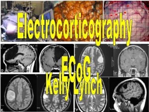
Electrocorticography ECo G ECo G Recording electrical signal


- Slides: 16

Electrocorticography ECo. G

ECo. G ? ? ? • Recording electrical signal in brain • How? • Placing electrodes on surface of the brain • Craniotomy or Burr holes • Searching what? • Epileptic zones • What is that? • Epileptic seizers

Why not EEG? ? ? Resource : http: //meg. aalip. jp/vs. EEGE. html

History at a glance • 1930 – Dr. Wilder Penfield developed “Montreal Procedure” to detect “auras” • 1948 – Rudolf M. Hess conducted first scalp EEG • 1949 – Hugo Krayenbuhl and Hess first attempt ECo. G • 1950 – Herbart Jasper and Dr. Wilder combined both to develop ECo. G method

Functions • Electrode placement • Electrodes • Monitoring • Signal acquirer • g. USBamp • Filtering

Clinical usage • Where this is being used? • Pre surgical planning • Evaluating the success of the surgery • For research aspects • Flexible in stimulating and recording signal » but • Limited field of view and influence of anesthetic

Limitations • • • Spontaneous brain activities Seizures are rarely recorded Needs quick decisions Limited sampling time Limited field of view Influence of anesthetics

Two forms • Extra-operative ECo. G • Intra-operative ECo. G www. thechildren. com/departments-and-staff/departments/department-of-neurophysiology

Two forms… (ctd) • Extra-operative ECo. G • Before a resectioning • EEG and MRI is also used » Structural lesion • Identifying exact place

Two forms… (ctd) • Intra-operative ECo. G • While resectioning • Monitor alleged tissue • Localizing critical zones

Research applications • Brain Computer Interface • Neural signal interface • Hybrid invasive Reference - http: //kin 450 -neurophysiology. wikispaces. com/Brain-Computer+Interface

Specifications • Typical time for an investigation? ü 40 min • Investigating time period? ü 1 - 2 weeks • Temporal resolution? ü 5 ms • Spatial resolution? ü 1 cm

Safety concerns • Invasive • Minor mistakes but Major cost

Risk Assessment • Maintenance Strategy Worksheet • • • Clinical function Physical risk Problem avoidance probability Incident history Manufacturer requirement

Any questions?

 Secuencia gradiente de eco
 Baseband signal and bandpass signal
 Digital signal as a composite analog signal
 Classification of signals
 Baseband signal and bandpass signal
 Recording log
 Zone bit recording
 Process recording example
 Analyzing and recording transactions
 Acoustic era
 Rough sketch and final sketch
 Posting journal entries to ledger
 Maintenance of records and reports
 Siprec recording
 When to use partial interval recording
 Litchfield recording studio
 Permanent product recording aba
