
Electrocardiography Application Normal and Abnormal Heart Rhythms Reading
Electrocardiography: Application, Normal and Abnormal Heart Rhythms Reading assignment: �CTVT pp: 933 -944 Stop at equine emergency nursing �VTDRG pp:
Applications of the Electrocardiogram �Acute onset of ______ �Shock �Fainting or _______ �Monitoring during and after _____ (monitors depth of anesthesia as well as ________ monitoring). �All cardiac ________. �_________ that is found on thoracic radiographs �___________ (with hx of heart issues or for all pets ages 7+
Applications of ECG continued �________ �Evaluating effect of cardiac drugs �Pericardiocentesis �Systemic diseases �________ disturbances ◦ What electrolytes do you think may be associated with ECG abnormalities?
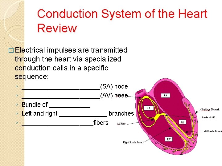
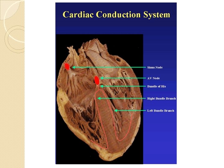
Conduction System of the Heart Review � Electrical impulses are transmitted through the heart via specialized conduction cells in a specific sequence: ◦ ◦ ◦ ____________(SA) node ____________(AV) node Bundle of ______ Left and right _______ branches ___________fibers
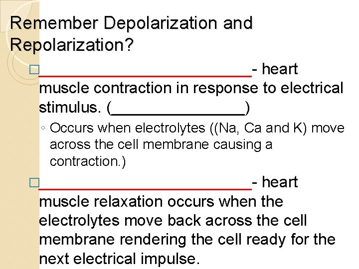
Remember Depolarization and Repolarization? �____________- heart muscle contraction in response to electrical stimulus. (________) ◦ Occurs when electrolytes ((Na, Ca and K) move across the cell membrane causing a contraction. ) �____________- heart muscle relaxation occurs when the electrolytes move back across the cell membrane rendering the cell ready for the next electrical impulse.
Five Physiologic Properties of Cardiac Muscle �Automaticity �Excitability �Refractory Period �Conductivity �Contractility
Five Physiologic Properties of Cardiac Muscle 1. Automaticity ◦ _____ node is the primary pacemaker of the heart. ◦ Any cells of conduction system can initiate their own impulses under right circumstances. ◦ The further down in the conduction system, the _________ the rate of automaticity 2. Excitability ◦ Cardiac muscle is excited when the electrical stimulus reduces the _______ potential to the ________ potential ◦ The degree of the resting potential within the cell determines its excitability and obeys the “_____-or_____” principle.
3. Refractory Period ◦ Heart muscle will not respond to external stimuli during its period of contraction (_______ period in between beats) 4. Conductivity ◦ Activation of an individual muscle cell produces _______ in the neighboring muscle cell. ◦ Conduction velocity varies in the different portions of the specialized conduction system and muscle fibers ◦ Velocity is _______ in the Purkinje Fibers and _______ in the mid-portion of the AV node. ◦ Activation sequence is so arranged that the ________ mechanical efficiency is provided from each corresponding contraction
5. Contractility ◦ Occurs in response to ________ current ◦ The ECG only measures the _______ for contraction - not the actual contraction itself. ◦ ECG is the tool of choice for measuring ___________.
Major Confusion!!! � Reading an ECG and deciphering what it is telling you can be confusing. � ECGs provide a graphical representation of the _______ impulses of the heart. � Electrical impulses are what make the heart contract. � This contraction is a result of ____________and ____________.
Electrocardiogram �Definition: Graphic recording of electrical potentials produced by heart muscle during the different _______of the cardiac cycle. �The information recorded on the EKG represents the heart’s _________ activity. �Most of the information on the EKG represents electrical activity of contraction of the __________.
A Little Vocabulary � __________ : A wave that arises from sources other than the heart. � Cardiac _________: Any electrical activity of the heart that differs from that of a healthy, awake patient. Can be inoccuous or life-threatening. � ____________ : Movement from one side to the other. � __________: Occurring in an abnormal location/position. � ________ : An electrical pathway created by the connection of electrodes. Provides a particular view of the electrical activity of the heart.
Solid Ground �Having the ECG machine well grounded is an important factor for obtaining an accurate reading. �Placing the patient on a rubber mat and ensuring that the machine is plugged into a grounded outlet as well as checking the ECG machine for loose wires or cracked insulation on wires are steps that can be taken for a good ground.
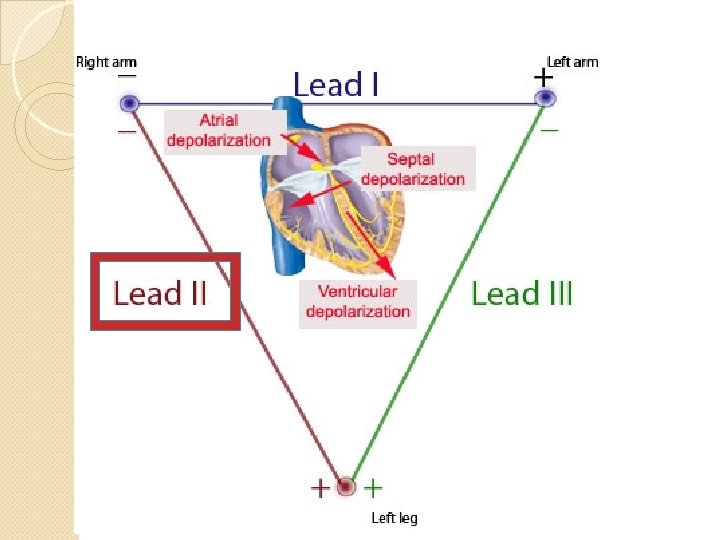
ECG Leads �Are the paths that measure the electrical activity of the heart �Leads are _______ or ______. (Settings on ECG machine determine which lead you are recording. ) �The most common leads or paths of the heart are I, II, and III.
Leads � Selecting specific leads on the ECG machine will cause specific cables to become negative or positive poles. � Lead I: causes right forelimb to become a negative pole and left forelimb to become a positive pole. � ***** Lead II: causes right forelimb to become negative and left rear limb to become positive. (Most commonly used) � Lead III: the left hind limb become a positive pole and the left forelimb becomes a negative pole.
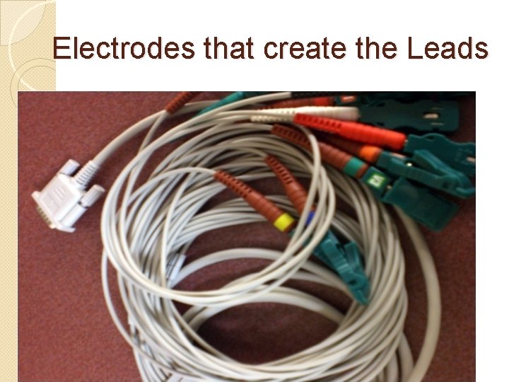
Electrodes that create the Leads
The Electrodes �There are 4 electrodes that are used in veterinary medicine: �______, ___________and ______ �The electrodes are placed on the patient in the _______ locations EVERY time.
Placing the Electrodes Correctly �In veterinary medicine, the following is the standard for applying the electrodes to all patients for ECG monitoring: ◦ ◦ White electrode: __________ limb Black electrode: __________ limb Green electrode: __________ limb Red electrode: __________ limb �“Clouds over grass and smoke over fire. ”
Color-Coded Cables Placement �Black and white on front limbs ◦ Placed at the __________ region �Green and red on back limbs ◦ Placed at the __________ region �_________, electrode gel, or ECG pads can be used as conduction agents for more accurate readings. If your patient has long hair, it should be shaved at the site of cable placement in order to achieve adequate conduction.
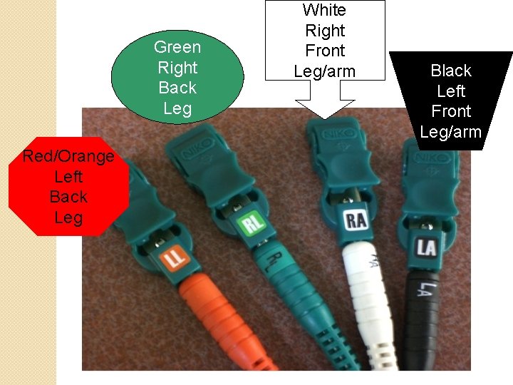
Green Right Back Leg Red/Orange Left Back Leg White Right Front Leg/arm Black Left Front Leg/arm
Leads Continued… �_________ deflection on the ECG-produced when electrical impulses travel towards a _________electrode. �_________ deflection on the ECG-produced when impulses travel toward a ___________electrode. �_________: produced when there is no electrical spread through the heart. �***We only need to use lead II to assess arrhythmias in animals.

Electrocardiograph used in SX
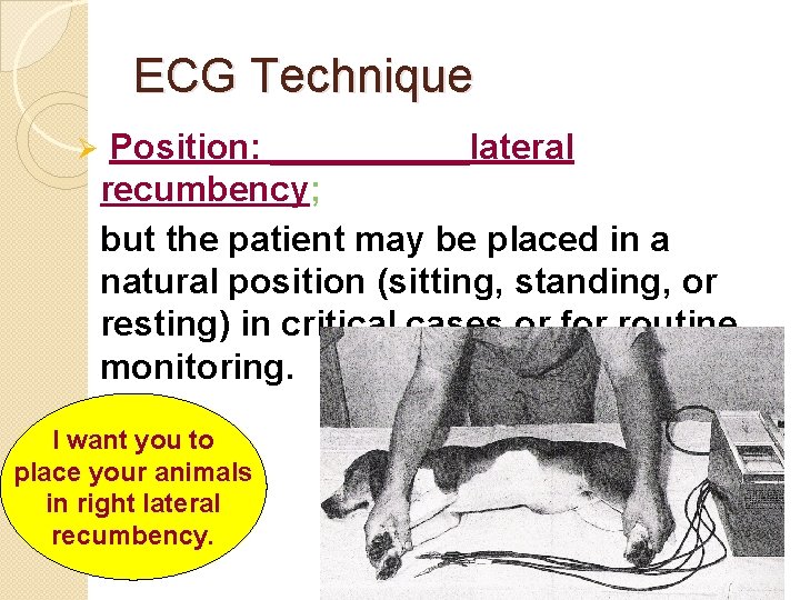
ECG Technique Ø Position: _____lateral recumbency; but the patient may be placed in a natural position (sitting, standing, or resting) in critical cases or for routine monitoring. I want you to place your animals in right lateral recumbency.
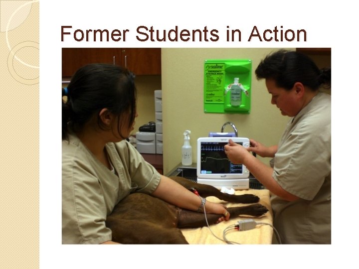
Former Students in Action
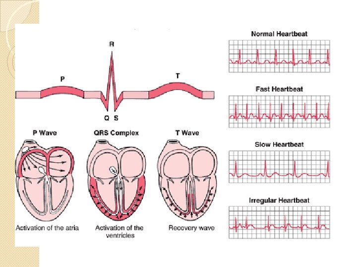
Wave Forms �Connection of the amplifier to selected body parts (usually the limbs) with wires (known as electrodes) and to a recorder (electrocardiograph), provides a characteristic wave form. �The wave form is a recording of the electrical activity of the heart. This wave form recording is the electrocardiogram (ECG). �Each event has a distinctive waveform.
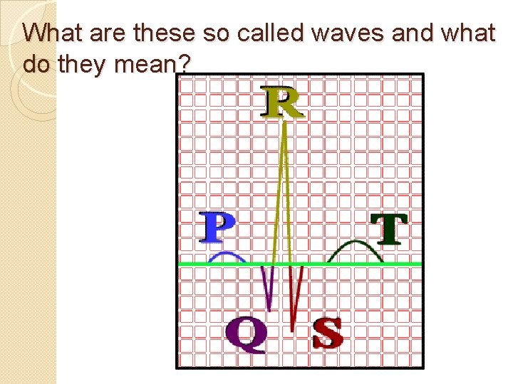
What are these so called waves and what do they mean?
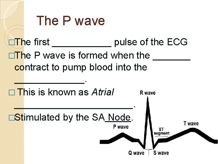
The P wave �The first ______ pulse of the ECG �The P wave is formed when the _______ contract to pump blood into the _______. � This is known as Atrial ___________. �Stimulated by the SA Node.
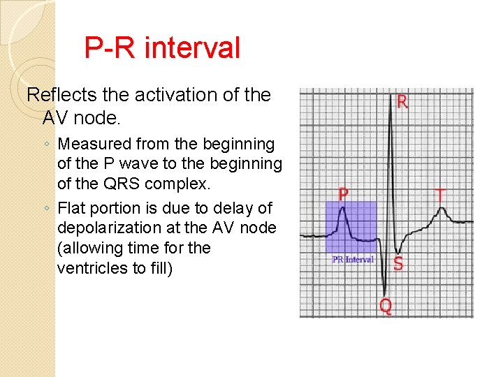
P-R interval Reflects the activation of the AV node. ◦ Measured from the beginning of the P wave to the beginning of the QRS complex. ◦ Flat portion is due to delay of depolarization at the AV node (allowing time for the ventricles to fill)
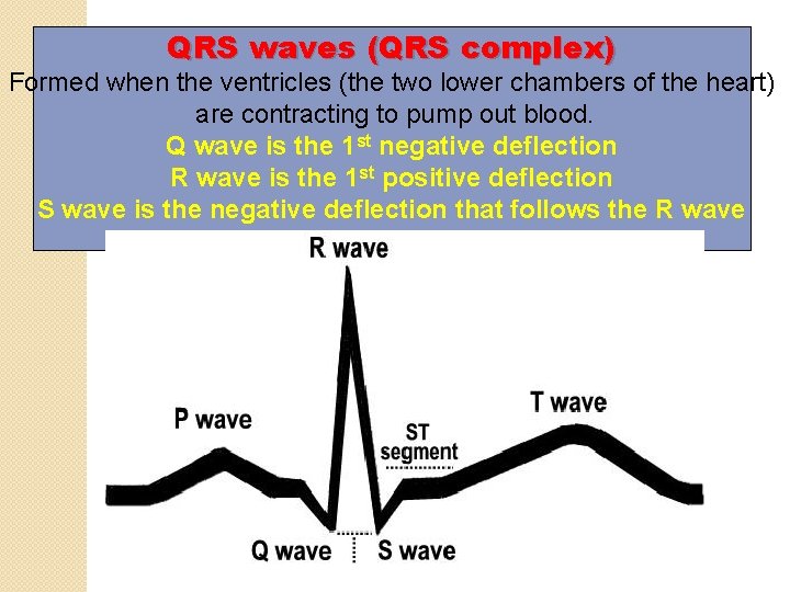
QRS waves (QRS complex) Formed when the ventricles (the two lower chambers of the heart) are contracting to pump out blood. Q wave is the 1 st negative deflection R wave is the 1 st positive deflection S wave is the negative deflection that follows the R wave
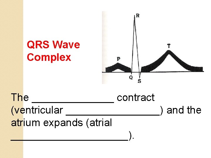
QRS Wave Complex The _______ contract (ventricular ________) and the atrium expands (atrial __________).
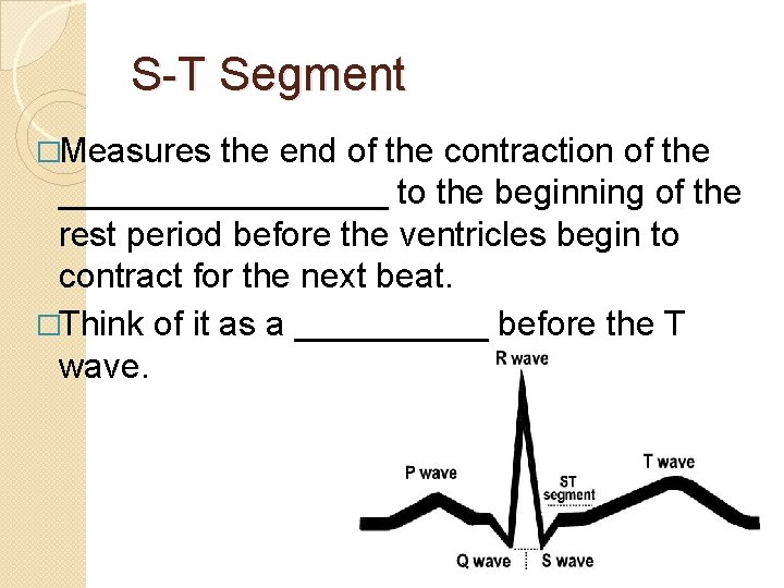
S-T Segment �Measures the end of the contraction of the _________ to the beginning of the rest period before the ventricles begin to contract for the next beat. �Think of it as a _____ before the T wave.
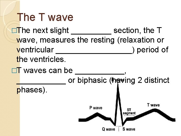
The T wave �The next slight _____ section, the T wave, measures the resting (relaxation or ventricular _________) period of the ventricles. �T waves can be ______, ______ or biphasic (having 2 distinct phases).
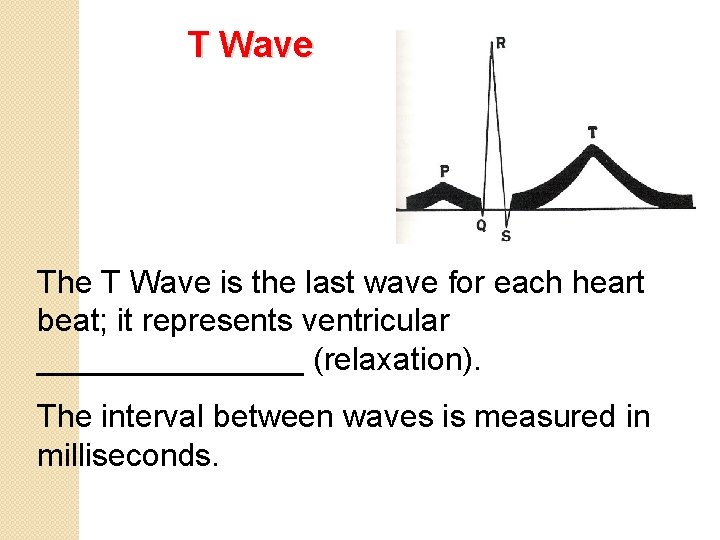
T Wave The T Wave is the last wave for each heart beat; it represents ventricular ________ (relaxation). The interval between waves is measured in milliseconds.
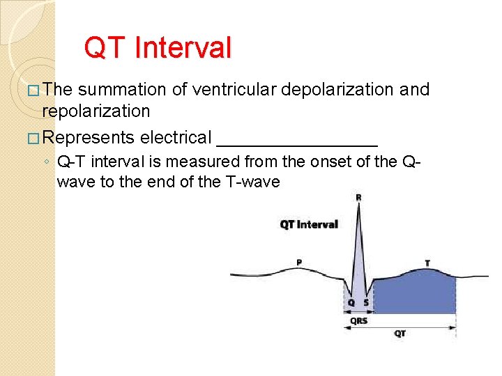
QT Interval � The summation of ventricular depolarization and repolarization � Represents electrical ________ ◦ Q-T interval is measured from the onset of the Qwave to the end of the T-wave
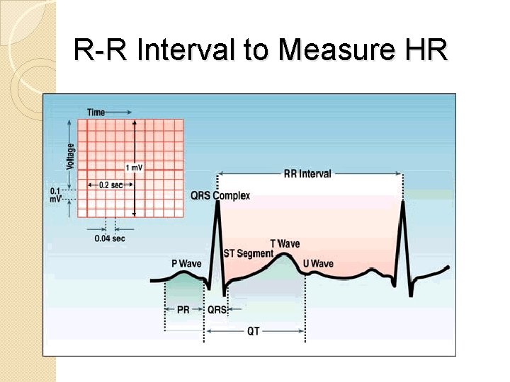
R-R Interval to Measure HR
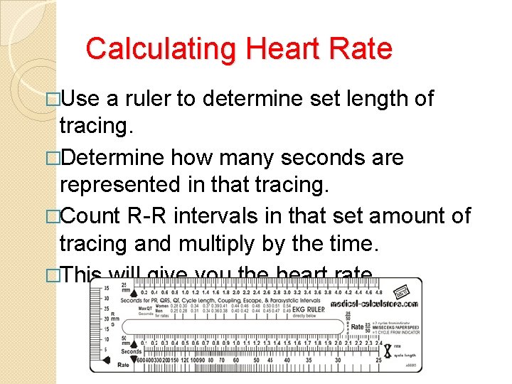
Calculating Heart Rate �Use a ruler to determine set length of tracing. �Determine how many seconds are represented in that tracing. �Count R-R intervals in that set amount of tracing and multiply by the time. �This will give you the heart rate.
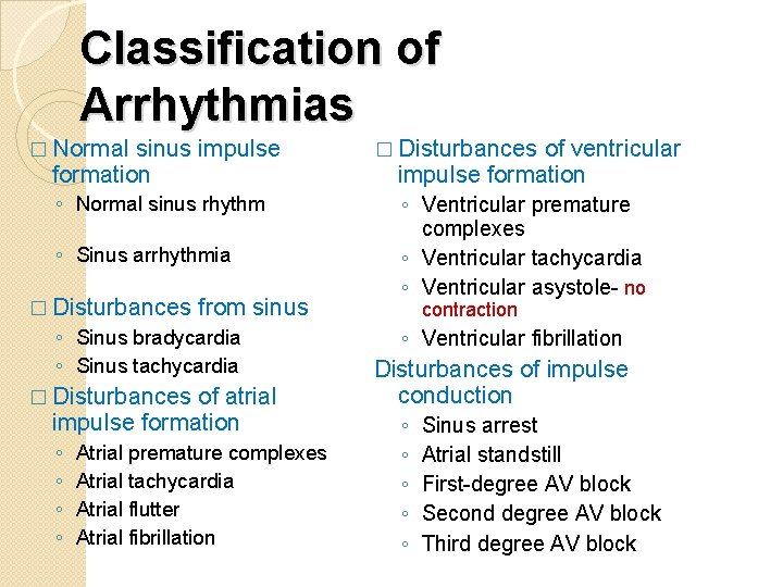
Classification of Arrhythmias � Normal sinus impulse formation ◦ Normal sinus rhythm ◦ Sinus arrhythmia � Disturbances from ◦ Sinus bradycardia ◦ Sinus tachycardia sinus � Disturbances of atrial impulse formation ◦ ◦ Atrial premature complexes Atrial tachycardia Atrial flutter Atrial fibrillation � Disturbances of ventricular impulse formation ◦ Ventricular premature complexes ◦ Ventricular tachycardia ◦ Ventricular asystole- no contraction ◦ Ventricular fibrillation Disturbances of impulse conduction ◦ ◦ ◦ Sinus arrest Atrial standstill First-degree AV block Second degree AV block Third degree AV block
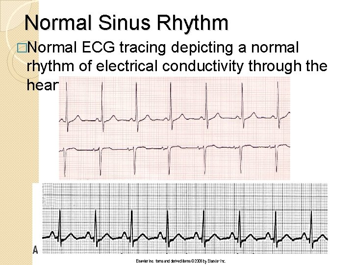
Normal Sinus Rhythm �Normal ECG tracing depicting a normal rhythm of electrical conductivity through the heart
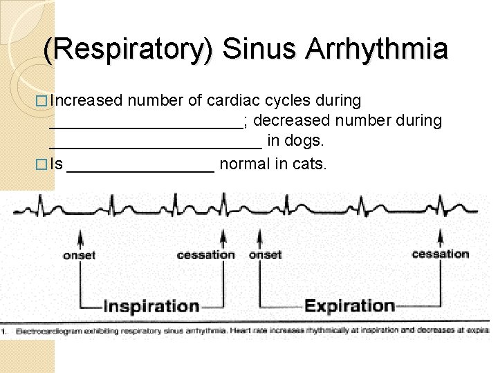
(Respiratory) Sinus Arrhythmia � Increased number of cardiac cycles during ___________; decreased number during ____________ in dogs. � Is ________ normal in cats.
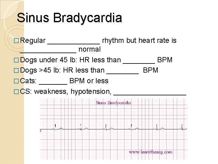
Sinus Bradycardia � Regular _______ rhythm but heart rate is _______ normal � Dogs under 45 lb: HR less than ____ BPM � Dogs >45 lb: HR less than ____ BPM � Cats: _______ BPM or less � CS: weakness, hypotension, _________
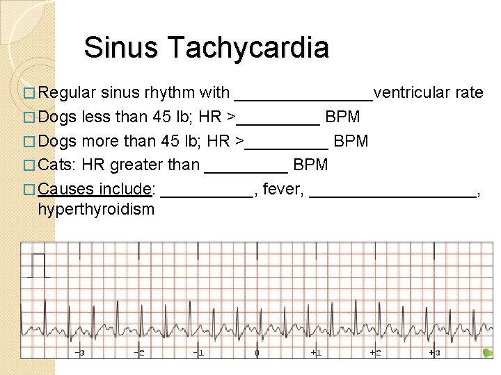
Sinus Tachycardia � Regular sinus rhythm with ________ventricular rate � Dogs less than 45 lb; HR >_____ BPM � Dogs more than 45 lb; HR >_____ BPM � Cats: HR greater than _____ BPM � Causes include: _____, fever, _________, hyperthyroidism
Atrial Premature Complexes � Premature atrial impulses originating from atrial site other than ____ node � Seen in dogs and cats with ______enlargement, electrolyte disturbances, drug reactions, congenital heart disease, and ______ � Premature P wave causes a heartbeat sooner than it should be � QRS complexes are ________ unless the P wave is so immature that it overlaps to varying degrees
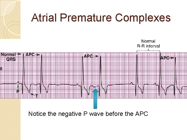
Atrial Premature Complexes Notice the negative P wave before the APC
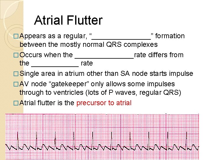
Atrial Flutter � Appears as a regular, “________” formation between the mostly normal QRS complexes � Occurs when the ________rate differs from the ______ rate � Single area in atrium other than SA node starts impulse � AV node “gatekeeper” only allows some impulses through to ventricles (lots of P waves, regular QRS) � Atrial flutter is the precursor to atrial ________
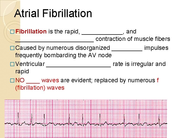
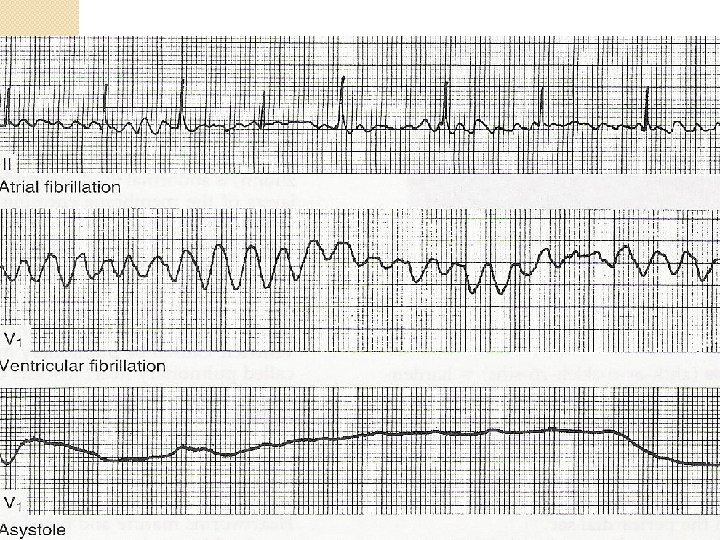
Atrial Fibrillation � Fibrillation is the rapid, ______, and ____________ contraction of muscle fibers � Caused by numerous disorganized _____ impulses frequently bombarding the AV node � Ventricular __________ rate is irregular and rapid � NO ____ waves are evident; replaced by numerous f (fibrillation) waves
Ventricular Premature Complexes (VPCs) �“_______beats” - cardiac impulses initiated within the _________ instead of the sinus node � Ventricle discharges before the arrival of the next anticipated impulse from the ______ node. (Takes a shortcut) � Can occur at any rate but pose a greater danger with _________. � Associated with ________ defects, cardiomyopathy, GDV, drug reactions, cardiac neoplasia, ______, acidosis, hyperthyroidism, hypokalemia � Also referred to as Premature Ventricular Complexes or PVCs.
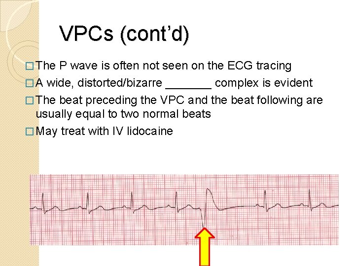
VPCs (cont’d) � The P wave is often not seen on the ECG tracing � A wide, distorted/bizarre _______ complex is evident � The beat preceding the VPC and the beat following are usually equal to two normal beats � May treat with IV lidocaine
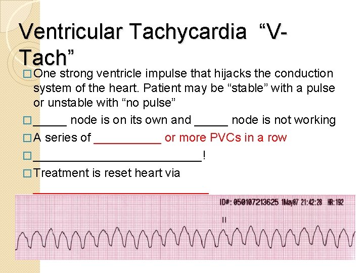
Ventricular Tachycardia “VTach” � One strong ventricle impulse that hijacks the conduction system of the heart. Patient may be “stable” with a pulse or unstable with “no pulse” � _____ node is on its own and _____ node is not working � A series of _____ or more PVCs in a row � _____________! � Treatment is reset heart via _____________
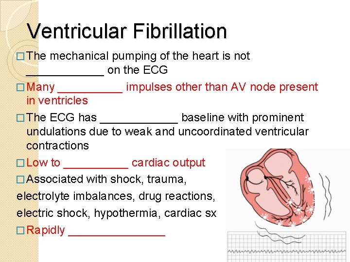
Ventricular Fibrillation � The mechanical pumping of the heart is not ______ on the ECG � Many _____ impulses other than AV node present in ventricles � The ECG has ______ baseline with prominent undulations due to weak and uncoordinated ventricular contractions � Low to _____ cardiac output � Associated with shock, trauma, electrolyte imbalances, drug reactions, electric shock, hypothermia, cardiac sx � Rapidly ________
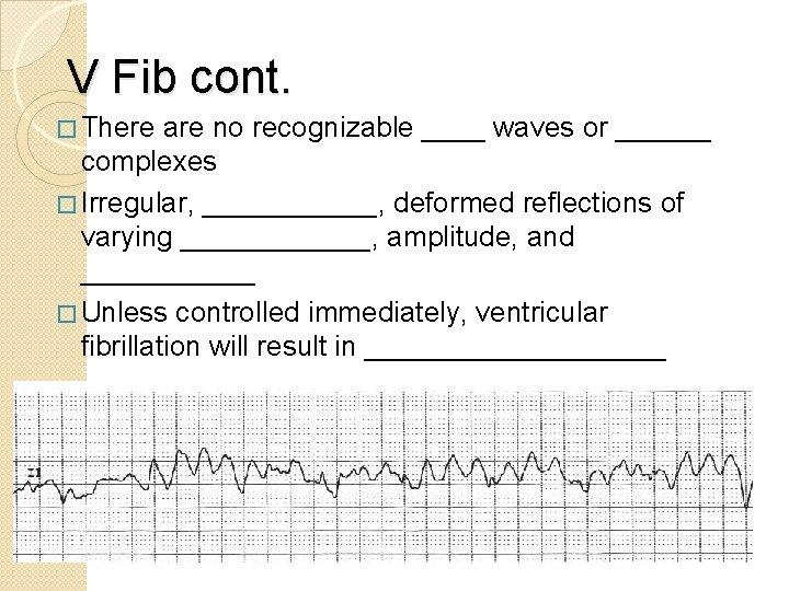
V Fib cont. � There are no recognizable ____ waves or ______ complexes � Irregular, ______, deformed reflections of varying ______, amplitude, and ______ � Unless controlled immediately, ventricular fibrillation will result in __________.
Conduction Issues �_______ Standstill �Heart ______ ◦ First degree ◦ Second degree ◦ Third degree �_________
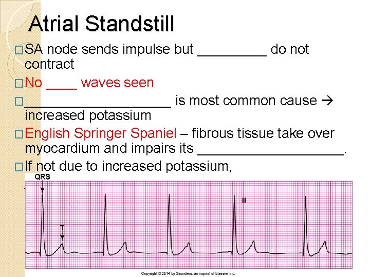
Atrial Standstill �SA node sends impulse but _____ do not contract �No ____ waves seen �__________ is most common cause increased potassium �English Springer Spaniel – fibrous tissue take over myocardium and impairs its __________. �If not due to increased potassium, _________ is warranted.
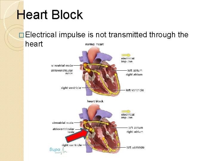
Heart Block � Electrical heart impulse is not transmitted through the
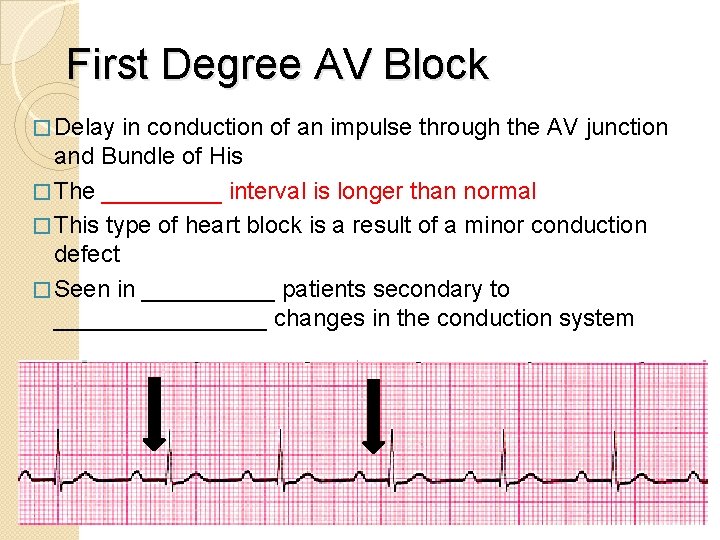
First Degree AV Block � Delay in conduction of an impulse through the AV junction and Bundle of His � The _____ interval is longer than normal � This type of heart block is a result of a minor conduction defect � Seen in _____ patients secondary to ________ changes in the conduction system
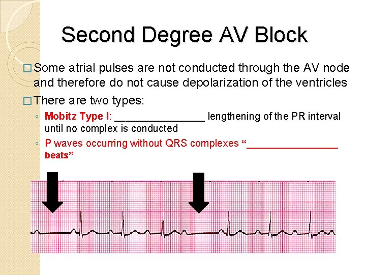
Second Degree AV Block � Some atrial pulses are not conducted through the AV node and therefore do not cause depolarization of the ventricles � There are two types: ◦ Mobitz Type I: ________ lengthening of the PR interval until no complex is conducted ◦ P waves occurring without QRS complexes “_________ beats”
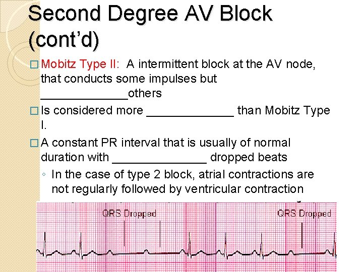
Second Degree AV Block (cont’d) � Mobitz Type II: A intermittent block at the AV node, that conducts some impulses but _______others � Is considered more _______ than Mobitz Type I. � A constant PR interval that is usually of normal duration with _______ dropped beats ◦ In the case of type 2 block, atrial contractions are not regularly followed by ventricular contraction ◦ 2 or more dropped QRS in a row
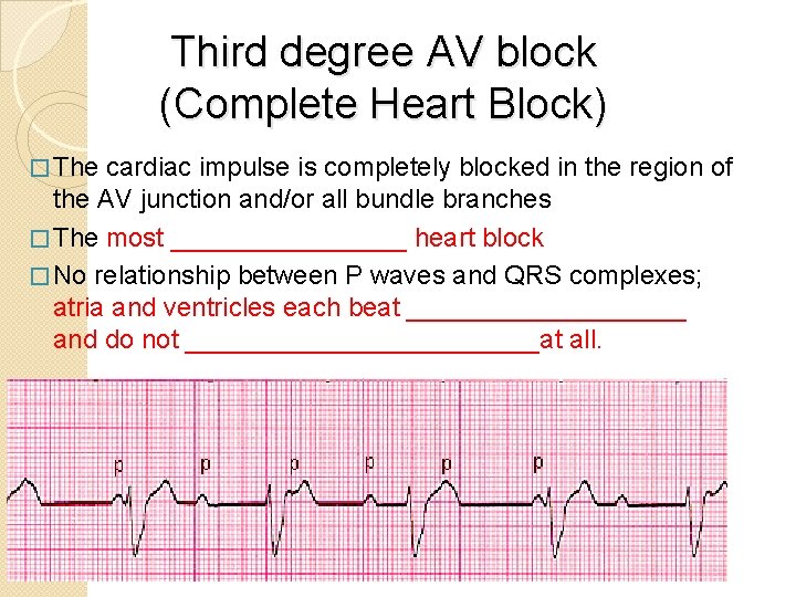
Third degree AV block (Complete Heart Block) � The cardiac impulse is completely blocked in the region of the AV junction and/or all bundle branches � The most ________ heart block � No relationship between P waves and QRS complexes; atria and ventricles each beat __________ and do not ____________at all.
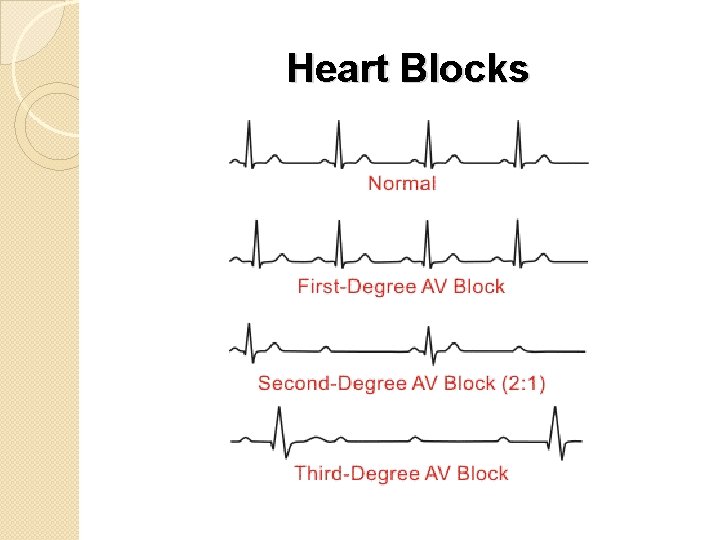
Heart Blocks
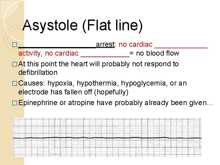
Asystole (Flat line) � __________arrest: no cardiac _______ activity, no cardiac ______= no blood flow � At this point the heart will probably not respond to defibrillation � Causes: hypoxia, hypothermia, hypoglycemia, or an electrode has fallen off (hopefully) � Epinephrine or atropine have probably already been given…
DONE with ECG/Arrhythmias!
- Slides: 64