Electrocardiograph What is mean the electrocardiogram ECG is


- Slides: 41

Electrocardiograph

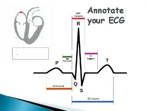
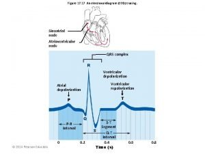
What is mean the electrocardiogram? (ECG) is a diagnostic tool that measures and records the electrical activity of the heart in detail. The ECG is composed of six waves three of these waves form complex and between these waves three are two intervals and one segment

The importance of ECG From the ECG tracing, the following information can be determined: -the heart rate -the heart rhythm -whethere are “conduction abnormalities” (abnormalities in how the electrical impulse spreads across the heart) -whethere has been a prior heart attack -whethere may be coronary artery disease -whether the heart muscle has become abnormally thickened

What is an action potential? An action potential is the point at which the cells become electrically activated and in the case of cardiac myocytes contract, what we know is that flow of ions As an increasing amount of positive sodium ions enter from outside of the cell the voltage becomes increasingly positive and the cell begins to depolarize once the voltage reaches a critical value it is triggered to produce an action potential (action potential results in firing of the SA node

The sinus node depolarizes on average just over once every second causing the heart to bet at rate of between 60 to 70 times every minute The cells then return to their resting state via a process of depolarization during this time (+)charged K+ ions are actively pumped out of the cell hence the inside of the cell becomes less (+) and the resting state is restored.

Electricity travels through the heart Sinoatrial node AV node Bundle of His Bundle Branches Purkinje fibers

Electricity travels through the heart The electricity starts in the sino-atrial node (acronym SAN. ) The SAN is a group of cells in the right atria. These cells start an electrical impulse. This electrical impulse travels through the atria making them contract. This motion is called 'atrial systole'. Once electrical impulse goes through the atrio-ventricular node (acronym AVN. ) The AVN. . makes the impulse decrease

Going slower through the AVN making impulses get to the ventricles at a later time. That is what makes the ventricular systole occur after atrial systole. Once going through the AVN occurs, the electrical impulse goes through the conduction system of the ventricle. This makes the electrical impulse travel to the ventricles. The first part of the conduction system is the bundle of His

Electricity travels through the heart Once the bundle goes through the ventricle muscle it is separate from it. Afterwards the bundle of His divides into bundle branches that are from left to right. The left bundle branch travels to the left ventricle. The right bundle branch travels to the right ventricle. At the end of the bundle branches, the electrical impulse goes into the ventricular muscle. This is what makes ventricle contraction take place and makes. ventricular systole

The electrocardiograph leads: There are twelve conventional ECG lead are: -extremity leads-six in number. -chest leads –six in number.

The limb leads An electrode is placed on each of three limbs namely right arm, left arm and left leg. There are 1 - standard limb leads. 2 -augmented limb leads.

1 - standard limb leads: Obtain a graph of the electrical forces as recorded between two limbs at a time. Also called bipolar leads. because one limb carries positive electrode and other limb carries a negative electrode. There are three standard limb leads. lead Positive negative I LA RA II LL RA III LL LA

2 -augmented limb leads: Obtain a graph of the electrical forces as recorded from one limb at a time. Also called unipolar leads. There are three augmented limb leads: lead a. VR a. VL a. VF Positive RA LA LL

The limb leads:


The chest leads V 1: right 4 th intercostals space right of sternal V 2: left 4 th intercostals space left of sternal V 3: halfway between V 2 and V 4: left 5 th intercostals space, mid-clavicle line V 5: horizontal to V 4, anterior axillary's line. V 6: horizontal to V 5, mid-axillary's line.

The chest leads: .

The ECG normal values:

how to read the ECG: P-wave *Represents atrial depolarization-at the time of the electrical impulse from the (SA) node to spread throughout the atria musculature. -Height: not exceed 2 to 2. 5 mm. -Duration: 0. 06 to 0. 11 second.

Are the P waves…. Too tall >2. 5 mm Too wide>0. 08 s Inverted - right atrial enlargement -left atrial enlargement. -Av Junction rhythm. -normal in a. VR. -coronary sinus rhythm (lead II, III, and a. VF) -left atrial rhythm (leads II, III , a. VF) -arm lead reversal.

Absent inus arrest and sinoatrial block. -atrial fibrillation. -atrial flutter. - hyperkalaemia.

P-R interval: *P-R interval represent the time it take impulse to travel from the atria through the AV node , bundle of branch to the purkinje fibers. -extends from the beginning of the p wave to the beginning of the QRS complex. -duration: 0. 12 to 0. 20 seconds.

Is are the PR interval: Too long -first degree AV block. -hypercalcaemia. -hyperkalaemia. -hypokalaemia. -drug effects. Too short hasten AV conduction -AV junctional rhythm.

-wolf –parkinsin white syndrome. ype I second degree AV block. -third degree AV block. -multifocal tachycardia

QRS complex: Represents ventricle depolarization. the QRS complex consist of 3 waves: 1 -the. Q wave : is always located at the beginning of the QRS complex. 2 - the R wave : is always the first positive deflection. 3 -the S wave , the negative deflect follows the R wave.

-height: no greater than 25 mm. -duration : no longer than 0. 10 seconds.

Are the QRS complexes… Too big -left ventricle hypertrophy. -myocardial infarction. -right ventricle hypertrophy. -wolf parkinson white. as lung disease (emphysema) -pt is obese.

-pericardial effusion. Too wide &right bundle branch block -hyperkalemia. -none ventricular conduction delay -aberrant conduction.

Varying in size Absent A systole during cardiac arrest pericardial effusion -ventricle standstill.

QT interval: Represents the time necessary for ventricle depolarization and repolarization. Extend from the beginning of the QRS complex to the end of T wave. Duration: o. 35 -o. 43 second.

Is the QT interval: Too long Too short -cardiomyopathy (hypertrophic -hypocalcaemia -hypothyroidism -MI -myocarditis. -hypercalcamia. -drug effects e. g. digoxin

T wave: Represents the repolarization of the ventricles. on rare occasions, a U wave can be seen following the T wave. the U wave reflects the repolarization of the his purkinje fibers. Hiegt: 5 mm or less in leads I, II, and III, 10 mm or less in leads. V 1 -V 6.

Are the T wave: Too tall too small -hyperkalaemia. -MI. -hypokalemia. -hypothyroidism. -pericardial effusion.

Inverted -hyperventilation. -hypothyroidism. -left bundle branch block (lead V 1 -V 4) -Mitral valve prolepses. -myocardial ischaemia. -myocardial infarction. -pulmonary embolism(V 1 -V 3

-right bundle branch block (leads V 1 -V 3) -subarachnoid haemorrhag. -pacemaker rhythm.

S-T segments Represents the end of the ventricl depolarization and the beginning of ventricle repolarization. -extend from the end of S wave to the beginning of the T wave.

Are the ST segments: -early repolarisation. -left bundle branch block. -left ventricular aneurysm. -MI -pericarditis. Elevated

ects. hypertrophy. - MI. entricular. -myocardial ischemia.

Calculate heart Rate 3 sec Option 1 l Count the # of R waves in a 6 second rhythm strip, then multiply by 10 Interpretation? 9 x 10 = 90 bpm •

R wave Option 2 l Find a R wave that lands on a bold line. • Count the # of large boxes to the next R wave. If the second R wave is 1 large box away the rate is 300, 2 boxes - 150, 3 boxes - 100, 4 boxes - 75, etc. (cont) •

3 1 1 0 5 0 7 6 5 0 0 0 5 0 0 Memorize the sequence: • 300 - 150 - 100 - 75 - 60 - 50 Interpretation? Approx. 1 box less than 100 = 95 bpm
 Guyton ecg
 Từ ngữ thể hiện lòng nhân hậu
 Trời xanh đây là của chúng ta thể thơ
 Tư thế ngồi viết
 Voi kéo gỗ như thế nào
 Ví dụ về giọng cùng tên
 Thể thơ truyền thống
 Sự nuôi và dạy con của hổ
 đại từ thay thế
 Thế nào là hệ số cao nhất
 Diễn thế sinh thái là
 Vẽ hình chiếu vuông góc của vật thể sau
 Thế nào là mạng điện lắp đặt kiểu nổi
 Mật thư tọa độ 5x5
 Lời thề hippocrates
 Vẽ hình chiếu đứng bằng cạnh của vật thể
 Thang điểm glasgow
 Quá trình desamine hóa có thể tạo ra
 Khi nào hổ con có thể sống độc lập
 Dạng đột biến một nhiễm là
 điện thế nghỉ
 Các châu lục và đại dương trên thế giới
 Biện pháp chống mỏi cơ
 Bổ thể
 Phản ứng thế ankan
 101012 bằng
 Thiếu nhi thế giới liên hoan
 Vẽ hình chiếu vuông góc của vật thể sau
 Chúa sống lại
 Một số thể thơ truyền thống
 Sơ đồ cơ thể người
 Cong thức tính động năng
 Thế nào là số nguyên tố
 Tỉ lệ cơ thể trẻ em
 đặc điểm cơ thể của người tối cổ
 Các châu lục và đại dương trên thế giới
 ưu thế lai là gì
 Thẻ vin
 Môn thể thao bắt đầu bằng chữ f
 Tư thế ngồi viết
 Bàn tay mà dây bẩn
