Electrical Signaling Resting Membrane Potential Action Potential PostSynaptic

Electrical Signaling Resting Membrane Potential Action Potential Post-Synaptic Potential

Signal Processing Neurons send and receive electrical & chemical signals

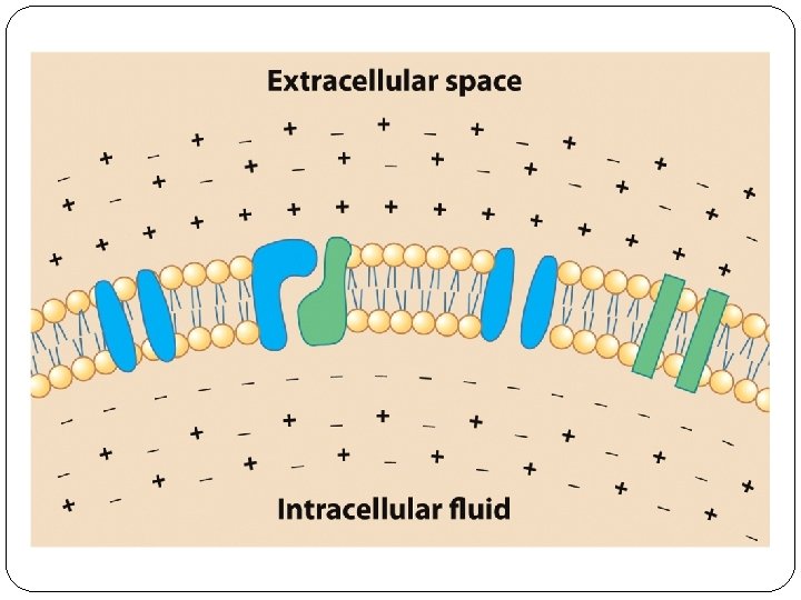
Resting Membrane Potential Difference in electrical charge across membrane when neuron is at rest
 �Developed a mathematical model for how electrical signals (")
Hodgekin & Huxley ( 1952) �Developed a mathematical model for how electrical signals ( action potentials) are produced and transmitted down the axon. �Describes flow of ions across membrane

Hodgekin & Huxley Expt
")
Results �Inside of neuron negatively charged �Differential distribution of ions ( electrically charged particles)

Background & Vocabulary of Electrical Signals �Polarized: difference in electrical charge between the inside and the outside of the cell �Ions—electrically charged molecules �Anions are negatively charged �Cations are positively charged �Ions are dissolved in intracellular fluid, or cytoplasm, and are separated from the extracellular fluid by the cell membrane, a lipid bilayer

Ion: Electrically charged molecule

�Electrons: negatively charged �Proton: positively charged �Total number of electrons is not equal to number of protons

Ions differentially distributed across cell membrane

Figure 3. 2 The Distribution of Ions Inside and Outside of a Neuron

Reasons for Differential Distribution of Ions �Selective permeability: �Diffusion �Electrostatic Force �Sodium-potassium pump
 move freely �Sodium Ions kept outside �WHY? ?")
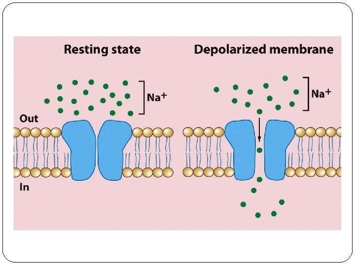
1. Selective Permeability �Potassium ions (K+) move freely �Sodium Ions kept outside �WHY? ? ? �Ion channels: Open for K+, Closed for NA+

Figure 3. 2 The Distribution of Ions Inside and Outside of a Neuron
 kept")
Ion Channels Regulate Flow of Ions � Gated Channels: Sodium Ions ( Na+) kept outside � Selectively Permeable Channels: Potassium ions ( K+) concentrated inside (we’ll see why soon) �Nodes Of Ranvier: location of Channels on unmyelinated parts of axon

2. Diffusion �Ions move from a high concentration to a low concentration in order to create equilibrium

Diffusion & Ions �Na+: �Concentrated outside �Can’t diffuse inside because channels are closed �K+: �Concentrated inside �Moves outside, but other force moves K+ back in

Figure 3. 3 Ionic Forces Underlying Electrical Signaling in Neurons

3. Electrostatic Pressure �Ions of similar charges repel �Ions of opposite charge attract

Electrostatic Force & Ions �Na+: �attracted to the inside of the neuron �but can’t enter due to closed channels �K +: �are attracted to inside of neuron �can enter freely

4. Sodium-Potassium Pump �“Leaky” channels: permit a few Na+ ions to enter �Na+/K+ Pump: exchanges 3 Na+ ions from inside for them for 2 K+ ions from outside

Resting Membrane Potential Summary �Polarization: inside of axon is negative relative to outside �Na+ ions located outside ; K+ ions inside �Na+ ions under pressure to move inside due to: �Diffusion- concentration gradient �Electrostatic force- attraction to oppositely charged area �But, Na+ ions kept outside by: �Selectively permeable membrane ( Na+ gates closed) �Sodium-Potassium-Pump ( actively pumps out Na+

Action Potential Rapid reversal of RMP, that is transmitted down axon towards next neuron

Changes in Electrical Potential Lead to Action Potential � Neurons receive chemical signals from nearby neurons �Excitatory signals depolarize the cell membrane (i. e. , reduce polarization) �Make inside less negative �Inhibitory signals hyperpolarize the cell (i. e. , increase polarization) �Make inside more negative

All inputs integrated at Axon Hillock

If Net Inputs Inhibitory: No Action Potential

If Net Inputs Excitatory, Action Potential Triggered

Action Potential �rapid reversal of inside potential conveyed down the axon to the next neuron �Triggered when membrane reaches the threshold �Critical level of depolarization—about – 40 m. V �The membrane potential reverses and the inside of the cell becomes positive

Figure 3. 5 The Effects of Hyperpolarizing and Depolarizing Stimuli on a Neuron

Local vs. Action Potentials

Local potential �Graded Potential: Size of change in electrical potential coded by strength of input stimulus �The greater the stimulus, the greater the change in membrane potential �Size diminishes as it moves away from the point of stimulation �Occurs at dendrites

Action Potential �All-or-none Potential: �The neuron fires at full amplitude or not at all —the size (amplitude) is independent of stimulus size �Rate Law: Information is encoded in changes in the number of action potentials—with increased stimulus strength more are produced, but the size is the same


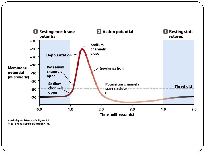
Steps in Action Potential �Depolarization: gradual entry of positive ions due to input from other neurons �Threshold: critical level of depolarization; causes all voltage-gated Na+ gates to open �Sodium-Influx: Na+ ions rush in �pushed by diffusion & Electrostatic forces �Membrane potential inside reverses; becomes positively charged �Potassium-Efflux: K+ ion channels open, allowing K+ to leave

�Peak: point at which Na+ channels close �Repolarization: K+ still leaving axon, leads to membrane potential returning to negative state �Hyperpolarization: Potassium channels close: membrane slightly more negative than RMP �Sodium-Potassium Pump Kicks in: return to RMP

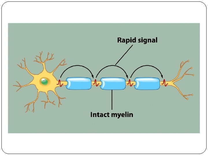
Action Potentials Spread Along Axon �Propagation: process in which Depolarization travels along an axon like a wave �Uni-directional: Action potentials always move away from the cell body to the terminal buttons. �Saltatory Conduction: Action potential regenerated at node of ranvier- rapid conduction
")
Axons (Part 2)

Multiple Sclerosis

What Happens at End of Axon?

Chemical Signaling Synapse Neurotransmitters Neuropharmacology

Electrical Signals into Chemical Signals

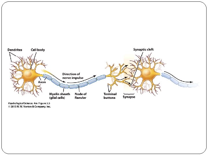
Parts of a Synapse • Presynaptic membrane—on the axon terminal of the presynaptic neuron • Synaptic cleft—a gap that separates the membranes • Postsynaptic membrane—on the dendrite or cell body of the postsynaptic neuron

Steps in Synaptic Transmission 1. Action potential arrives at the presynaptic axon terminal 2. Ca++ Influx: Voltage-gated calcium channels in the terminal membrane open and calcium ions (Ca 2+) enter 3. Exocytosis: Release of Neurotransmitter 1. Calcium ions cause synaptic vesicles fuse with the presynaptic membrane 2. Vesicles rupture, releasing transmitter into the synaptic cleft

4. Receptor Binding: Transmitters bind to specific postsynaptic receptor molecules, causing the opening of ion channels and leading to an EPSP or IPSP 5. Post-synaptic Potential: EPSPs or IPSPs spread toward the postsynaptic axon hillock—if threshold is reached, an action potential will occur

4. Termination of signal: Synaptic transmission is rapidly stopped 4. Re-uptake 5. Enzymatic Breakdown 5. Pre-synaptic modulation: Transmitter may activate presynaptic receptors, decreasing transmitter release

Post-synaptic Potentials

Figure 4. 2 The Versatility of Neurotransmitters

Neurotransmitters Bind With Specific Post. Synaptic Receptors �Receptors are linked to specific ion channels �Channels open when neurotransmitter binds to receptor �Ions move across membrane of dendrites, producing local potentials �EPSP �IPSP
 �Produces a small local depolarization �Makes inside more positive �Results")
Excitatory postsynaptic potential (EPSP) �Produces a small local depolarization �Makes inside more positive �Results from Na+ or Ca++ entering the cell
 �Produces a small hyperpolarization �Makes cell more negative inside �Results")
Inhibitory postsynaptic potential (IPSP) �Produces a small hyperpolarization �Makes cell more negative inside �Results from: �chlorine ions (Cl–) entering the cell �Potassium ions (K+) leaving the cell

IPSPs and EPSPs are local potentials Summed together in integration at hillock

From Brain to Behavior

1. Why don’t we act our dreams?

Motor Neurons are Hyperpolarized during REM

REM Sleep Behavior Disorder �When Patricia Becker noticed a man crouching in the corner while she was in a public restroom, she felt threatened and concerned. In an act of self-defense, she decided to jump on him. That's when she woke up – alone in her bedroom, bloody and bruised.

If IPSPs are missing, motor neurons activated

Signals to muscles now result in behavior

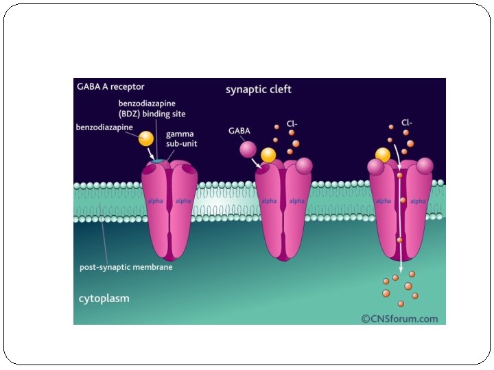
2. Why does Valium decrease Anxiety?
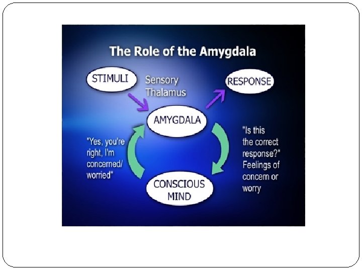

GABA Receptors on Amygdala

Valium binds to GABA Receptors

Binding Lets in more Cl- ions Produces IPSPs, hyperpolarizes amygdala neurons

3. Brain Anatomy & Autism

Smaller at birth; then larger

MRI Results

Rapid growth leads to abnormal connections �"During this period of important learning and plasticity, when the brain is experiencing the world and deciding how to construct itself, it's growing too fast in the infant with autism. �Without the guidance of experience and learning, the brain may be creating abnormal connections that make it very hard for autistic children to make sense of the world they live in. ” • Eric Courchesne

Axonal Disorganization in White Matter

More Folds, Thicker cortex

Abnormal Cortical Layers

More Dendritic Spines

Implications � Regions affected are those that mediate social, emotional, communication, and language functions. �Cortical patches found in temporal & frontal cortex � dendritic spines in temporal lobes �Overconnectivity may lead to overstimulation; epilepsy � Explains why early interventions can be effective: �the developing brain may have a chance to rewire its connections

Mini-Review

1. Which structure is involved in emotional processing? 1. Basal Ganglia 2. Limbic System 3. Thalamus 4. Cerebellum; Pons; Medulla 5. Wernicke’s Area

1. Which structure is involved in emotional processing? 1. Basal Ganglia 2. Limbic System 3. Thalamus 4. Cerebellum; Pons; Medulla 5. Wernicke’s Area

2. Which glia provides myelin for the brain? 1. Astrocytes 2. Microglia 3. Oligodendrocytes 4. Schwann cells

2. Which glia provides myelin for the brain? 1. Astrocytes 2. Microglia 3. Oligodendrocytes 4. Schwann cells

3. Nodes of Ranvier are: �areas in the terminal containing neurotransmitter �areas on the post-synaptic membrane where neurotransmitters bind �areas on the axon where gated ion channels are located �Areas on the pre-synaptic terminal that move neurotransmitter back into the terminal

3. Nodes of Ranvier are: �areas in the terminal containing neurotransmitter �areas on the post-synaptic membrane where neurotransmitters bind �areas on the axon where gated ion channels are located �Areas on the pre-synaptic terminal that move neurotransmitter back into the terminal

4. During the Resting Membrane Potential, the inside of the neuron is _____ relative to the outside & Na +ions are concentrated _______ � 1. negatively charged; outside the neuron � 2. negatively charged; inside the neuron � 3. positively charged; outside the neuron � 4. positively charged; inside the neuron

4. During the Resting Membrane Potential, the inside of the neuron is _____ relative to the outside & Na +ions are concentrated _______ � 1. negatively charged; outside the neuron � 2. negatively charged; inside the neuron � 3. positively charged; outside the neuron � 4. positively charged; inside the neuron

The Rising Phase of the Action Potential � 1. Is due to K+ ions rushing in � 2. Is due to Na+ ions rushing in � 3. Is due to K+ ions rushing out � 4. Reflects rapid depolarization of the membrane � 5. Both 2 & 4

5. The Rising Phase of the Action Potential � 1. Is due to K+ ions rushing in � 2. Is due to Na+ ions rushing in � 3. Is due to K+ ions rushing out � 4. Reflects rapid depolarization of the membrane � 5. Both 2 & 4

6. IPSPs � 1. are graded potentials � 2. reflect hyperpolarization of the membrane � 3. are caused by CL- ions entering the neuron � 4. All of the above

6. IPSPs � 1. are graded potentials � 2. reflect hyperpolarization of the membrane � 3. are caused by CL- ions entering the neuron � 4. All of the above

7. Neurotransmitter release � 1. Is triggered by K+ influx during the Action Potential � 2. Can be increased or decreased by drugs � 3. causes IPSPs or EPSPs at receptors � 4. Both 2 & 3 � 5. All of the above

7. Neurotransmitter release � 1. Is triggered by K+ influx during the Action Potential � 2. Can be increased or decreased by drugs � 3. causes IPSPs or EPSPs at receptors � 4. Both 2 & 3 � 5. All of the above

Neurotransmitters

Figure 4. 3 “Soups” versus “Sparks”: The First Neurotransmitter
: first transmitter to be discovered �applied a")
Otto Loewi: Identification of acetylcholine �Acetylcholine (Ach): first transmitter to be discovered �applied a stimulus to slow the heart �collected the surrounding fluid �Applied fluid to a second heart �it also slowed, showing the presence of a chemical signal

Drugs and the Brain Drugs produce their effects by altering ongoing electrical or chemical processes

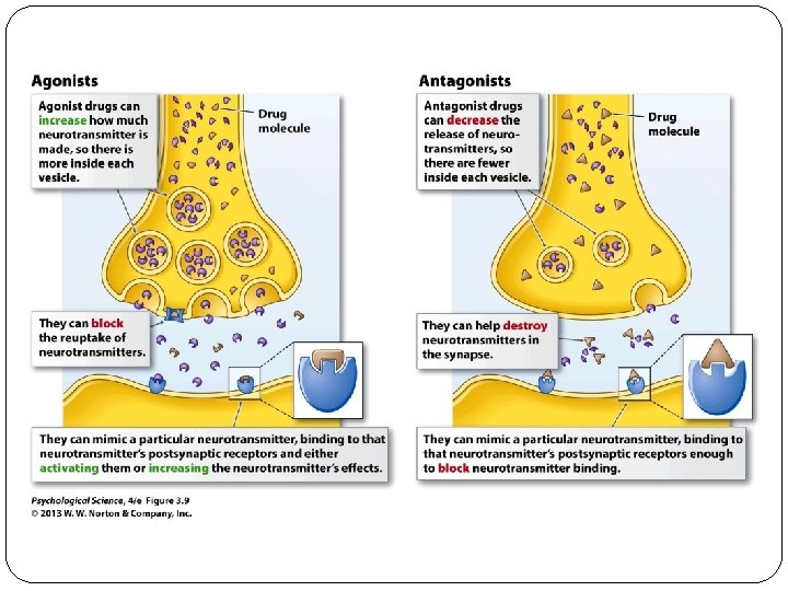
Drugs alter Neurotransmitter effects �Agonists: enhance the actions of neurotransmitters �Antagonists: inhibit the actions of neurotransmitters

Drugs can alter pre-synaptic processes �Pre-synaptic processes: �Conduction down Axon �Transmitter production �Transmitter release �Transmitter clearance: enzymes; transporters

Figure 4. 7 Drug Effects on Presynaptic Mechanisms

Drugs which affect conduction

�Novocaine �Blocks Na+ channels on pain axons �TTX �Blocks ALL Na+ channels in body

Drugs which Affect Release of Neurotransmitters

�Botox �Blocks release of Acetylcholine �Prevents muscles from contacting

Meth: Releases Dopamine

Drugs which Affect Transmitter clearance

Cocaine �Blocks Re-uptake of Dopamine at transporters �Prolongs effect of dopamine at receptor
 that break down acetylcholine")
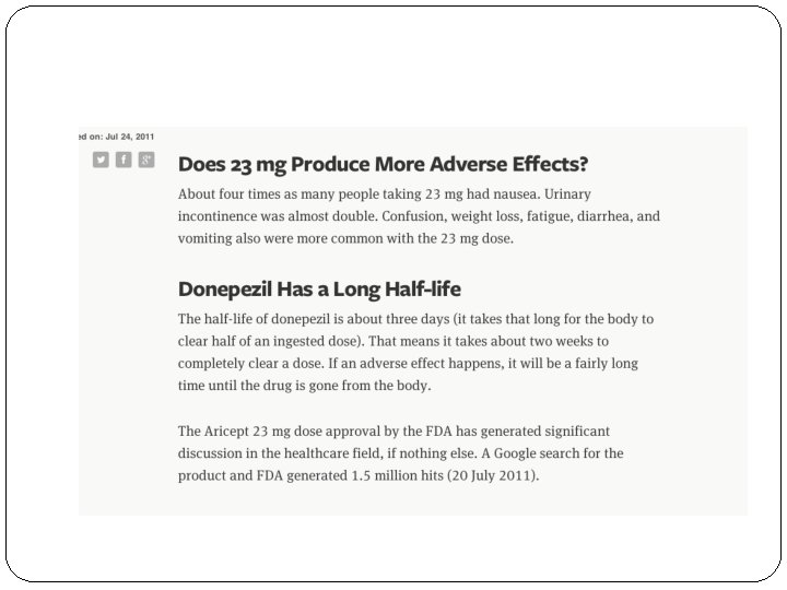
Aricept: blocks enzyme (acetylcholinesterase) that break down acetylcholine

End Result? ? �Aricept is a member of ACHE Inhibitor drugs �Aricept leads to increased levels of Acetylcholine �It is an ACH AGONIST

The Dose is the Poison

Sarin: ACHE Inhibitor

What happens when ACH is uncontrolled? �Initial symptoms following exposure to sarin are a runny nose, tightness in the chest and constriction of the pupils. �Difficulty breathing, nausea, drooling �Continued loss of control of bodily functions: vomiting, defecation, urination �Muscle Twitching, jerking, convulsive spasms

Treatment?

Block ACH receptors �Atropine: ACH Receptor Antagonist

Drugs can alter post-synaptic processes �Post-synaptic processes: �Receptors �Intracellular post-synaptic processes

Figure 4. 8 Drug Effects on Postsynaptic Mechanisms

Drugs which Mimic or Block Neurotransmitters at Receptors

Drugs that fit on ACH Receptors �Atropine : blocks ACh receptors �antagonist �Nicotine: mimics ACh and acts like the transmitter �agonist

Hallucinogens & Serotonin �LSD and other hallucinogens are structurally similar to serotonin �Agonists at post- synaptic serotonin receptors

Drug vs. Transmitter Effects �Neurotransmitters have localized effects, within one particular circuit �Drugs can affect multiple neurotransmitter receptors simultaneously- global effects

- Slides: 122