Electrical Control Cardiac muscles are specialized muscle tissue


 located between")


- Slides: 12
Electrical Control • Cardiac muscles are specialized muscle tissue • When it contracts, it does so in waves • The heart also coordinates all of its contractions into a steady rhythm of both atria contracting & then both ventricles contracting
• The contractions begin at specific places in the heart • The sinoatrial node (SA node) is a group of specialized cardiac muscle cells in the right atrium • The SA node generates an electrical pulse that moves outward throughout the rest of the atrial cells (like a wave in a pond)
• When the electrical pulse reaches the atrioventricular node (AV node) located between the atria & ventricles, the pulse is relayed to the ventricular cells causing them to contract • The ventricular contraction is a fraction of a second after the atrial contraction
• A heartbeat has two phases: – 1. Systole: occurs when the ventricles contract closing the AV valves & opening the SL valves (lub) – 2. Diastole: occurs when the ventricles relax closing the SL valves & opening the AV valves (dub)
• This creates the characteristic ‘lub-dub’ sound of a human heartbeat • If one of the valves does not close completely, then some blood is allowed to move backwards in the heart - this is called a murmur
Blood Vessels • A persons pulse is a series of pressure waves within an artery caused by the contraction of the left ventricle • When the blood surges through the arteries, the elastic walls of the vessels expand stretch • The average pulse rate is 70 -90 beats per minute (bpm)
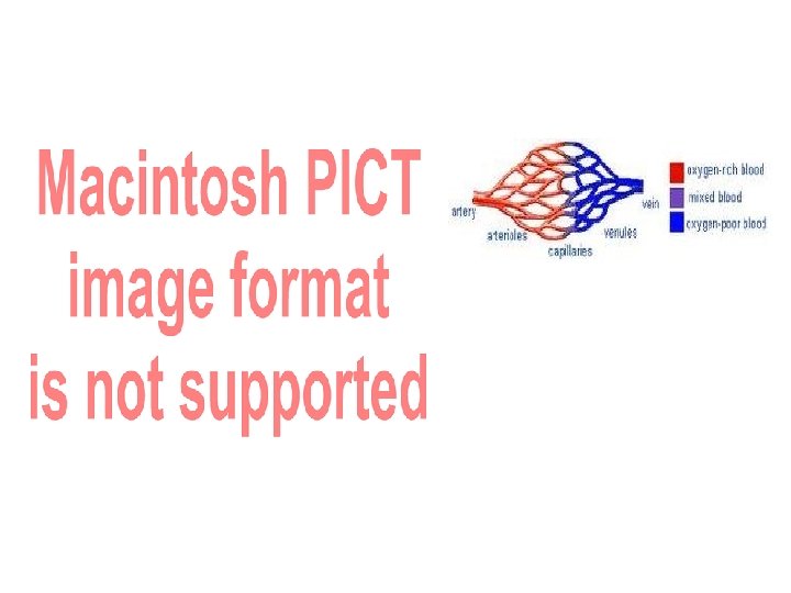
The Arteries • The large muscular vessels that carry blood away from the heart are called arteries • The walls of the arteries have three layers: • 1. A smooth endothelial layer • 2. A middle layer of smooth muscle • 3. Outer layer of connective tissue for strength
• As the arteries move away from the heart, they split into smaller & smaller vessels • First arteries become arterioles, arterioles then capillaries • A capillary is so tiny that blood cells must move through it in single file!
The Veins • After gas exchange occurs between the cells & the capillaries it is time to begin the journey back to the heart • The capillaries, now carrying O 2 poor blood turn into veinules • Several veinules will merge to become larger veins
• Veins continue to merge to form the very large vein, the Vena Cava • Veins & the Vena Cava have small valves inside to prevent the blood from moving backwards
THE END!!