Electrical circuits and nerve muscle preparation The Main


 nerve fiber but is")


- Slides: 12
Electrical circuits and nerve muscle preparation
• The Main Parts of the Nerve Cell • The nerve cell may be divided on the basis of its structure and function into three main parts: • (1) the cell body, also called the soma; • (2) numerous short processes of the soma, called the dendrites; and, • (3) the single long nerve fiber, the axon.
• The Synapse: The junction between an axon and the next cell with which it communicates is called the synapse. Information proceeds from the cell body unidirectionally over the synapse, first along the axon and then across the synapse to the next nerve or muscle cell.
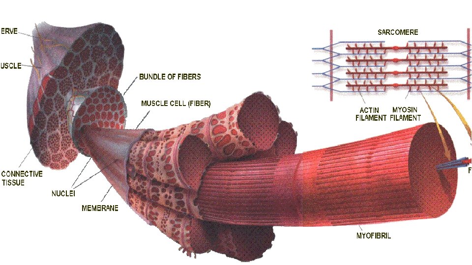
• MUSCLE CELL • There are three types of muscles in the body: • - smooth muscle, • - striated muscle (skeletal muscle), and • - cardiac muscle. • Smooth muscles are involuntary (i. e. , they cannot be controlled voluntarily). Their cells have a variable length but are in the order of 0. 1 mm. Smooth muscles exist, for example, in the digestive tract, in the wall of the trachea, uterus, and bladder. The contraction of smooth muscle is controlled from the brain through the autonomic nervous system. • Striated muscles, are also called skeletal muscles because of their anatomical location, are formed from a large number of muscle fibers, that range in length from 1 to 40 mm and in diameter from 0. 01 to 0. 1 mm. Each fiber forms a (muscle) cell and is distinguished by the presence of alternating dark and light bands. This is the origin of the description "striated, " as an alternate terminology of skeletal muscle.
• The striated muscle fiber corresponds to an (unmyelinated) nerve fiber but is distinguished electrophysiologically from nerve by the presence of a periodic transverse tubular system (TTS), a complex structure that, in effect, continues the surface membrane into the interior of the muscle. Propagation of the impulse over the surface membrane continues radially into the fiber via the TTS, and forms the trigger of myofibrillar contraction. The presence of the TTS affects conduction of the muscle fiber so that it differs (although only slightly) from propagation on an (unmyelinated) nerve fiber. Striated muscles are connected to the bones via tendons. Such muscles are voluntary and form an essential part of the organ of support and motion. • Cardiac muscle is also striated, but differs in other ways from skeletal muscle: Not only is it involuntary, but also when excited, it generates a much longer electric impulse than does skeletal muscle, lasting about 300 ms. Correspondingly, the mechanical contraction also lasts longer. Furthermore, cardiac muscle has a special property: The electric activity of one muscle cell spreads to all other surrounding muscle cells, owing to an elaborate system of intercellular junctions.
Mechanism of muscle contraction
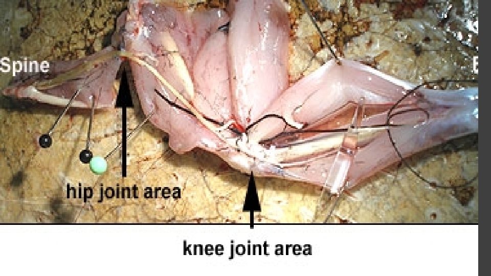
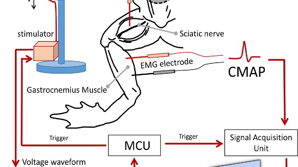
the sciatic nerve isolated from a frog leg • The sciatic nerve is a large bundle of many nerve fibers. The fibers come out between vertebrae at the caudal end of the vertebral column. The nerve of the frog is dissected from its origin at the spinal cord as 3 -4 bundles of the sciatic plexus, all the way to the gastrocnemius muscle. There the nerve divides into two branches, one to each head of the muscle. As we proceed distally along the nerve, from the plexus, we find fibers leaving the main trunk and entering muscles and skin. Therefore, having fewer fibers, the distal end of the nerve is of smaller diameter than the proximal end at the plexus. The fibers that normally carry impulses from the central nervous system (CNS) outward to the periphery to muscles and skin are efferent or motor. Impulses normally move toward the CNS along afferent or sensory fibers from peripherally located sensory receptors in muscle and skin. • Afferent fibers are dendrites, and efferent fibers are axons, but any peripheral nerve fiber of unspecified nature is commonly called an axon. If the nerve is stimulated at the proximal end, impulses travel in the normal direction (orthodromically) in the efferent fibers but antidromically in the afferent fibers. That is, impulse propagation can be induced to proceed equally well in either direction along a nerve fiber. Advantage is taken of this fact in the sciatic nerve taken out of a frog leg. • The sciatic nerve is dissected free from the leg of a frog and is immersed in a bath of physiological solution. The bath is provided with electrodes for initiating and recording action potentials.
Preparation of frog nerve.
Dissecting out the Frog Sciatic Nerve