Electrical Activity of the Heart The Body as

Electrical Activity of the Heart

The Body as a Conductor This is a graphical representation of the geometry and electrical current flow in a model of the human thorax. The model was created from MRI images taken of an actual patient. Shown are segments of the body surface, the heart, and lungs. The colored loops represent the flow of electric current through the thorax for a single instant of time, computed from voltages recorded from the surface of the heart during open chest surgery.

Electrical Currents This figure illustrates the voltage distribution of a body surface potential map (BSPM) on the right. The torso on the left has been opened to reveal electrical current paths within the torso volume at one instant in time within a heart beat. Here again, red indicates positive voltage, blue indicates negative voltage, and green indicates voltages of approximately zero.

Polarized

Action Potential ✦ Two micro-electrodes ✦ One outside the cell, one inside the cell ✦ ✦ Difference between the two (-90 m. V inside) The cell is said to be ‘polarized’

Depolarized

Action Potential Skeletal Cardiac

Ions Ion Extra- Intra- Na 140 10 K 4 135 Ca 2 0. 1

Inside Outside

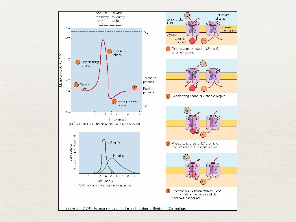
Action Potential ✦ Phase 0 ✦ Stimulation of the myocardial cell ✦ Influx of sodium ✦ The cell becomes depolarize ✦ Inside the cell = +20 m. V

Action Potential ✦ Phase 1 ✦ ✦ ✦ Ions ✦ Influx of sodium ✦ Efflux of potassium Partial repolarization Phase 2 ✦ ✦ Ions ✦ Influx of sodium ✦ Efflux of potassium ✦ Influx of calcium Plateau

✦ Action Potential Phase 3 ✦ ✦ ✦ Ions ✦ Influx of sodium ✦ Efflux of potassium* ✦ Influx of calcium Repolarization (slower process than depolarization) Phase 4 ✦ ✦ Interval between repolarization to the next action potential Pumps restore ionic concentrations

Ion 0 1 2 3 4 Na influx pump efflux* pump influx pump K Ca

Action Potential & Mechanical Contraction
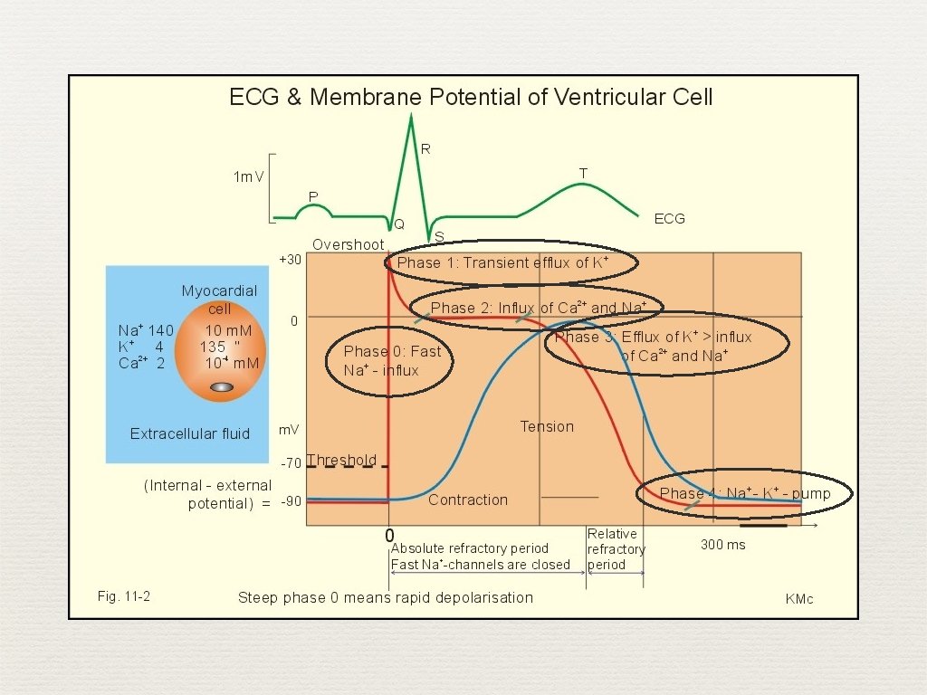

Action Potential ✦ Fast and slow response ✦ ✦ ✦ Fast response is more negative Greater up-slope, amplitude, and overshoot Slow conduction increases rhythm disturbances
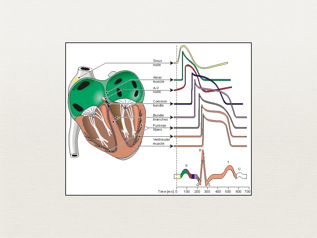
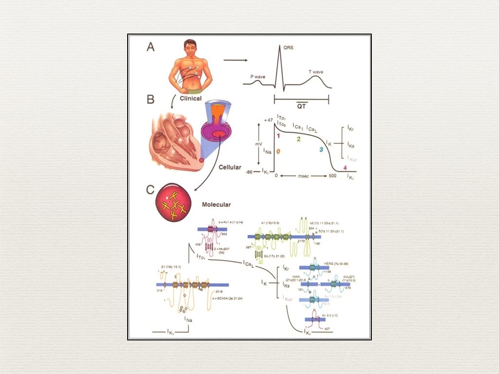

Action Potential ✦ ECG records depolarization and repolarization ✦ Atrial depolarization ✦ Ventricular depolarization ✦ ✦ Atrial repolarization Ventricular repolarization

ECG Complexes

ECG Complexes Note: No atrial ST segment or T wave due to low amplitude

ECG Complexes

ECG Complexes

ECG Paper ✦ Small boxes = 1 mm ✦ Large boxes = 5 mm ✦ Small boxes = 0. 04 seconds ✦ Large boxes = 0. 20 seconds ✦ Paper speed = 25 mm / sec

ECG Paper

ECG Paper ✦ Standardization mark ✦ 10 mm vertical deflection = 1 m. Volt

ECG Paper ✦ Standardization marks ✦ Double if ECG is too small ✦ Half is ECG is too large Top: Low amplitude complexes in an obese women with hypothyroidism Bottom: High amplitude complexes in a hypertensive man
 ✦ recorded in mm ✦")
ECG Description ✦ ECG description ✦ ✦ amplitude (voltage) ✦ recorded in mm ✦ positive or negative or biphasic width (duration)

✦ ECG Waves P wave ✦ ✦ atrial depolarization ✦ ≤ 2. 5 mm in amplitude ✦ < 0. 12 sec in width PR interval (0. 12 - 0. 20 sec. ) ✦ time of stimulus through atria and AV node ✦ prolonged interval = first-degree heart block

ECG Waves ✦ ✦ QRS Ventricle depolarization ✦ Q wave: when initial deflection is negative ✦ R wave: first positive deflection ✦ S wave: negative deflection after the R wave

ECG Waves ✦ QRS ✦ ✦ May contain R wave only May contain QS wave only Small waves indicated with small letters (q, r, s) Repeated waves are indicated as ‘prime’

ECG Waves ✦ QRS ✦ width usually 0. 12 second or less

ECG Waves ✦ RR interval ✦ interval between two consecutive QRS complexes

ECG Waves ✦ ✦ J point: ✦ end of QRS wave ✦ beginning of ST segment ✦ beginning of ventricular repolarization ✦ normally isoelectric (flat) ✦ changes, elevation or depression, may indicate pathological condition

ECG Waves

ECG Waves ✦ T wave ✦ part of ventricular repolarization ✦ asymmetrical shape ✦ usually not measured ✦ normally upright in lead II

ECG Waves ✦ QT interval ✦ from beginning of QRS to the end of the T wave ✦ ventricular repolarization ✦ length varies with heart rate (table 2. 1) RR (sec) HR (bpm) QT (sec) 1. 00 60 0. 43 0. 40 150 0. 27

ECG Waves ✦ Rate Corrected QT Interval ✦ QTc = QT divided by square root of RR ✦ normal is less than or equal to 0. 44 sec.

ECG Waves ✦ ✦ Long QT interval ✦ certain drugs ✦ electrolyte distrubances ✦ hypothermia ✦ ischemia ✦ infarction ✦ subarachnoid hemorrhage Short QT interval ✦ drugs or hypercalcemia

ECG Waves ✦ U Wave ✦ last phase of repolarization ✦ small wave after the T wave ✦ not always seen ✦ significance is not known
 Count the number")
Heart Rate Calculation ✦ ✦ Count boxes (for regular rhythm HR) Count the number of large boxes between two consecutive QRS complexes. Divide 300 by that number ✦ ✦ 300 ÷ 4 = 75 Count the small boxes. Divide 1500 by that number ✦ 1500 ÷ 20 = 75

Heart Rate Calculation most accurate 1500 divided by the take time to calculate number of small boxes only use with regular between two R waves rhythms quick 300 divided by the not too accurate number of large boxes only use with regular between two R waves rhythm less precise 10 multiplied by the use with irregular number of R waves in 6 rhythms seconds very quick 1 lg sq = 300 bpm 2 lg sq = 150 bpm 3 lg sq = 100 bpm 4 lg sq = 75 bpm 5 lg sq = 60 bpm 6 lg sq = 50 bpm

Heart Rate Calculation R-R interval is two large squares. The rate is 300/2=150

Heart Rate Calculation ✦ Count the number of cardiac cycles in 6 seconds and multiple this by 10. (Figure 2. 15)

Heart Rate Calculation ✦ Count the number of cardiac cycles in 10 seconds and multiple this by 6. Irregular rhythm with 21 R-R intervals in 10 seconds. The rate is 21 x 6=126.

The ECG as a Combination of Atrial and Ventricular Parts ✦ Atrial ECG = P wave ✦ Ventricular ECG = QRS-T waves ✦ Normally, sinus node paces the heart and P wave precedes QRS ✦ ✦ P-QRS-T Sometimes, atria and ventricles paced separately (e. g. complete heart block)

ECG in Perspective 1. ECG recording of electrical activity not the mechanical function 2. ECG does not depict abnormalities 3. ECG does not record all the heart’s electrical activity

Questions ✦ End of chapter, questions 1 -6.
- Slides: 49