
Elbow dysplasia June 4 th 2012 Sweden ZAMP
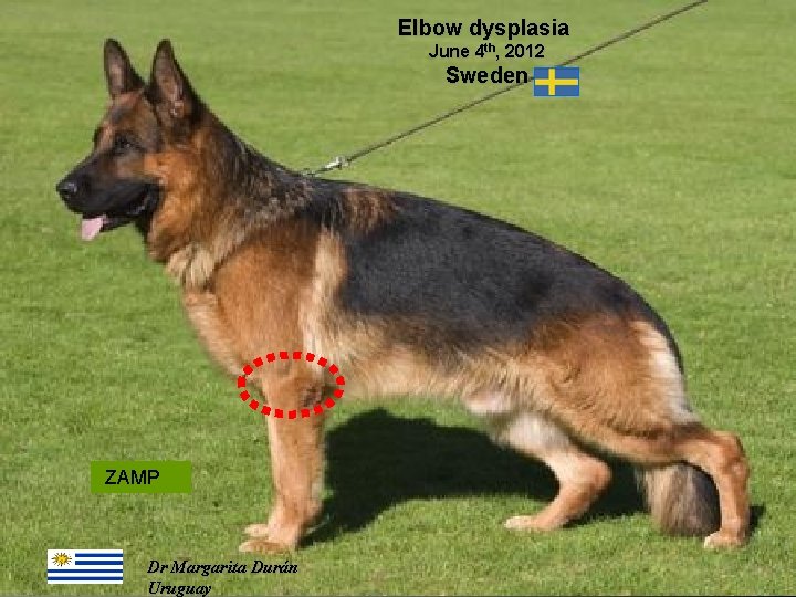
Elbow dysplasia June 4 th, 2012 Sweden ZAMP 03/10/2020 Dr Margarita Durán Uruguay
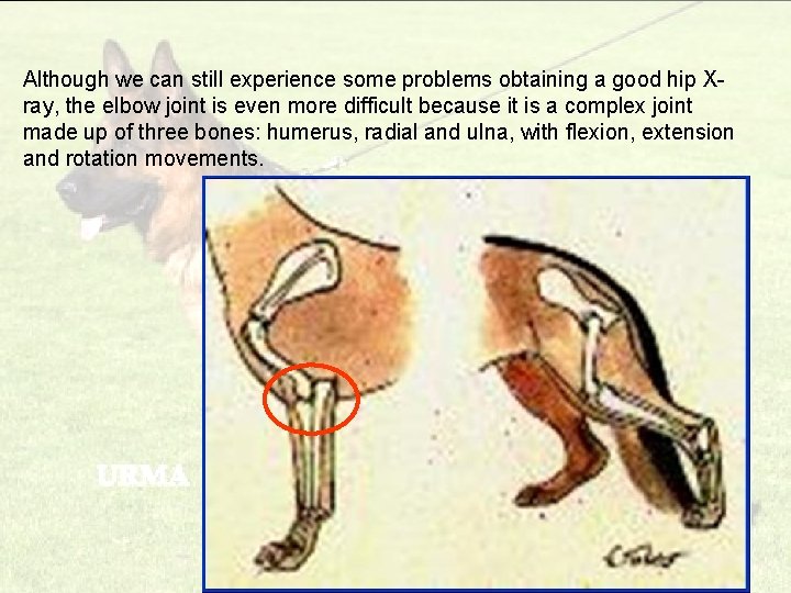
Although we can still experience some problems obtaining a good hip Xray, the elbow joint is even more difficult because it is a complex joint made up of three bones: humerus, radial and ulna, with flexion, extension and rotation movements. 03/10/2020
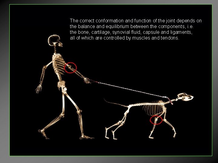
The correct conformation and function of the joint depends on the balance and equilibrium between the components, i. e. the bone, cartilage, synovial fluid, capsule and ligaments, all of which are controlled by muscles and tendons.
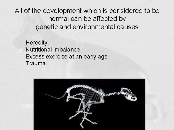
All of the development which is considered to be normal can be affected by genetic and environmental causes Heredity Nutritional imbalance Excess exercise at an early age Trauma.
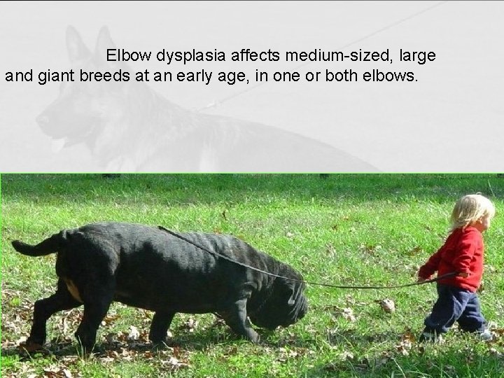
Elbow dysplasia affects medium-sized, large and giant breeds at an early age, in one or both elbows.
• Originally, the term “elbow dysplasia” covered generalised osteoarthrosis of the elbow joint with Ununited Anconeal Process (UAP) • UAP is currently one of the various components covered by the term “elbow dysplasia”. The IEWG (International Elbow Working Group) includes: • • Ununited Anconeal Process (UAP), Fragmented Coronoid Process (FCP), Osteochondrosis (OC), Articular incongruence.
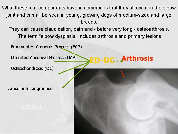
What these four components have in common is that they all occur in the elbow joint and can all be seen in young, growing dogs of medium-sized and large breeds. They can cause claudication, pain and - before very long - osteoarthrosis. The term “elbow dysplasia” includes arthrosis and primary lesions Fragmented Coronoid Process (FCP) Ununited Anconeal Process (UAP) Osteochondrosis (OC) Articular Incongruence ED-DC Arthrosis
AETIOLOGIES Genetic and Environmental Influences • The population of pure-bred dogs is commonly genetically closed, with high levels of consanguinity. • Only a few members of the breed (champions) are used for reproduction. This then creates a bottleneck and the genetic heterogeneity is reduced, increasing the incidence of genetic diseases and the chance of carrying the risk of elbow dysplasia factor or any other genetic disease. 03/10/2020
• The selection of dysplasia-free dogs increases the percentage of descendants who are free of it in the population. • An unknown number of genes is involved and may come from both the father and the mother.

Nutritional imbalance • Nutrition has the greatest impact upon skeletal development • Food with a high level of calcium, vitamin D and minerals cause endochondral ossification problems and can lead to the appearance of primary lesions. • Unbalanced diets or excessive food can also produce an unfavourable effect. 03/10/2020
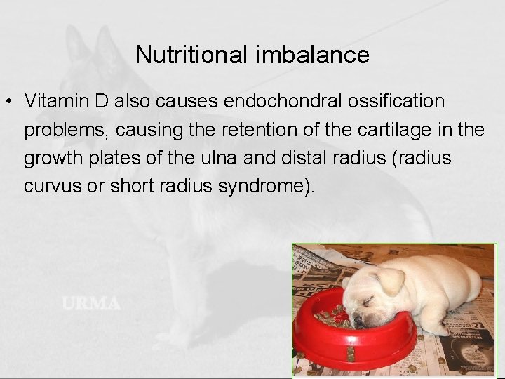
Nutritional imbalance • Vitamin D also causes endochondral ossification problems, causing the retention of the cartilage in the growth plates of the ulna and distal radius (radius curvus or short radius syndrome). 03/10/2020
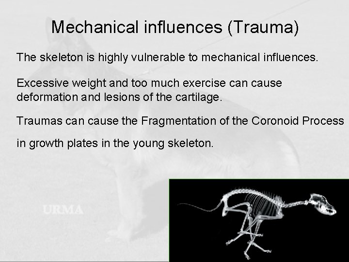
Mechanical influences (Trauma) • The skeleton is highly vulnerable to mechanical influences. Excessive weight and too much exercise can cause deformation and lesions of the cartilage. Traumas can cause the Fragmentation of the Coronoid Process in growth plates in the young skeleton. 03/10/2020
Clinical Signs Pain Rigid Movements Too much weight on the other limbs Various degrees of claudication 03/10/2020
Diagnosis Clinical signs, confirmed by radiological examination. Diagnosis by a qualified professional. Based upon the IEWG (International Elbow Working Group) protocol 03/10/2020
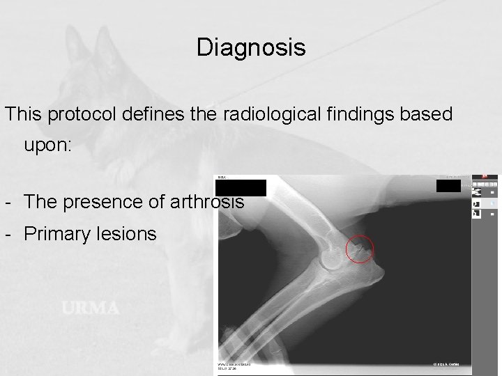
Diagnosis This protocol defines the radiological findings based upon: - The presence of arthrosis - Primary lesions 03/10/2020
Radiographic technique • Minimum age for the official check: 12 months. • X-rays of both elbows are taken separately. • Mark the “Right” and “Left” sides. • High-speed rare earth screens. • The ray must be collimated in order to obtain a high-quality image. • In the lateral projection, the elbow must be flexed at 45º. • Concentric superimposition of the humeral condyles.
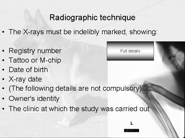
Radiographic technique • The X-rays must be indelibly marked, showing: • • Full details Registry number Tattoo or M-chip Date of birth X-ray date (The following details are not compulsory). Owner's identity The clinic at which the study was carried out
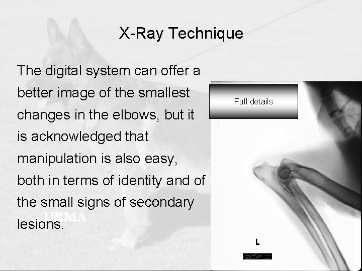
X-Ray Technique The digital system can offer a better image of the smallest changes in the elbows, but it is acknowledged that manipulation is also easy, both in terms of identity and of the small signs of secondary lesions. Full details
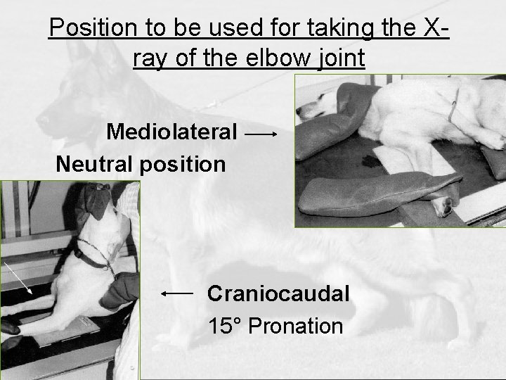
Position to be used for taking the Xray of the elbow joint Mediolateral Neutral position Craniocaudal 15° Pronation
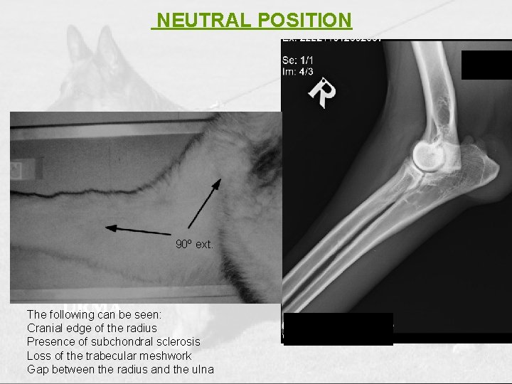
NEUTRAL POSITION 90º ext. The following can be seen: Cranial edge of the radius Presence of subchondral sclerosis Loss of the trabecular meshwork Gap between the radius and the ulna
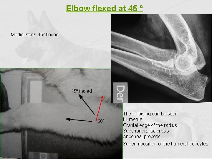
Elbow flexed at 45 º Mediolateral 45º flexed 90º The following can be seen: Humerus Cranial edge of the radius Subchondral sclerosis Anconeal process Superimposition of the humeral condyles
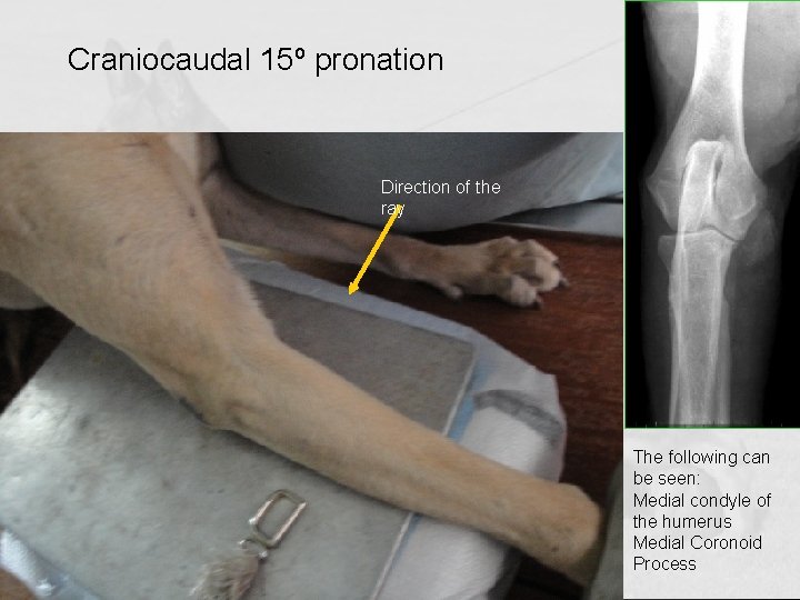
Craniocaudal 15º pronation Direction of the ray The following can be seen: Medial condyle of the humerus Medial Coronoid Process
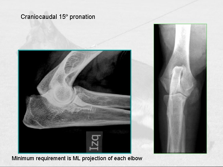
Craniocaudal 15º pronation Minimum requirement is ML projection of each elbow
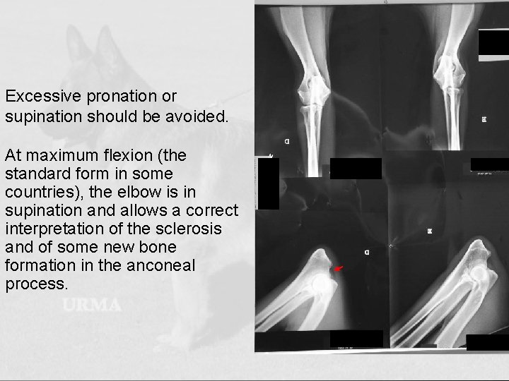
Excessive pronation or supination should be avoided. At maximum flexion (the standard form in some countries), the elbow is in supination and allows a correct interpretation of the sclerosis and of some new bone formation in the anconeal process.
Interpretation of the film: The radiological findings vary depending upon the aetiology, the breed, the severity and the age of the dog. The diagnosis of elbow dysplasia is based upon the presence of primary lesions and/or arthrosis 03/10/2020
Secondary lesions (arthrosis) are the main criterion in the classification programme Grade 0 NORMAL: There are no signs of arthrosis Borderline: Minimal changes on the dorsal edge of the anconeal process, or minimal subchondral sclerosis. Grade I SLIGHT: Osteophytes of lessthan 2 mm. Grade II MEDIUM: Osteophytes from 2 to 5 mm. Grade III SEVERE: Osteophytes of more than 5 mm. 03/10/2020
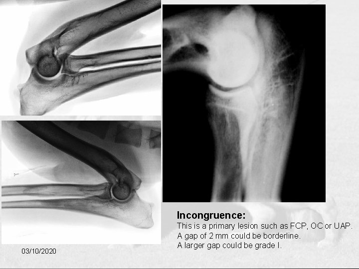
Incongruence: 03/10/2020 This is a primary lesion such as FCP, OC or UAP. A gap of 2 mm could be borderline. A larger gap could be grade I.
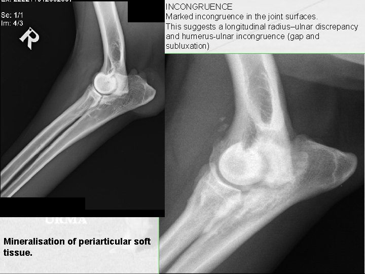
INCONGRUENCE Marked incongruence in the joint surfaces. This suggests a longitudinal radius–ulnar discrepancy and humerus-ulnar incongruence (gap and subluxation) Mineralisation of periarticular soft tissue. 03/10/2020
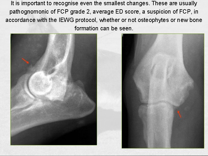
It is important to recognise even the smallest changes. These are usually pathognomonic of FCP grade 2, average ED score, a suspicion of FCP, in accordance with the IEWG protocol, whether or not osteophytes or new bone formation can be seen.
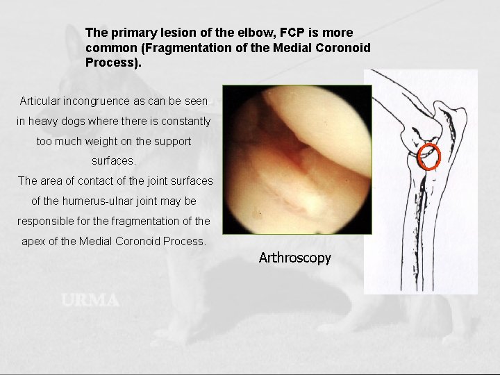
The primary lesion of the elbow, FCP is more common (Fragmentation of the Medial Coronoid Process). Articular incongruence as can be seen in heavy dogs where there is constantly too much weight on the support surfaces. The area of contact of the joint surfaces of the humerus-ulnar joint may be responsible for the fragmentation of the apex of the Medial Coronoid Process. Arthroscopy 03/10/2020
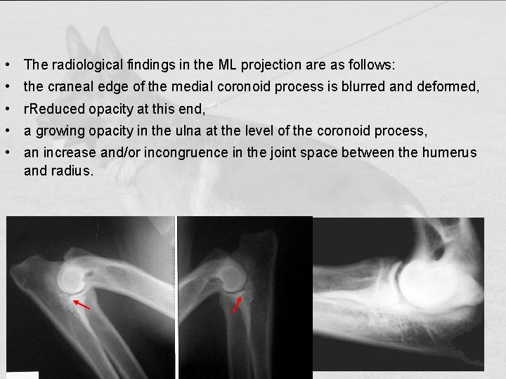
• The radiological findings in the ML projection are as follows: • the craneal edge of the medial coronoid process is blurred and deformed, • r. Reduced opacity at this end, • a growing opacity in the ulna at the level of the coronoid process, • an increase and/or incongruence in the joint space between the humerus and radius.
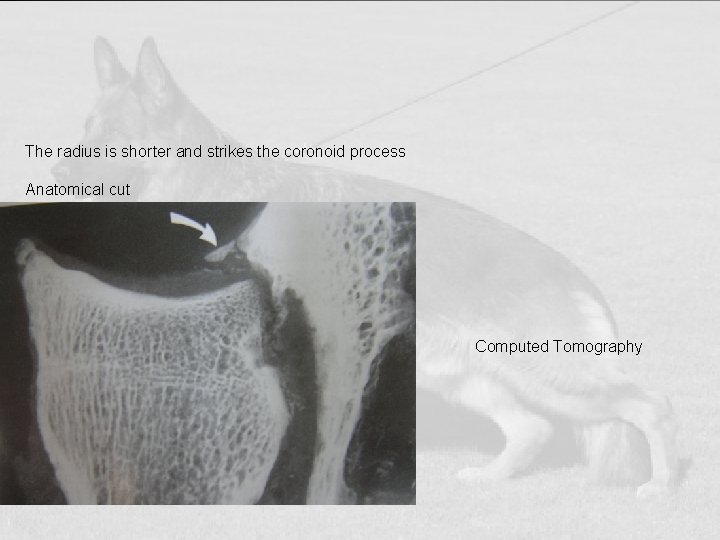
The radius is shorter and strikes the coronoid process Anatomical cut Computed Tomography
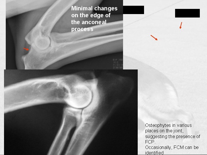
Minimal changes on the edge of the anconeal process Osteophytes in various places on the joint, suggesting the presence of FCP. Occasionally, FCM can be identified
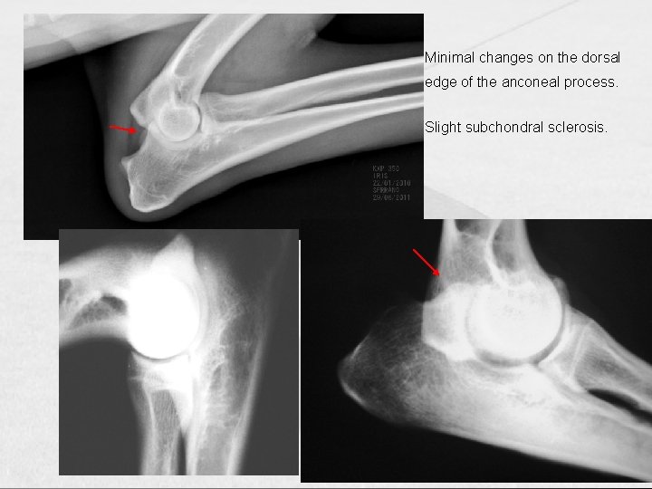
Minimal changes on the dorsal edge of the anconeal process. Slight subchondral sclerosis.
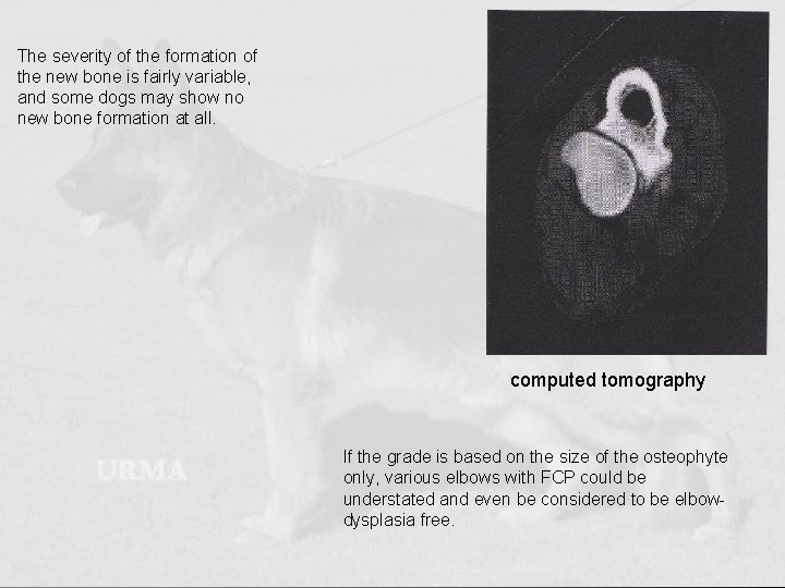
The severity of the formation of the new bone is fairly variable, and some dogs may show no new bone formation at all. computed tomography If the grade is based on the size of the osteophyte only, various elbows with FCP could be understated and even be considered to be elbowdysplasia free. 03/10/2020
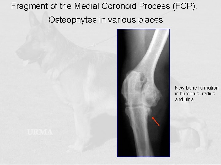
Fragment of the Medial Coronoid Process (FCP). Osteophytes in various places New bone formation in humerus, radius and ulna.
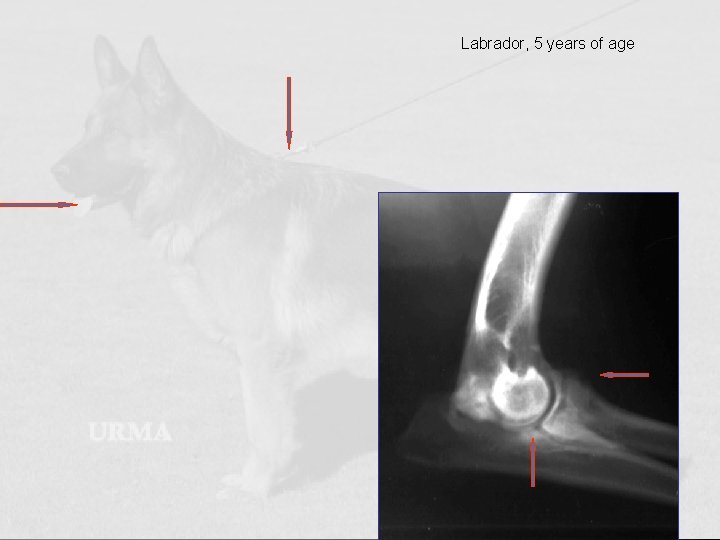
Labrador, 5 years of age 03/10/2020
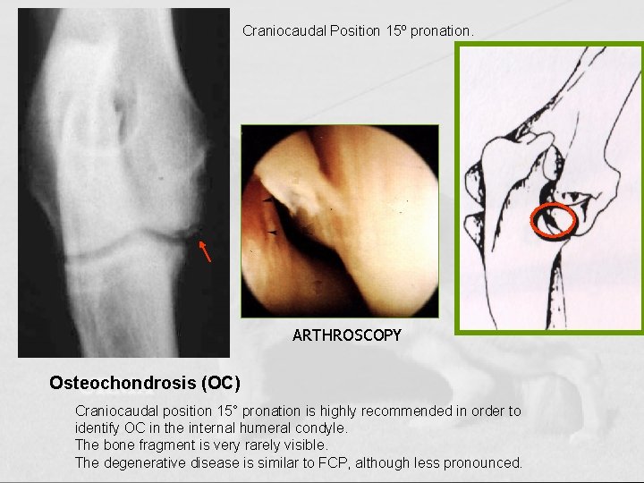
Craniocaudal Position 15º pronation. ARTHROSCOPY Osteochondrosis (OC) Craniocaudal position 15° pronation is highly recommended in order to identify OC in the internal humeral condyle. The bone fragment is very rarely visible. 03/10/2020 The degenerative disease is similar to FCP, although less pronounced.
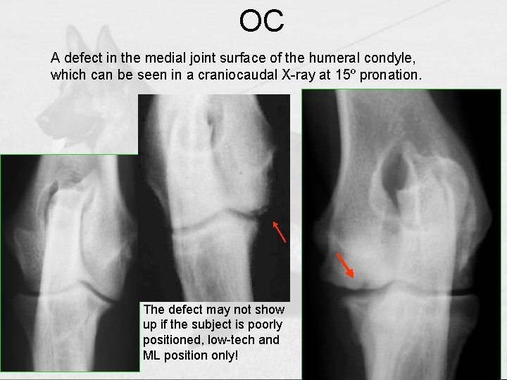
OC A defect in the medial joint surface of the humeral condyle, which can be seen in a craniocaudal X-ray at 15º pronation. 03/10/2020 The defect may not show up if the subject is poorly positioned, low-tech and ML position only!
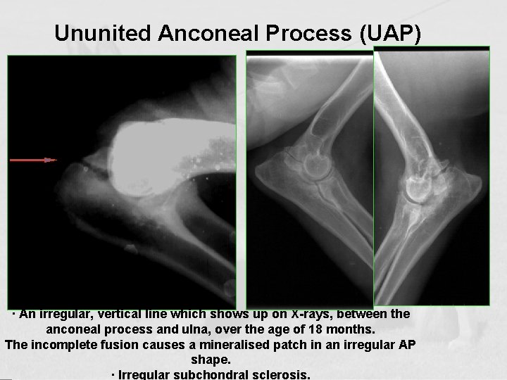
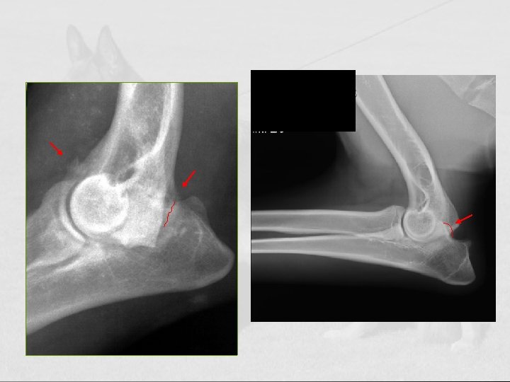
Ununited Anconeal Process (UAP) Growth line · An irregular, vertical line which shows up on X-rays, between the anconeal process and ulna, over the age of 18 months. The incomplete fusion causes a mineralised patch in an irregular AP 03/10/2020 shape. · Irregular subchondral sclerosis.
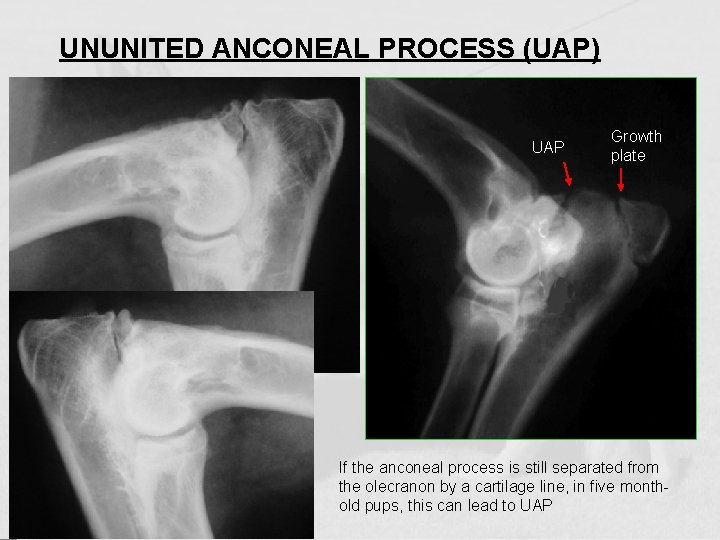
UNUNITED ANCONEAL PROCESS (UAP) UAP Growth plate If the anconeal process is still separated from the olecranon by a cartilage line, in five monthold pups, this can lead to UAP
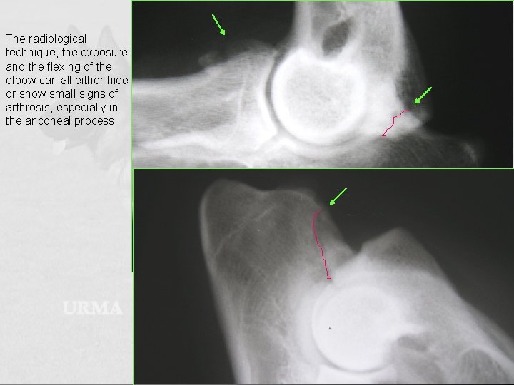
The radiological technique, the exposure and the flexing of the elbow can all either hide or show small signs of arthrosis, especially in the anconeal process
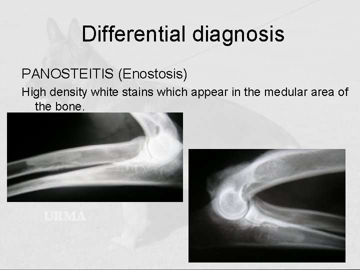
Differential diagnosis PANOSTEITIS (Enostosis) High density white stains which appear in the medular area of the bone.
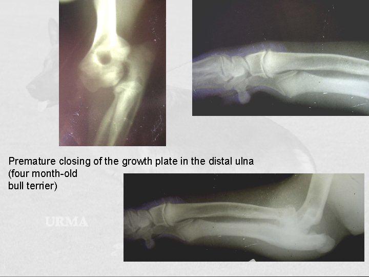
Premature closing of the growth plate in the distal ulna (four month-old bull terrier)
SCORING The radiological findings are assessed in accordance with the severity of the arthrosis and/or with the presence of primary lesions according to the IEWG protocol. Degree 0 Normal elbow joint NORMAL joint. There are no signs of incongruence, sclerosis or arthrosis. Borderline: Can be classified as: Presence of subchondral sclerosis. Grade 1: SLIGHT arthrosis. Subchondral sclerosis, or 2 mm gap between radius and ulna or 1 osteophyte measuring less than 2 mm. Grade 2: MEDIUM arthrosis: Osteophytes measuring from 2 to 5 mm. Grade 3: SERIOUS arthrosis “l ED” Primary Lesions. Osteophytes measuring more than 5 mm or “l ED” incongruence, UAP, FCP, OCD

03/10/2020 bvcm, . jhgfdsf ghj cn
- Slides: 49