
EKG Rounds Rebecca BurtonMac Leod R 4 Emerg

EKG Rounds Rebecca Burton-Mac. Leod R 4, Emerg Med July 20 th, 2006
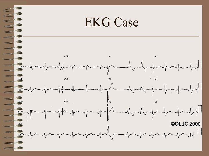
EKG Case
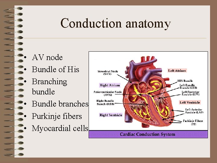
Conduction anatomy • AV node • Bundle of His • Branching bundle • Bundle branches • Purkinje fibers • Myocardial cells
Bundle branch blocks • RBBB—transmission is delayed or fails to conduct along right bundle branch • LBBB—transmission is delayed or fails to conduct along left bundle branch • LAFB—most common type of intraventricular conduction defect • LPFB—very rare!
Terminology • Bifascicular block—conduction defect in RBB and either LAF or LPF – Does not include RBBB and LBBB combination, as this is termed 3 rd degree AV block • Trifascicular block—as above, with 1 st degree AV block (prolonged PR)
Conduction • Consequence of BBB is that ventricle must await depolarization by opposite ventricle • Activation proceeds on cell-to-cell basis • Results in much slower activation along normal pathways
RBBB • Causes: – In children—surgical repair of VSD’s is most common cause; cardiomyopathy, myocarditis, CHF, hereditary causes (Brugada syndrome), muscular dystrophy – In adults—normal variant, RVH or strain (ex: PE), CAD
RBBB PE • What will you hear on physical examination? – Persistently split S 2
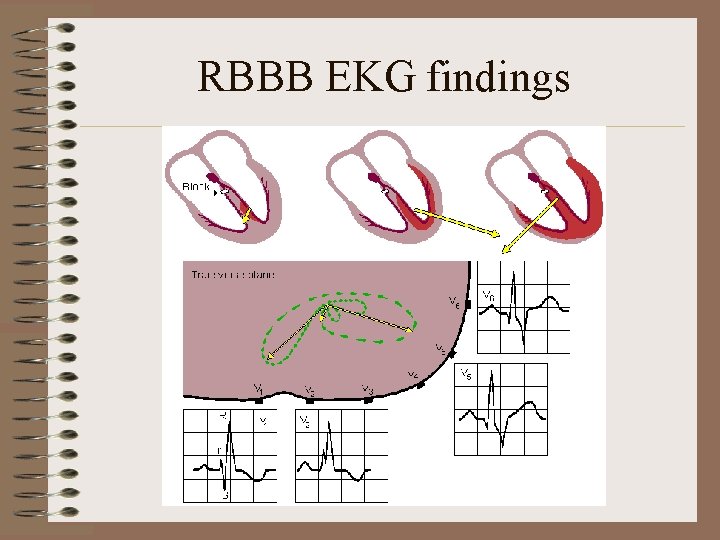
RBBB EKG findings
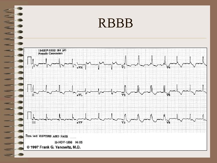
RBBB
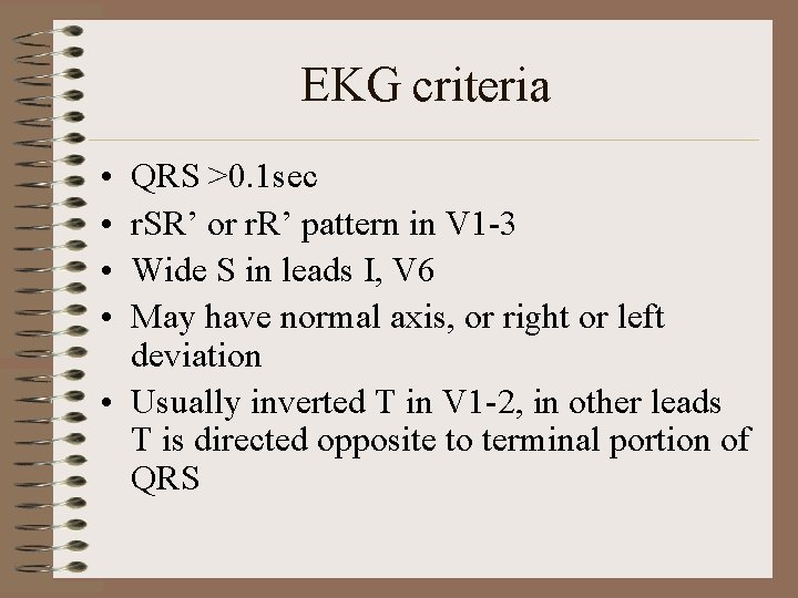
EKG criteria • • QRS >0. 1 sec r. SR’ or r. R’ pattern in V 1 -3 Wide S in leads I, V 6 May have normal axis, or right or left deviation • Usually inverted T in V 1 -2, in other leads T is directed opposite to terminal portion of QRS
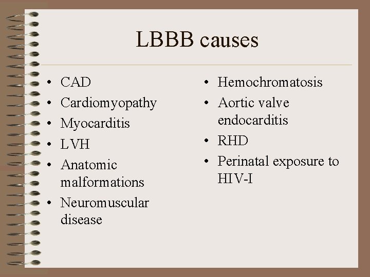
LBBB causes • • • CAD Cardiomyopathy Myocarditis LVH Anatomic malformations • Neuromuscular disease • Hemochromatosis • Aortic valve endocarditis • RHD • Perinatal exposure to HIV-I
LBBB PE • What heart sound changes will you hear on auscultation? – Absent or diminished S 1, reverse split S 2
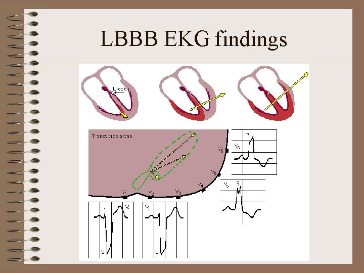
LBBB EKG findings
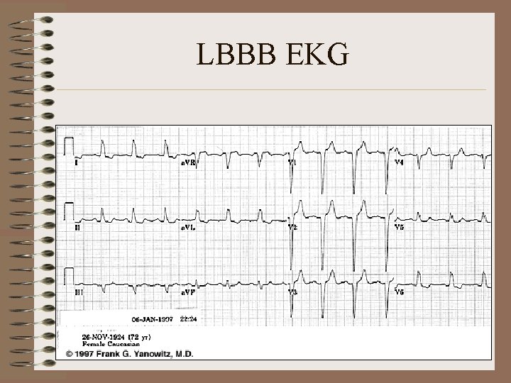
LBBB EKG
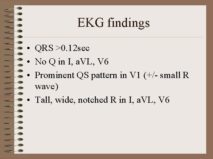
EKG findings • QRS >0. 12 sec • No Q in I, a. VL, V 6 • Prominent QS pattern in V 1 (+/- small R wave) • Tall, wide, notched R in I, a. VL, V 6
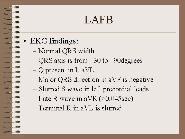
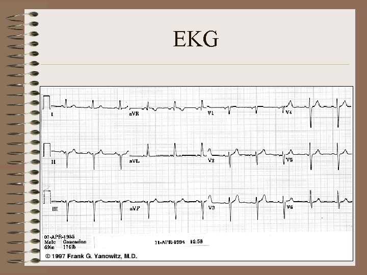
LAFB • EKG findings: – Normal QRS width – QRS axis is from – 30 to – 90 degrees – Q present in I, a. VL – Major QRS direction in a. VF is negative – Slurred S wave in left precordial leads – Late R wave in a. VR (>0. 045 sec) – Terminal R in a. VL is slurred
EKG
LPFB Ddx • Must first exclude other causes of right axis!!! – Cor pulmonale – Pulmonary heart disease – Pulmonary hypertension, etc.
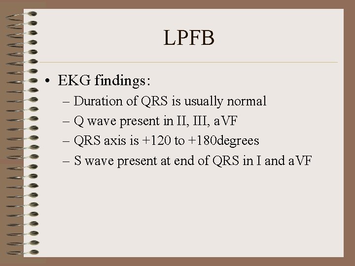
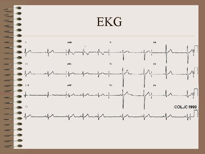
LPFB • EKG findings: – Duration of QRS is usually normal – Q wave present in II, III, a. VF – QRS axis is +120 to +180 degrees – S wave present at end of QRS in I and a. VF
EKG
Tough scenarios with BBB • RVH • LVH • MI
MI ?
MI ?
MI ?
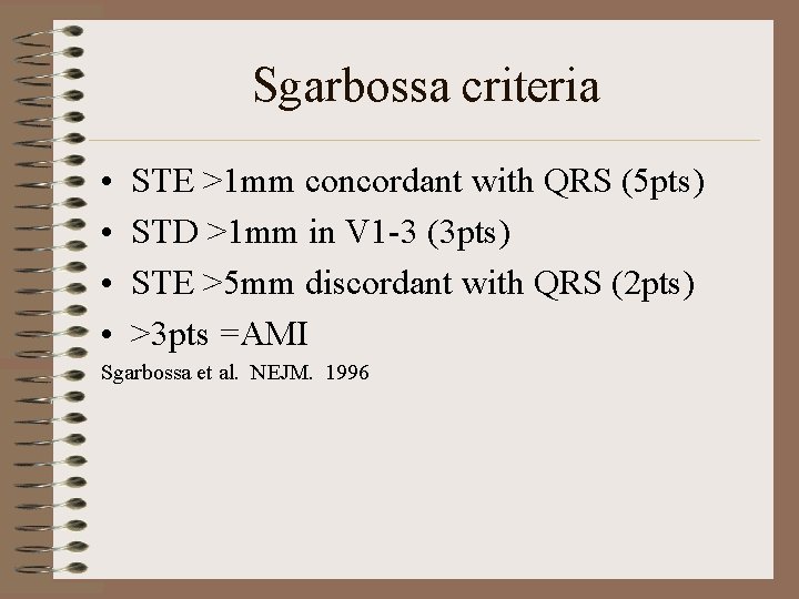
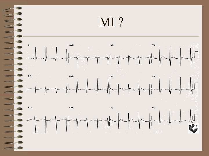
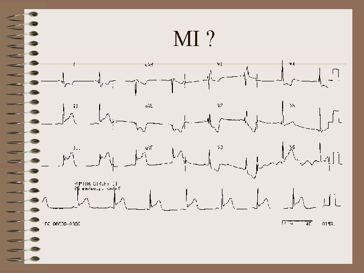
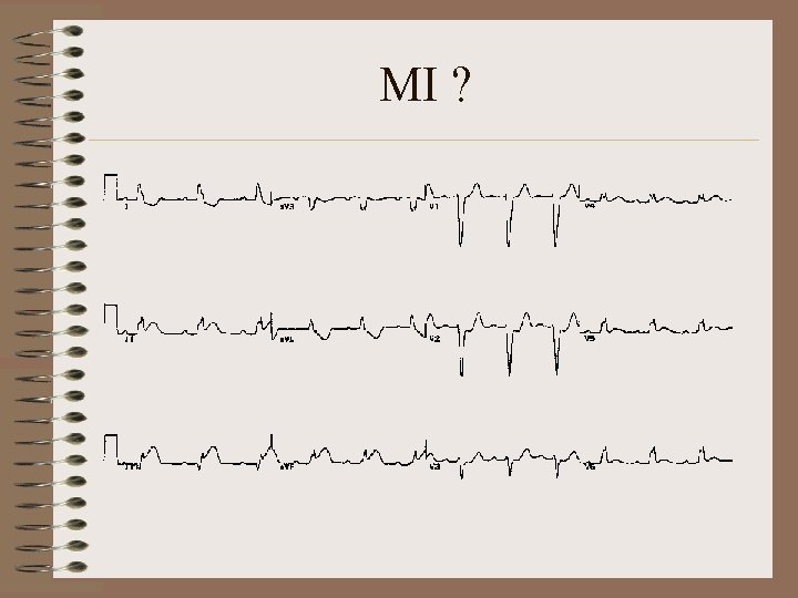
Sgarbossa criteria • • STE >1 mm concordant with QRS (5 pts) STD >1 mm in V 1 -3 (3 pts) STE >5 mm discordant with QRS (2 pts) >3 pts =AMI Sgarbossa et al. NEJM. 1996
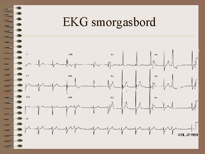
EKG smorgasbord
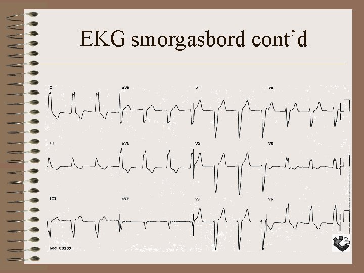
EKG smorgasbord cont’d
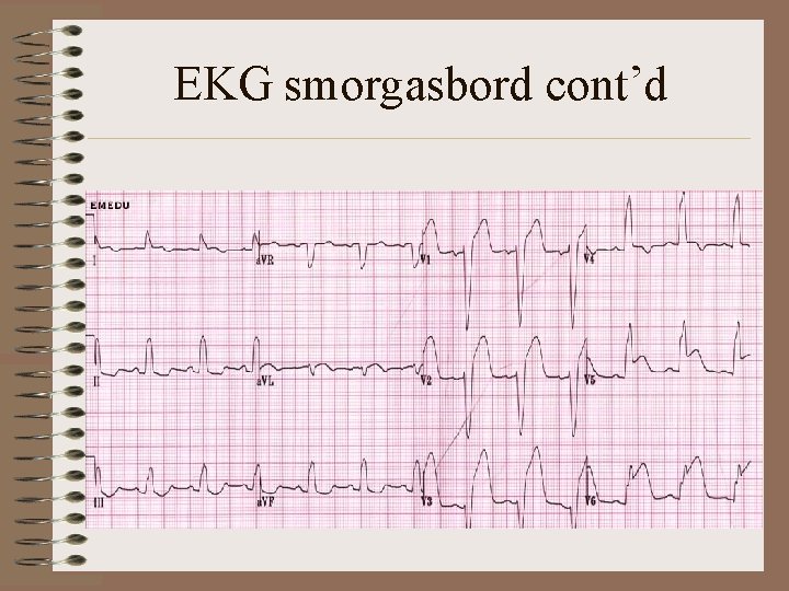
EKG smorgasbord cont’d
EKG smorgasbord cont’d
EKG smorgasbord cont’d
EKG smorgasbord cont’d

Questions ?
- Slides: 33