EKG MONITORING OVERVIEW Dr Jennifer Lucy CCMA Prep


 lead must be positioned anywhere above")


- Slides: 30
EKG MONITORING OVERVIEW Dr. Jennifer Lucy CCMA Prep 2017
Anatomy of 12 lead EKG
12 Lead EKG placement
Chest Leads 6 chest leads are labelled as V leads and are numbered from V 1 -V 6 Positioned in specific positions on the rib cage. Find the 2 nd Intercostal space below the collar bone. From this position, run your fingers downward across the next rib, and the next one. The space you are in is the 4 th intercostal space. Where this space meets the sternum is the position for V 1 should be positioned at the 4 th Intercostal space to the right of the sternum. V 2 is to be placed at the 4 th Intercostal space to the left of the sternum and V 3 midway between V 2 and V 4 must be then positioned at the 5 th Intercostal space at the midclavicular line V 5 at the anterior axillary line at the same level as V 4. V 6 is to be placed at the maxillary line at the same level as V 4 and V 5.
Peripheral Limb Leads Peripheral leads: RL (right leg) lead must be positioned anywhere above the ankle and below the torso. RA (right arm) lead is to be placed anywhere between the shoulder and the elbow, LL (left leg) lead should be positioned anywhere above the ankle and below the torso, and LA (left arm) lead anywhere between the shoulder and the elbow. It is very important that the ECG leads are placed correctly since incorrect placement can lead to a false diagnosis of infarction or negative changes on the ECG.
5 Lead Telemetry Lead Placement This is the most common type of continuous monitoring Used in Telemetry to monitor cardiac rhythm while patients are in hospital
Pneumonic to remember placement White Clouds go right over the Green fields Black Smoke over red fire Brown chocolate lies close to the heart
5 Lead placement
Holter Monitors Holter monitor is a small non-invasive ambulatory, portable ECG machine used to record the heart’s electrical activity in a 24– 72 hour period It correlates any symptoms (chest pain, palpitations) the patient experiences with the heart’s electrical activity at that time Record arrhythmias that occur, providing diagnostic information about the type of arrhythmia, how long it lasts, and what might trigger it Determine how well anti-arrhythmia medications are working
Important to know The box records and stores continuous heart rhythm data transmitted by the electrodes. The Record Box will indicate it is working by displaying the time and a “recording” message on the screen Once the Holter Monitor is attached to the patient it must stay on the patient for the prescribed time, 24 -72 hrs. Patient is allowed to go about normal activities of daily living, however they must avoid getting the Holter Monitor wet, therefore no showers or baths Patient should record activities such as having a bowel movement or eating as this can affect reading
Holter Monitor
3 Lead Monitor In these leads, one limb carries a positive electrode and the other limb, a negative one. The three limb electrodes, I, II and III form a triangle (Einthoven's Equilateral Triangle), at the right arm (RA), left arm (LA) and left leg (LL).
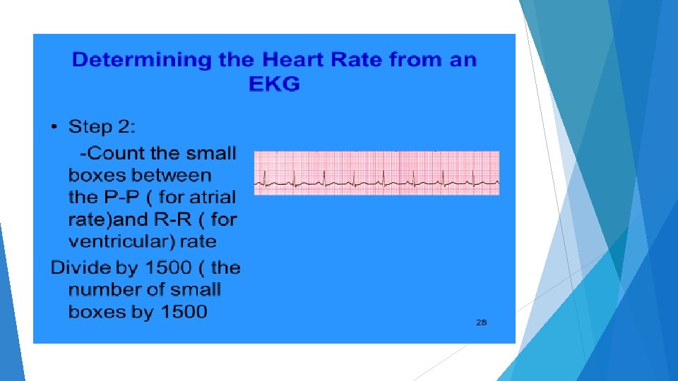
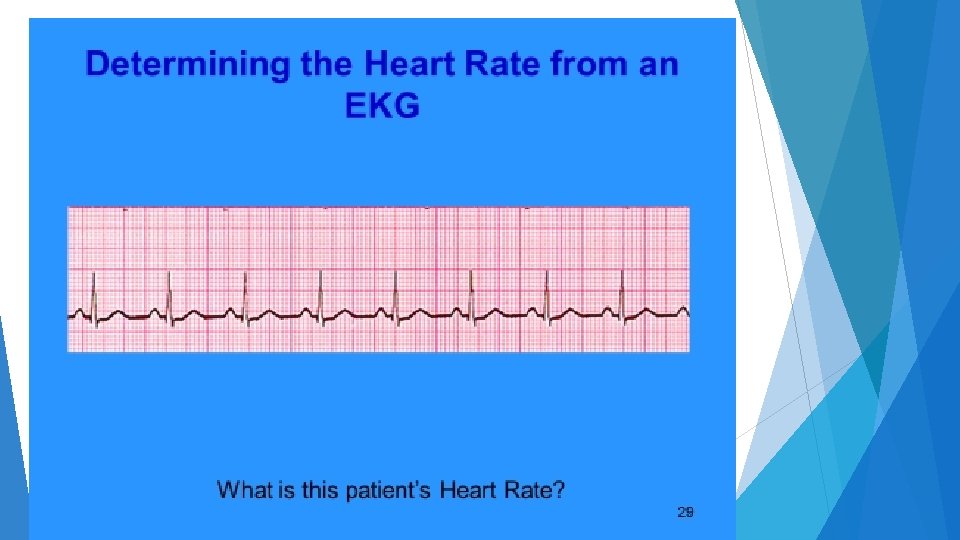
Counting Heart Rates Regular Rythyms
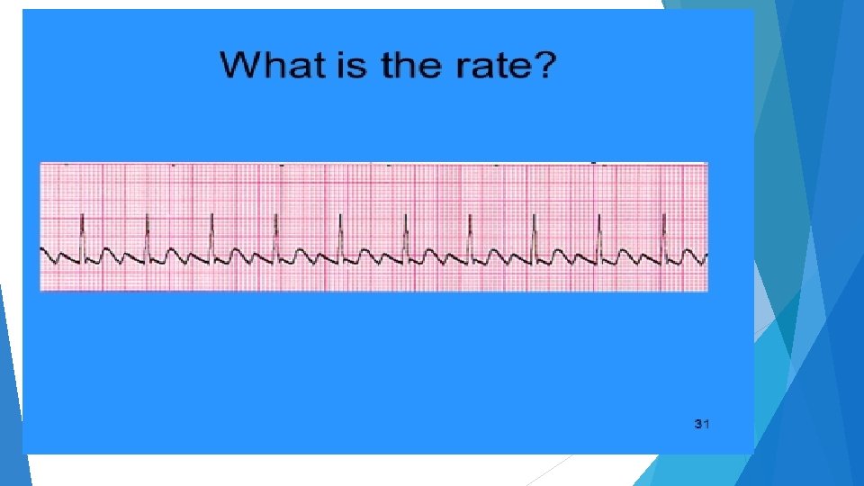
Counting Irregular Rates
Rhythm Check ASYSTOLE
ATRIAL FLUTTER
Second degree block
ATRIAL FIBRILLATION
VENTRICULAR FIBRILLATION
SINUS RHYTHYM
VENTRICULAR TACHYCARDIA
IDIOVENTRICULAR RHYTHYM
SINUS TACHYCARDIA
DETERMINING IF NORMAL OR ABNORMAL Is it rhythm regular? Is there a p wave for every qrs complex? Is the p wave upright? Is there a QRS for every complex? What is the PR interval ? Should be. 12 -. 20 ( 3 to 5 large boxes) What is the QRS Duration? Should be less that 0. 12 ( 3 boxes) What is the rate? Below 60 Bradycardia Above 100 Tachycardia
What does the Complex mean? P wave? Atrial depolarization QRS COMPLEX Ventricular Depolarization ST segment Initial repolarization T wave Ventricular Repolarization