
EKG Conduction Abnormalities Part II Sandra Rodriguez M
EKG Conduction Abnormalities Part II Sandra Rodriguez, M. D. January 15, 2008
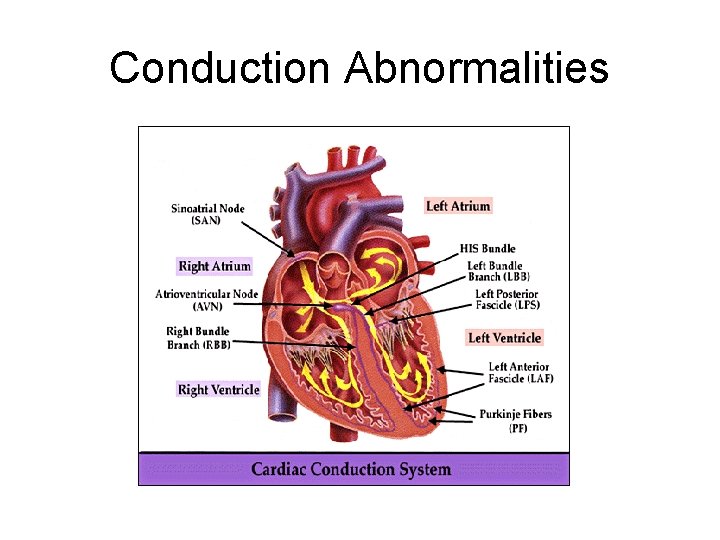
Conduction Abnormalities
Sino-atrial exit block Type 1 • PP intervals gradually shorten until a pause occurs (i. e. , the blocked sinus impulse fails to reach the atria). • The pause duration is less than the two preceding PP intervals. • The PP interval following the pause is greater than the PP interval just before the pause.
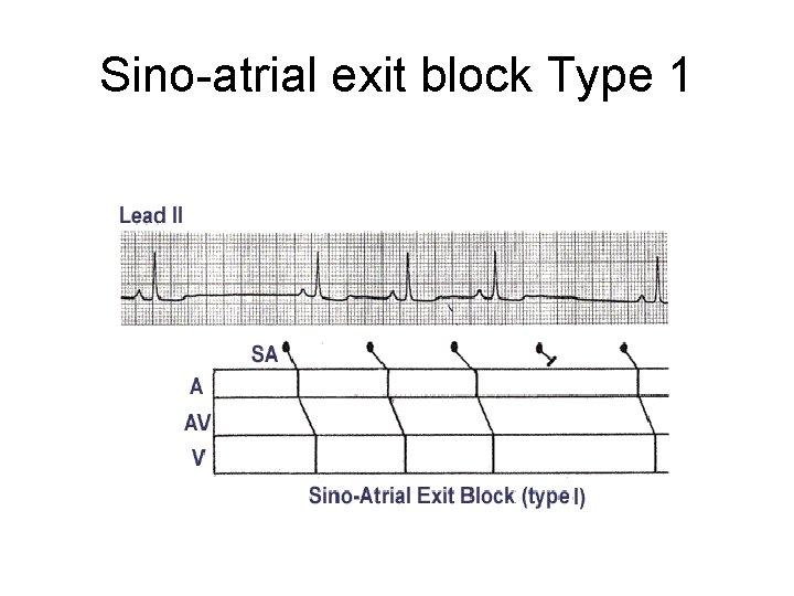
Sino-atrial exit block Type 1
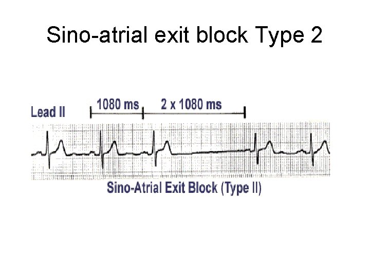
Sino-atrial exit block Type 2 • PP intervals fairly constant (unless sinus arrhythmia present) until conduction failure occurs. • The pause is approximately twice the basic PP interval.
Sino-atrial exit block Type 2
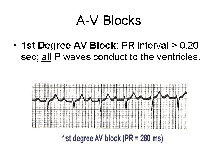
A-V Blocks • 1 st Degree AV Block: PR interval > 0. 20 sec; all P waves conduct to the ventricles.
A-V Block 2 nd degree, type I • In "classic" Type I (Wenckebach) AV block the PR interval gets longer (by shorter increments) until a nonconducted P wave occurs. • The RR interval of the pause is less than the two preceding RR intervals, and the RR interval after the pause is greater than the RR interval before the pause.
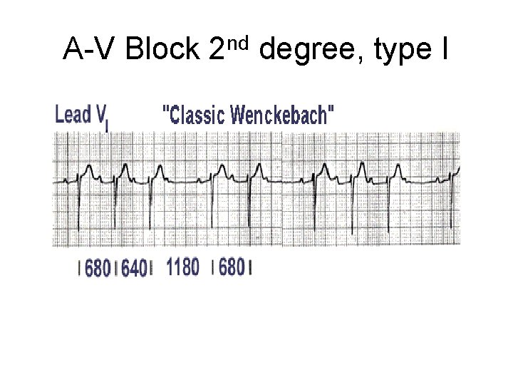
A-V Block 2 nd degree, type I
A-V Block 2 nd degree, type II • In Type II (Mobitz) AV block the PR intervals are constant until a nonconducted P wave occurs. • There must be two consecutive constant PR intervals to diagnose Type II AV block (i. e. , if there is 2: 1 AV block we can't be sure if its type I or II). • The RR interval of the pause is equal to the two preceding RR intervals.
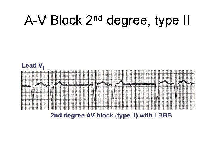
A-V Block 2 nd degree, type II
A-V Block 3 rd degree • Usually see complete AV dissociation because the atria and ventricles are each controlled by separate pacemakers. • Narrow QRS rhythm suggests a junctional escape focus for the ventricles with block above the pacemaker focus, usually in the AV node. • Wide QRS rhythm suggests a ventricular escape focus (i. e. , idioventricular rhythm).
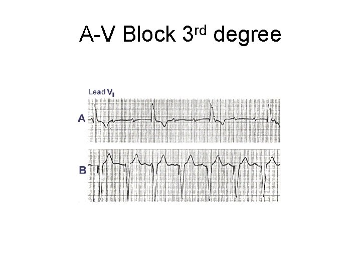
A-V Block 3 rd degree
AV Dissociation • Not synonymous with 3 rd degree AV block, although AV block is one of the causes. • May be complete or incomplete. • In complete AV dissociation the atria and ventricles are always independent of each other. • In incomplete AV dissociation there is either intermittent atrial capture from the ventricular focus or ventricular capture from the atrial focus.
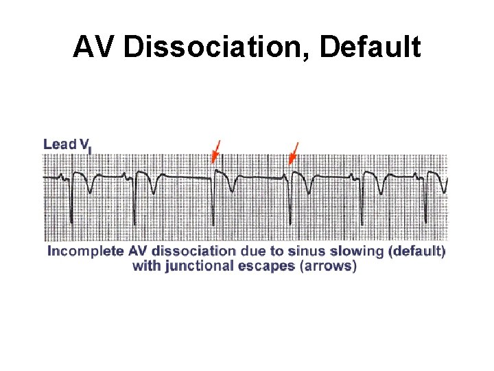
AV Dissociation, Default
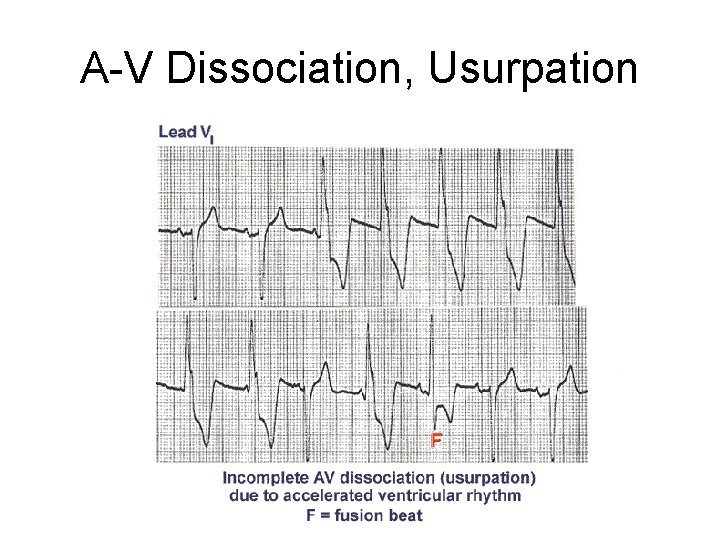
A-V Dissociation, Usurpation
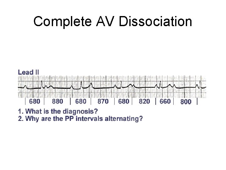
Complete AV Dissociation
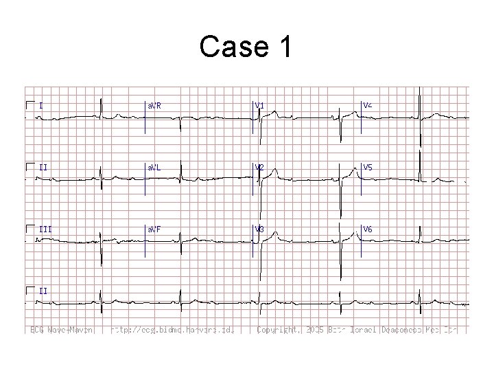
Case 1
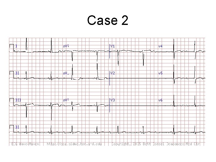
Case 2
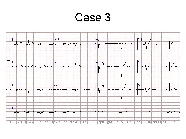
Case 3
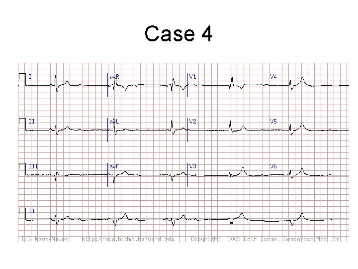
Case 4
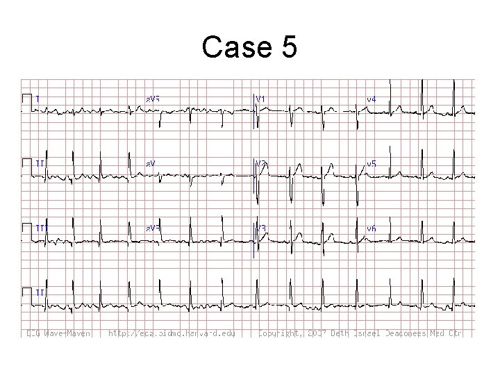
Case 5
- Slides: 22