EKG AUTORHYTHMICITY Figure 21 11 The Cardiac Cycle


- Slides: 8
EKG AUTORHYTHMICITY
Figure 21. 11 The Cardiac Cycle Start Atrial systole begins: Atrial contraction forces a small amount of additional blood into relaxed ventricles. Atrial systole ends, atrial diastole begins 0 800 msec 100 msec Ventricular diastole—late: All chambers are relaxed. Ventricles fill passively. le l ar systo tri cu Cardiac cycle Ven tricul ar d ias to l e Atrial sy stole Atr ial dia sto le Ventricular systole— first phase: Ventricular contraction pushes AV valves closed but does not create enough pressure to open semilunar valves. 370 msec Ventricular systole— second phase: As Ventricular diastole—early: As ventricles relax, pressure in ventricles drops; blood flows back against cusps of semilunar valves and forces them closed. Blood flows into the relaxed atria. ventricular pressure rises and exceeds pressure in the arteries, the semilunar valves open and blood is ejected.
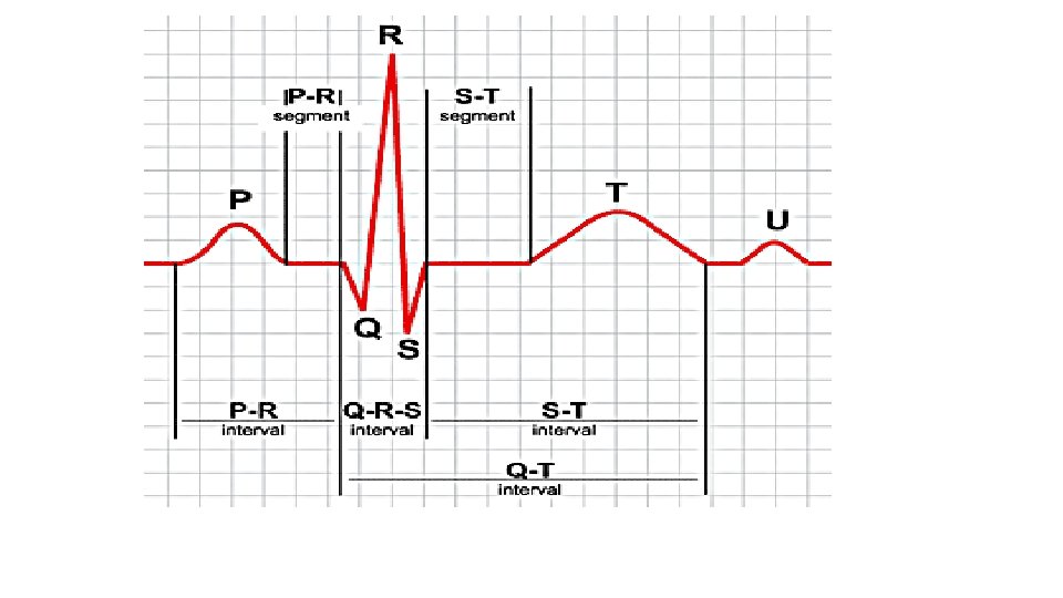
EKG • Electrokardiogram- device to record the action potential of cardiac muscle summation • • • Cannot detect force of contraction Cannot detect Blood Pressure Can detect abnormal heart rates/ rhythems Can detect abnormal conduction pathways Can detect hypertrophy and atrophy and relative position of damage • P Wave- Action potential depolarization of atrial myocardium • Causes Atrial contraction • QRS Complex- Ventricular depolarization • Causes onset of ventricular contraction • Also atrial repolarization masked by QRS signal • T Wave- repolarization before ventricular relaxation • U wave – repolarization of the purkinje fibers • PQ/PR Interval-. 16 sec- atria contract and relax • QT Interval-. 3 sec- ventricles contract and relax • 1 Cardiac Cycle- from onset muscle contraction to next
Autorhythmicity of Cardiac Muscle • Action potentials in heart without external stimuli 1. 2. 3. 4. • • • After each action potential the membrane potential returns to its resting membrane potential Unstable slow ion channels open and cause depolarization This causes fast channels to open and increase depolarization When depolarization reaches threshold--action potential happens more often in SA Node because more slow channels Plateau Phase- Prolonged period of depolarization, separates contractions in the heart Heart has long action potential so the heart will rest between contractions and not tetanic contractions Absolute refractory period – CM insensitive to further stimuli
Figure 21. 11 The Cardiac Cycle Start Atrial systole begins: Atrial contraction forces a small amount of additional blood into relaxed ventricles. Atrial systole ends, atrial diastole begins 0 800 msec 100 msec Ventricular diastole—late: All chambers are relaxed. Ventricles fill passively. le l ar systo tri cu Cardiac cycle Ven tricul ar d ias to l e Atrial sy stole Atr ial dia sto le Ventricular systole— first phase: Ventricular contraction pushes AV valves closed but does not create enough pressure to open semilunar valves. 370 msec Ventricular systole— second phase: As Ventricular diastole—early: As ventricles relax, pressure in ventricles drops; blood flows back against cusps of semilunar valves and forces them closed. Blood flows into the relaxed atria. ventricular pressure rises and exceeds pressure in the arteries, the semilunar valves open and blood is ejected.
The Cardiac Cycle ANIMATION The Heart: Cardiac Cycle ANIMATION The Cardiac Cycle: Part 1 ANIMATION The Cardiac Cycle: Part 2 ANIMATION The Cardiac Cycle: Part 3 ANIMATION The Cardiac Cycle: Part 4 ANIMATION The Cardiac Cycle: Part 5