Effects of Ventral and Dorsal CA 1 Subregional

Effects of Ventral and Dorsal CA 1 Subregional Lesions on Trace Fear Conditioning J. L. Rogers, M. R. Hunsaker, R. P. Kesner

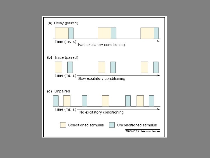
Role of Hippocampus in Pavlovian Fear Conditioning • Complete lesions produce deficits in contextual fear. (acquisition and consolidation) • Regulates when and where extinction memories are expressed. • Necessary for association if stimuli separated in time (trace). – Over time performance depends on prefrontal cortex (PFC).

Fear Conditioning Circuitry

Contextual Extinction Retrieval
 – 50% of volume – Preferential role –")
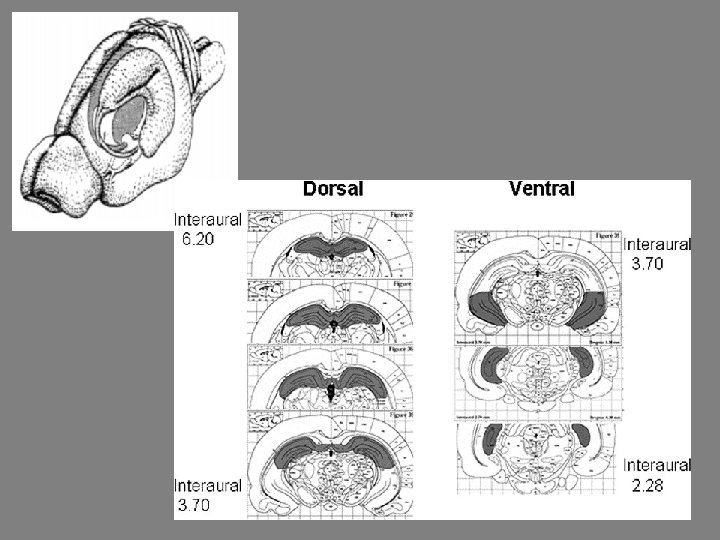
Hippocampus • Dorsal (posterior - primates) – 50% of volume – Preferential role – spatial learning • Ventral (Anterior – primates) – 50% of volume – Preferential role – anxiety behaviors

Anatomical Connections • Major input of visual-spatial information from primary sensory cortical areas is into dorsal subregion. (olfactory evenly distributed) • Ventral subregion projects to PFC, dorsal does not. • Ventral more closely connected to BNST, Amygdala, and Hypothalamic structures (HPA)
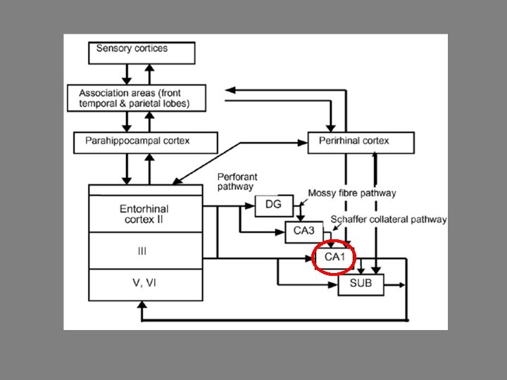

• CA 1 subregion involved in temporal pattern association and intermediate-term memory. • CA 1 pyramidal neurons increase activity during trace conditioning. • CA 1 NMDA receptors necessary for trace conditioning.

Aims • Determine if ventral and dorsal CA 1 subregion of hippocampus have different roles in trace fear conditioning.
 • Tested during light")
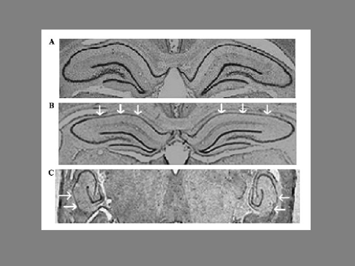
Methods • 24 Long-Evans rats (4 months, 300 -400 g) • Tested during light phase • Stereotaxic infusion of ibotenic acid into dorsal (n = 9) and ventral CA 1 (n =7) • Controls (n = 8) – vehicle infusions into CA 1

Ibotenic Acid • Toxin produced by Amanita muscaria and Amanita pantherina mushrooms • Excitatory Amino acid agonist • Damage to adjacent areas, fibers-of-passage, and damage to the vasculature are minimized.

Methodological Considerations • Excitotoxic lesions can cause over-excitation of “downstream” structures – dysfunction of other parts of the circuit. • Rats – hippocampus surface area ~1. 2 cm 2 , entire isocortex ~1. 5 cm 2. • Damage to amygdala and cortex. • Produce cell death – oxidative stress.

Apparatus • One used for conditioning and context acquisition • Another, without contextual cues or shock, used for retention testing for tone-trace

• Day 1 - Acquisition – 2 min – 15 trials tone-trace-shock » 32 s, 10 s, 2 s (0. 5 m. A) (72 s ITI) – Freezing measured during baseline, tone, trace • Day 2 – Context retention – (24 hrs later) Same chamber, 8 min. without tone, freezing measured every 8 s. • Day 3 – Tone-trace retention – (48 hrs later) Different chamber, 2 min preexposure with 15 tone trace combinations freezing measured every 8 s.

Context Acquisition

Context Test

Summary Context Results • All groups displayed freezing during ITI of acquisition phase. • Ventral CA 1 lesion group froze less than dorsal CA 1 lesion group and controls. • Dorsal CA 1 lesion group froze significantly less than controls. • Both involved in contextual retention , ventral more.

Trace Acquisition

Trace Test

Summary Trace Results • Dorsal and ventral CA 1 lesion groups not different from controls during acquisition. • Retention – ventral CA 1 froze less than controls and dorsal CA 1 lesion group, dorsal CA 1 lesion group was not different from controls.

Tone Acquisition

Tone Test

Summary Tone Results • During acquisition all groups froze during tone. • No significant main effect for groups during retention.
 lesions. – May")
Conclusions • Acquisition not disrupted by CA 1 (d or v) lesions. – May involve entire hippocampus (DG, CA 3) • Ventral CA 1 “more important” than dorsal CA 1 for retention of context and trace fear memory but dorsal involved. • “Mild” deficits to tone retention.
- Slides: 27