Ectopic ACTH syndrome EAS is rare but is

 is rare but is frequently a severe EAS should often")
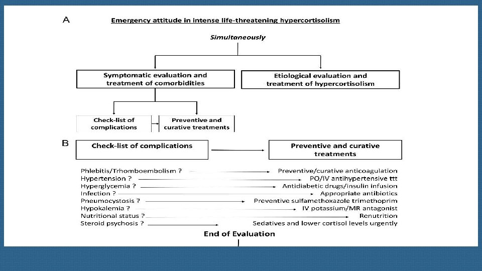
Ectopic ACTH syndrome (EAS) is rare but is frequently a severe EAS should often be considered as an endocrine condition. emergency requiring an emergency response both in terms of diagnostic procedures and therapeutic interventions. endocrinology teams in collaboration with specialized hormonal laboratory, modern imaging platforms and intensive care units.
 NETs in the cervical spine region EATs from the cervical region are mainly")
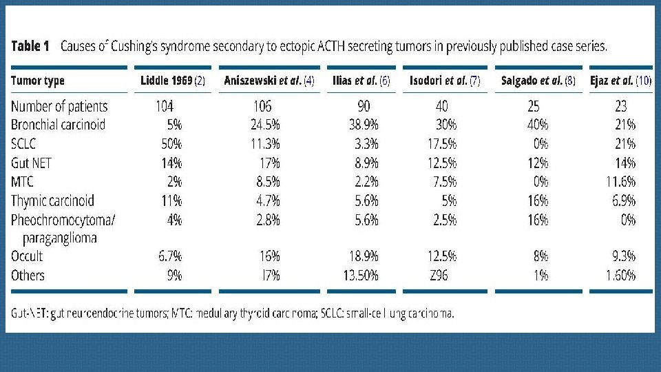
(A) NETs in the cervical spine region EATs from the cervical region are mainly represented by medullary thyroid cancer (MTC), although fewer than a hundred cases have been reported in the literature, accounting for less than 1% of EATs. Absence of detectable thyroid tumor in the presence of increased plasma CT concentrations indicate a gut ACTH and CT co-secreting tumor.
 Thoracic neuroendocrine tumors and carcinomas Most primary endocrine tumors responsible for EAS are")
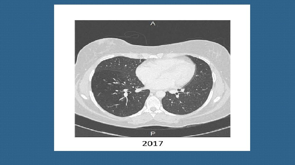
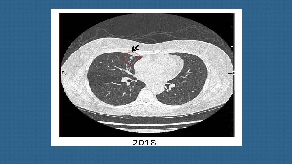
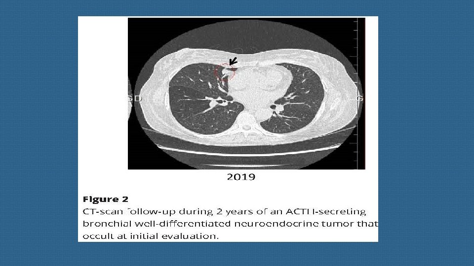
(B) Thoracic neuroendocrine tumors and carcinomas Most primary endocrine tumors responsible for EAS are located in the chest. Well-differentiated bronchial NETs account for the vast majority of ‘occult’ (radiologically invisible) forms, but their apparent frequency may decline as imaging techniques improve.
 Abdominal and retroperitoneal NETs Pancreatic NETs account for fewer than 15% of all")
(C) Abdominal and retroperitoneal NETs Pancreatic NETs account for fewer than 15% of all cases of EAS. Most of these are overt tumors that are readily visible on imaging studies. ACTH-secreting pheochromocytomas and paragangliomas generally also secrete catecholamines and metanephrines.
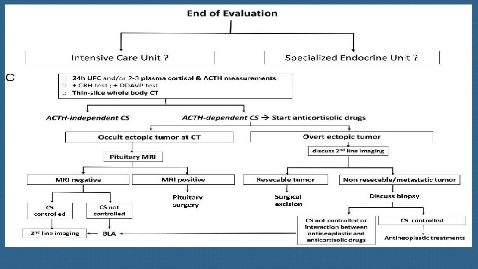

First-line imaging procedures Spiral thin-slice CT of the cervical-thoracic-abdominal and pelvic regions is essential , together with pituitary MRI, in severe ACTH-dependent Cushing’s syndrome. New technologies such as c 11 C-methionine PET have been proposed to visualize occult pituitary microadenomas, but these are still part of clinical research and are not compatible in this context of emergency. One of the main diagnostic issues is bronchial carcinoids which represent one of the main causes of EAS.

these richly vascularized tumors are notoriously difficult to detect due to their small size and their location being close to pulmonary vessels. a metanalysis published in 2015 suggests that, overall, approximatively 53% of EAT NET will be clearly revealed by CT, making this imaging method a valuable tool for diagnosis and patient management. When imaging shows no culprit NET, special attention should then be paid to sites such as the bronchi , small intestine and other much rarer sites involved in ectopic ACTH secretion.

we strongly suggest a second careful examination of the thoracic CT-scan, which is the main source of occult EATs, ideally by several radiologists, to limit the risk of false negatives. Elsewhere, false positives on CT have been described in 3. 6% of cases , a finding that emphasizes the importance of using an appropriate CT protocol and the experience of the radiologist to limit the frequency of thoracic lures.

Prophylaxis of Pneumocystis jirovecii pneumonia with sulfamethoxazoletrimethoprim is recommended for all patients with intense hypercortisolism. In a recent survey issued from the European Register on Cushing’s Syndrome (ERCUSYN) registry, infections were the most common cause of death during the 3 months following diagnosis, emphasizing the need for clinical vigilance at that time, especially in patients with intense hypercortisolism and diabetes mellitus.

On the basis of the available literature and our personal experience, we propose that in the context of severe EAS, the search for an EAT should begin with good-quality whole-body thin-slice CT performed by a dedicated expert radiologist aware of the difficulty in identifying these tumors. Functional imaging such as 68 Ga-PET/CT or 18 -FDG PET/CT should be considered in second line to detect occult tumors on CT or to reinforce the hypothesis of the neuroendocrine nature of small tumors detected by CT and also to contribute to the workup of a metastatic tumor.
- Slides: 19