ECTOPIA LENTIS BY ADREA R BENKOFF M D

ECTOPIA LENTIS BY ADREA R. BENKOFF M. D.

Case Presentation CC/HPI: 19 yo female presents with decreased VA in both eyes over the past year. Subsequently complaining that she is unable to drive. Denies head or facial trauma, diplopia, flashes, floaters, blindspots, or pain. POH: high/pathologic myopia, right esotropia (poor vision from young age OU) *current glasses: -7. 50 sph OD, -9. 75 sph OS Social Hx: Lives with family, one of 7 children. Currently in college, doing well academically, but socially limited due to poor vision. Meds: None Allergies: NKDA
 20/80 PH 20/50 -2 Cycloplegic")
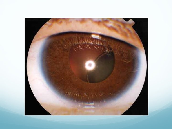
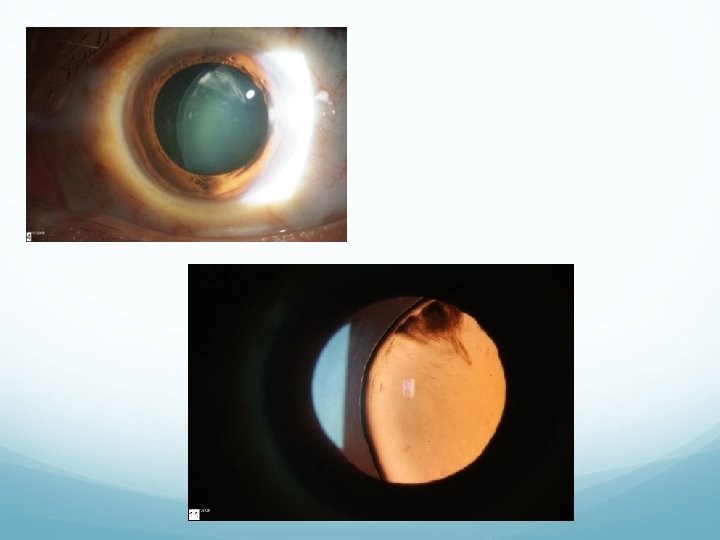
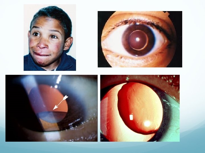
Exam SLE: Distance VA: cc 20/200 PH NI (squinting) 20/80 PH 20/50 -2 Cycloplegic Refract: -11. 50 sph 20/100 -11. 75 sph 20/60 -2 Tear film: wnl OU Lids/adnexa: wnl OU, no ptosis Conjunctiva/Sclera: white & quiet OU EOM: Full OU Muscle Balance: (6 meters) Right esotropia 10 diopters cc Cornea: clear OU Pupils: ERRL, no APD OU Iris: round, no NV OU CVF: full to count fingers OU Lens: inferonasal subluxation OU TA: 15 OU Capsule: clear OU Anterior Chamber: deep and quiet OU

Exam DFE: 1% Mydriacyl, 1% cyclogyl Vitreous: clear OU Cup/Disc: . 2 OU, sharp and pink OU Blood Vessels: normal caliber OU Macula: flat, wnl OU Periphery: flat, no hemorrhages, breaks, tears, or detachments OU

Differential Diagnosis Simple ectopia lentis Ectopia lentis et pupillae Marfan’s Syndrome Homocystinuria Weill-Marchesani Syndrome Hyperlysinemia Sulfite oxidase deficiency Traumatic lens subluxation Pseudoexfoliative disease Other: aniridia, Ehlers-Danlos Syndrome, congenital syphilis, chronic uveitis

Diagnosis Marfan’s Syndrome Ectopia lentis

Past Medical and Family History 50 20/30+ *aortic insufficiency 23 20/40 *scoliosis 19 20/100 20/60 -2 Marfan’s Syndrome 17 20/4020/30+ 16 20/70 20/50+ 13 20/50+ 20/50 -2
 put together the skeletal manifestations of")
Marfan’s Syndrome: Background 1896: Antoine Marfan (French Physician) put together the skeletal manifestations of the disease Classic triad: subluxed lenses, skeletal anomalies, and cardiovascular disease point mutation in chromosome 15 q, band 21 abnormal fibrillin Autosomal Dominant, prevalence 4 -6 per 100, 000 High penetrance 15% sporadic

Epidemiology Most frequent cause of inherited lens dislocation! Affects both sexes equally Found in all races

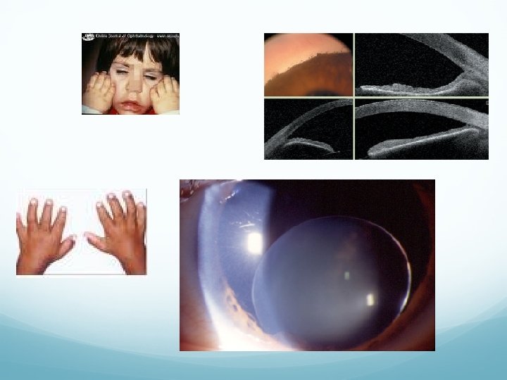
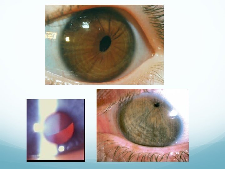
Ocular Features Ectopia lentis: subluxated/luxated lenses Superior or superotemporal lens displacement, bilateral and symmetric Zonules appear intact, but stretched/elongated or focally attenuated, broken or absent 50 -80% of patients exhibit this finding Minority of pts with ectopia lentis glaucoma Signs: progressive myopia or noncorneal astigmatism, phacodonesis, iridodonesis, asymmetry of AC depth between two eyes, positional variation in IOP
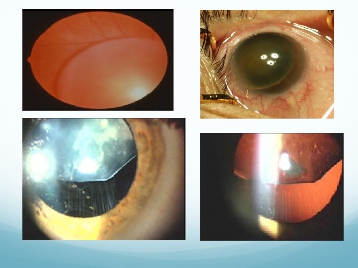

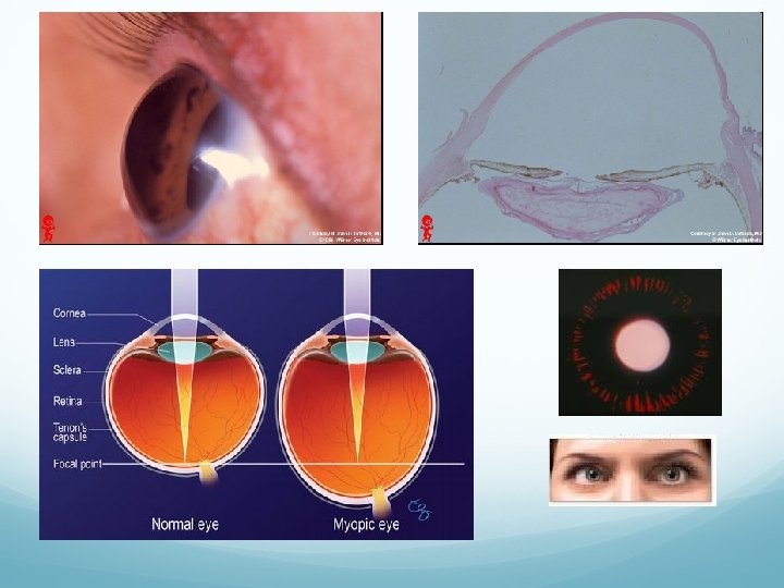
Other Ocular Features High axial myopia • Megalocornea, flat corneas High risk of lattice degeneration • Premature cataracts (30 -50 yo) Atrophic disease of the peripheral retina (thinning, pigmentary changes) Retinal detachment Microphakia • Hypoplasia of iris stroma and dilator muscle can have peripheral transillumination, and poor dilation with mydriatic drops Strabismus • Early vitreous syneresis Ptosis • Uveal colobomas

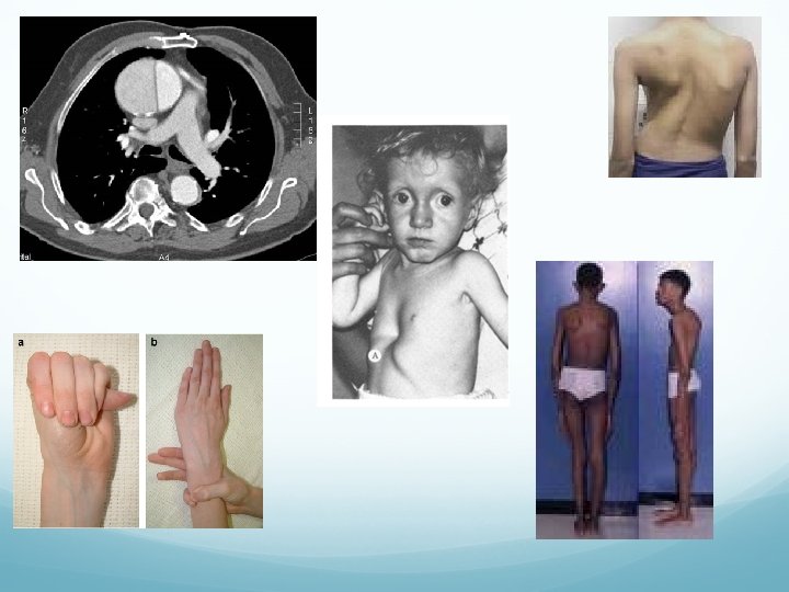
Systemic Features Marfanoid habitus Arachnodactyly Muscular hypoplasia & hypotonia joint laxity Pectus excavatum Scoliosis Aortic dilation dissecting aneurysm Valve disease: MVP, AI Decreased subcutaneous fat Lungs: cystic malformation, lobulation, emphysema Genitourinary system: ureteric strictures recurrent pyelonephritis
 FBN 1 gene mutation on chromosome 15 Limited use due")
Diagnosis Genetic testing: (fibrillin-1) FBN 1 gene mutation on chromosome 15 Limited use due to locus heterogeneity, large size of gene, and sporadic cases Clinical diagnosis: depends on major and minor signs as defined by Ghent nosology (1996) Unequivocally diagnoses or exclude Marfan in 86% of cases

Pathology Ectopia Lentis: Abnormal production, distribution, and attachment of zonules Chief component of zonules is fibrillin Zonular bundles are thin and scanty

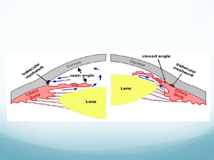
Pathology Secondary glaucoma: Lens dislocation into pupil or AC pupillary block iris bowing angle closure glaucoma Open angle: can present in childhood or adolescence Congenital anomalies of the AC angle Dense iris processes, incompletely developed angle Concave iris tissue Abnormal insertion of ciliary musculature into trabecular meshwork (chronic)

Pathology Retinal detachments: Combination of axial myopia, vitreous syneresis and abnormal vitreoretinal adhesions rhegmatogenous RD Subluxated/luxated lens capsule exerts traction on vitreous base peripheral tears

Management Ectopia lentis: Refractive aids: glasses, contact lenses, need reading add if subluxed lens Pharmacological pupil manipulation Surgical lens extraction: cataract formation, anisometropia or refractive error not otherwise correctable, impending dislocation, lens-induced glaucoma or uveitis Marfan pts have high rate of surgical complications
")
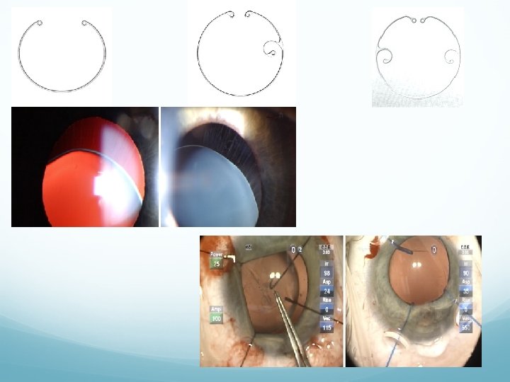
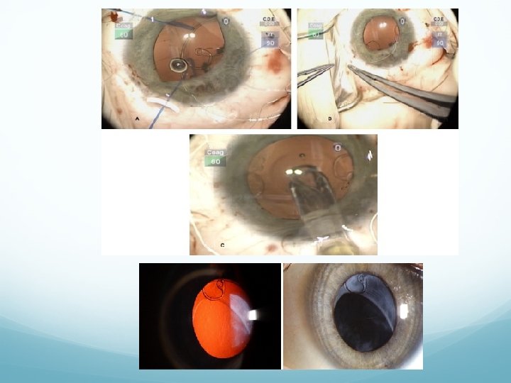
Surgical Management Lens extraction: phacoemulsification with PCIOL implantation Cionni ring (modified capsular tension ring) MCTR has a hook allowing for scleral fixation without violation of capsular bag Less likely to have vitreous loss operatively, can perform through 3. 0 mm incision rapid visual recovery Can achieve 20/40 or better post-op with Cionni ring Most common complication is PCO requiring YAG posterior capsulotomy

Management Glaucoma: Anterior lens dislocation: pupillary block with acute or chronic angle closure glaucoma Goniotomy or trabeculotomy for children Older kids and adults: medical management of IOP with mydriatics, and supine positioning Peripheral laser iridotomy Surgery: iridectomy, lens extraction if in AC, tube shunt placement

General Management Approximately 50% of patient’s with Marfan’s Syndrome are diagnosed by an ophthalmologist! Myopia is not adequately corrected by lenses Non-traumatic lens dislocation Slit-lamp exam, dilated fundus exam for every evaluation Annual exam is recommended until age 12 to prevent amblyopia If family Hx then initial exam at 3 -6 months of age Must work up the patient and refer for full medical (internist, cardiologist) and orthopedic evaluation (including immediate family)

Prognosis Ectopia Lentis: Depends on degree of lens subluxation and location Secondary complications of lens subluxation/luxation as discussed When lens completely dislocates, if posterior without complications then good prognosis Good VA with glasses or contact lenses

Familial lens subluxation/luxation 50 Inferior and inferonasal dislocation in vitreous 23 Inferotemporal OD Temporal OS 19 Inferonasally OU 17 Superonasal OD inferotemporal OS 16 Nasal OD Inferotemporal OS 13 Superonasal subluxation OU

Case Presentation Plan: Refer for surgical consultation OS Lensectomy with Cionni ring technique New refractive prescription for progressive myopia Follow up in 1 year

Traumatic Lens Dislocation Most common cause of acquired lens displacement Accounts for 22 -50% of lens subluxations Associated with cataracts and rhegmatogenous retinal detachment Zonules rupture presumably at lens attachments secondary to mechanical stretching Eye is compressed in ant-post direction (i. e. fist) and the distention of the globe in the medial-lateral plane ruptures the zonular fibers

Homocystinuria 2 nd most common cause of inherited lens dislocation AR, prevalence 1: 200, 000 Virtual absence of cystathionine b-synthetase Dx: increased serum homocystine, methionine, homocysteine Ocular abnormalities: ectopia lentis, RD Lens displacement: bilateral and symmetric, usually inferonasal Systemic features: fair skin, malar flush, pale coarse hair, poor peripheral circulation, 50% mental retardation, marfanoid habitus, osteoporosis, seizures Increase thromboembolic events, especially with anesthesia

Homocystinuria Pathology: abnormal zonular development due to cysteine deficiency Zonular fibers are made up of cysteine-rich glycoprotein Affected zonules are brittle and easily disrupted, with short disoriented fibrils Exam findings: spherophakic, scalloped lens border, lens dislocation into AC glaucoma Treatment: low methionine, high cysteine diet and vitamin supplementation with coenzyme pyridoxine decreased incidence of ectopia lentis Prognosis: favorable if treatment is started soon after birth

Weill-Marchesani Syndrome 3 rd most common cause of inherited lens dislocation AR, some heterozygote expression Dx: molecular genetic test for ADAMTS 10 Ocular abnormalities: ectopia lentis, microspherophakic lens, lenticular myopia, micro/megalocornea, cataracts, pupillary block Lens displacement: usually inferior but sometimes central Zonular bundles appear taut or lax and abnormally elongated Systemic features: brachymorphism-short stature, short, broad hands and feet, limited joint mobility, well-developed muscular appearance, thick skin, brachycephaly
 Treatment: Lensectomy")
Weill-Marchesani Syndrome Frequent episodes of pupillary block causing angle closure glaucoma (microphakia) Treatment: Lensectomy Glaucoma: mydriatics, laser iridotomy (can be prophylactic), iridectomy and miotics to prevent lens dislocation in AC Prognosis: loss of vision is earlier and more severe than other lens dislocation syndromes
 ectopia lentis Usually AD, most at birth with superotemporal lens displacement Some")
Simple (isolated) ectopia lentis Usually AD, most at birth with superotemporal lens displacement Some cases of delayed onset in 6 th/7 th decade with downward dislocation Gene defect localized to fibrillin gene on chromosome 15 Ocular findings: microspherophakic Deficiency of zonular fibers, poor aggregation of zonules still present, and some abnormally thick fibrils No associated systemic features

Ectopia Lentis et Pupillae AR, pupils are oval or slit-like with >. 5 mm nasal and downward eccentricity Ocular findings: lens is displaced in direction opposite of pupil displacement, usually bilateral and asymmetric Often atrophic irides that dilate poorly and with marked transillumination defects rapidly progressing cataracts, severe axial myopia, occasional RD Developmental defect of neuroectodermal layer results in failure of iris pigment epithelial cells to develop normal dilator muscle, presumably associated with poor secretion of zonular fibrils

Sulfite Oxidase Deficiency Rare AR, disease of sulfur metabolism Enzyme deficiency results in excess sulfite Ocular features: dislocated lens (nonspecific) Systemic features: (in first year of life) Poor feeding, severe neurologic abnormalities, seizures, myoclonus, and severe mental retardation Excess sulfite can destroy disulfide bonds, may cause lens dislocation since disulfide linkages are important for intramolecular bonds in fibrillin Generally poor prognosis for neurologic abnormalities

Hyperlysinemia Rare AR, defect in amino acid lysine metabolism Ocular findings: bilateral superior subluxation of lenses, strabismus, bilateral spherophakia Systemic findings: mental retardation, muscle hypotonia, convulsions Dx: increase plasma levels of lysine Ocular lens pathology unknown Tx: low protein diet may be helpful

Pseudoexfoliation Spontaneous subluxation in 5% of patients Signs: may see phacodonesis, iridodonesis may be absent due to relative immobility from pseudoexfoliative deposits in stroma and muscle Zonules break midstream or at ciliary body Zonules are infiltrated with pseudoexfoliative material and are fragile

Other causes of ectopia lentis Aniridia Ehlers-Danlos Syndrome Congenital syphilis Chronic uveitis
- Slides: 45