Echocardiography for Heart Failure PAR TNERS IN HEALTH


 Depth Gain -near -far -total Freeze Save (still)")


-shaped plane of a")


 Diastole (filling) In")


 Diagnosis Cardiomyopathy Significant Mitral Stenosis Other Valvular")


 Diagnosis Cardiomyopathy Significant Mitral Stenosis Other Valvular")


 Diagnosis Cardiomyopathy Significant Mitral Stenosis Other Valvular")


")


")


 Diagnosis Cardiomyopathy Significant Mitral Stenosis Other Valvular")


 Diagnosis Cardiomyopathy Significant Mitral Stenosis Other Valvular")


 Diagnosis Cardiomyopathy Significant Mitral Stenosis Other Valvular")


 Diagnosis Cardiomyopathy Significant Mitral Stenosis Other Valvular")


 Diagnosis Cardiomyopathy Significant Mitral Stenosis Other Valvular")


 Diagnosis Cardiomyopathy Significant Mitral Stenosis Other Valvular")


 Diagnosis Cardiomyopathy Significant Mitral Stenosis Other Valvular")


 Diagnosis Cardiomyopathy Significant Mitral Stenosis Other Valvular")


 Diagnosis Cardiomyopathy Significant Mitral Stenosis Other Valvular")

- Slides: 168
Echocardiography for Heart Failure PAR TNERS IN HEALTH CHRONIC CARE TRAINING FOR DISTR ICT H OSPITAL NURSES 2013
Unit Content Session I: Introduction to Echocardiography Session II: Group Practice – Echocardiography Examples Session III: Simulation Practical Session IV: Group Practice – Echocardiography Video Clips Session V: Hospital Wards Practical
Learning Objectives By the end of this module, participants will be able to: Understand the basic principles of echocardiography imaging Describe the important features and functioning of the echocardiography machine Describe the standard views used in echocardiography Describe the uses of each view in diagnosing a patient’s heart disease Generate a basic report and correctly assign a preliminary heart failure diagnostic classification using echocardiography Demonstrate proficiency in image acquisition for echocardiography
Echocardiography - Session I INTRODUCTION TO ECHOCARDIOGRAPHY
Learning Objectives By the end of this module, participants will be able to: Understand the basic principles of echocardiography imaging Describe the important features and functioning of the echocardiography machine Describe the standard views used in echocardiography Describe the uses of each view in diagnosing a patient’s heart disease Generate a basic report and correctly assign a preliminary heart failure diagnostic classification using echocardiography Demonstrate proficiency in image acquisition for echocardiography
Basic Principles of Echocardiography is a tool (just like a BP cuff and a stethoscope). Remember: the most powerful tool is your brain! Data obtained through an echo is integrated with the history and physical exam to help arrive at a diagnosis.
Basic Principles of Echocardiography is a type of ultrasound imaging test that uses high frequency sound waves Sound waves reflect (bounce) off of structures Tissues appear white Fluid (blood, effusion) appears black Tissue Blood
When should I use echocardiography? To determine if an echo is needed: 1 Obtain a thorough history 2 3 Perform a physical exam (including vital signs and auscultation) If history and physical exam indicate that patient could have heart failure, perform an echo
Diagnostic Categories of Heart Failure Cardiomyopathy Hypertensive Heart Disease Significant Mitral Stenosis Other Valvular or Congenital Disease (not MS) Isolated Right Heart Failure Pericardial Effusion
Learning Objectives By the end of this module, participants will be able to: Understand the basic principles of echocardiography imaging Describe the important features and functioning of the echocardiography machine Describe the standard views used in echocardiography Describe the uses of each view in diagnosing a patient’s heart disease Generate a basic report and correctly assign a preliminary heart failure diagnostic classification using echocardiography Demonstrate proficiency in image acquisition for echocardiography
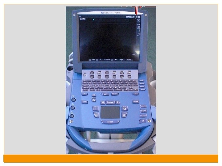
Sonosite Keyboard Power Patient Save (video) Depth Gain -near -far -total Freeze Save (still) 2 D
Machine Adjustments Gain: Similar to brightness; Can adjust near or far from transducer Depth: Adjust how deep into the patient the image will show (you want to see just enough of the structures of interest) Freeze: Will freeze the image Save: Saves a still image after freeze Save Clip: Saves a video recording of the image 2 D: Resets the image (if you made any changes; does not correct gain)
Learning Objectives By the end of this module, participants will be able to: Understand the basic principles of echocardiography imaging Describe the important features and functioning of the echocardiography machine Describe the standard views used in echocardiography Describe the uses of each view in diagnosing a patient’s heart disease Generate a basic report and correctly assign a preliminary heart failure diagnostic classification using echocardiography Demonstrate proficiency in image acquisition for echocardiography
Image Planes The image that is acquired represents one fan (triangle)-shaped plane of a three dimensional structure. As the probe is angled in different directions, a different image will be obtained.
Placing the Echo Probe Standard views are obtained from positioning the probe in different ways on the patient’s chest
Echo Axes Long axis: probe points towards the right shoulder Short axis: probe points at a 90 degree angle to the long axis 4 -chamber axis: probe points toward the back
Improving Image Quality Most of the views of the heart appear clearer if the patient is positioned on the left side. The probe should be placed in a space between the ribs to improve the image quality. Use enough ultrasound gel (but not too much!)
Improving Image Quality - Depth
Improving Image Quality - Depth
Echo Analysis: Standard Questions For each echo, ask yourself the following questions: 1. 2. 3. 4. 5. 6. 7. 8. 9. Which view is represented? Which chambers can be identified? Which valves are visualized? Is the LV function significantly abnormal? (EF ≤ 40%) Is the RV significantly dilated? (moderate to severe; ≥ 2 x aortic root size on parasternal long) Is significant mitral stenosis present? (moderate to severe ) Is a large pericardial effusion present? (≥ 2 cm) Is the IVC ≤ 1 cm in diameter? Is the image of good quality (clarity, depth, on-axis)?
Four Basic Echo Views Parasternal long axis Parasternal short axis Apical 4 -chamber Subcostal
1. Parasternal long axis Most useful view! Good for: LV systolic function RV size Mitral valve abnormalities Pericardial effusion
1. Parasternal long axis
2. Parasternal Short Axis Good for: LV systolic function RV size Pericardial effusion
2. Parasternal Short Axis
3. Apical 4 -Chamber Good for: LV systolic function Mitral valve abnormalities Pericardial effusion
3. Apical 4 -Chamber
4. Subcostal Good for: LV systolic function Pericardial effusion Best if the patient lies SUPINE
4. Subcostal
Subcostal IVC View Rotate probe 90 degrees from the standard subcostal view (aligned with the longitudinal axis of the patient – probe marker towards the head) The IVC can be seen entering the right atrium The IVC normally collapses with inspiration In some conditions, the IVC can be dilated (>2. 2 cm) Hypervolemia Constrictive pericarditis
Subcostal IVC view
Echo Views: Overview Parasternal Long Parasternal Short Apical 4 Chamber Subcostal LV systolic function RV size Mitral Stenosis Pericardial Effusion IVC size
Echo Views: Standard Questions For each view, ask yourself… 1. 2. 3. 4. 5. 6. 7. 8. 9. Which view is represented? Which chambers can be identified? Which valves are visualized? Is the LV function significantly abnormal? (EF ≤ 40%) Is the RV significantly dilated? (moderate to severe; ≥ 2 x aortic root size on parasternal long) Is significant mitral stenosis present? (moderate to severe ) Is a large pericardial effusion present? (≥ 2 cm) Is the IVC ≤ 1 cm in diameter? Is the image of good quality (clarity, depth, on-axis)?
Basic Echo Report Left Ventricle Systolic Function Normal Significantly Depressed Not well visualized Right Ventricle Size Normal Significantly Dilated Not well visualized Mitral valve Significant Stenosis Absent, no mitral stenosis Present, yes Not well visualized Pericardial Significant Effusion Absent, no large effusion Present, yes large effusion Not well visualized IVC Size Normal Dilated > 1 cm Not well visualized Other ________________
Learning Objectives By the end of this module, participants will be able to: Understand the basic principles of echocardiography imaging Describe the important features and functioning of the echocardiography machine Describe the standard views used in echocardiography Describe the uses of each view in diagnosing a patient’s heart disease Generate a basic report and correctly assign a preliminary heart failure diagnostic classification using echocardiography Demonstrate proficiency in image acquisition for echocardiography
Evaluating Structure and Function In this section, we will learn how to evaluate for normal and abnormal heart structure and function. The following 5 structures should be evaluated: 1. 2. 3. 4. 5. Systolic Function RV Size Mitral Valve abnormalities Pericardial effusion IVC size
1. Systolic Function The cardiac cycle has 2 phases: Systole (ejection) Diastole (filling) In many patients with heart failure, the systolic function may be impaired. This means the heart will have difficulty pushing the blood to the rest of the body. This is described as: Ejection Fraction and Fractional Shortening
Ejection Fraction: The fraction of the volume of blood which is pushed out of the heart with each beat is called the Ejection Fraction (EF) The Ejection Fraction is generally classified as: normal (EF>55%), mildly reduced (45 -55%) moderately to severely reduced (< 45%)
Fractional Shortening Diastole Systole In a cross-sectional view, the distance between the walls of the When the normal heart squeezes in systole, the ventricle as they move toward each other with each heartbeat is distance between walls narrows by only 25% called thethe fractional shortening.
Normal Systolic Function Ejection Fractional Shortening 55% 25% You will be asked only to ESTIMATE the fractional shortening and ejection fraction
Basic Echo Report Left Ventricle Systolic Function Normal Significantly Depressed Not well visualized Right Ventricle Size Normal Significantly Dilated Not well visualized Mitral valve Significant Stenosis Absent, no mitral stenosis Present, yes Not well visualized Pericardial Significant Effusion Absent, no large effusion Present, yes large effusion Not well visualized IVC Size Normal Dilated > 1 cm Not well visualized Other ________________
Normal Systolic Function
Significantly Depressed Systolic Function This finding is both NECESSARY and SUFFICIENT to assign the diagnostic category of CARDIOMYOPATHY
2. Right Ventricle Size Assessing right ventricular size: In many cases of heart failure, the right ventricle may appear enlarged on the parasternal long axis view. In ISOLATED RIGHT HEART FAILURE, Right Ventricle dilation may be the only abnormal finding.
Basic Echo Report Left Ventricle Systolic Function Normal Significantly Depressed Not well visualized Right Ventricle Size Normal Significantly Dilated Not well visualized Mitral valve Significant Stenosis Absent, no mitral stenosis Present, yes Not well visualized Pericardial Significant Effusion Absent, no large effusion Present, yes large effusion Not well visualized IVC Size Normal Dilated > 1 cm Not well visualized Other ________________
Normal RV Size Normally, the right ventricle is roughly the same size as the aortic root and the left atrium. If the RV is more than 2 x the size of the aorta, the RV is dilated.
RV Dilation – Parasternal Long Axis
RV Dilation – Short Axis In the short axis view, the LV usually appears as a circle If the RV is dilated, then it will push on the LV and cause the interventricular septum to become flat and the LV to be shaped like the letter “D”
3. Mitral Stenosis: Inflammation and scarring of the mitral valve caused by recurrent episodes of rheumatic fever Key echo findings (not all will be present) • Thickening of the mitral valve leaflets • Restriction and immobility of mitral valve (particularly the posterior leaflet) • “Elbow deformity” of the anterior leaflet is very common • Calcification of the valve– appears white on echo • In pure mitral stenosis, the left ventricle will appear small with a normal ejection fraction These features are best appreciated in the PARASTERNAL LONG AXIS view
Basic Echo Report Left Ventricle Systolic Function Normal Significantly Depressed Not well visualized Right Ventricle Size Normal Significantly Dilated Not well visualized Mitral valve Significant Stenosis Absent, no mitral stenosis Present, yes Not well visualized Pericardial Significant Effusion Absent, no large effusion Present, yes large effusion Not well visualized IVC Size Normal Dilated > 1 cm Not well visualized Other ________________
Normal Mitral Valve
Mitral Stenosis
4. Pericardial Effusion Pericardial effusions appear as an extra layer of fluid around the heart that appears black on the echo This can be seen best in the Subcostal, the Parasternal long axis view, and the Apical 4 -Chamber views A pericardial effusion of >2 cm on diastole in a patient in decompensated heart failure is an indication for immediate referral to the district hospital.
Basic Echo Report Left Ventricle Systolic Function Normal Significantly Depressed Not well visualized Right Ventricle Size Normal Significantly Dilated Not well visualized Mitral valve Significant Stenosis Absent, no mitral stenosis Present, yes Not well visualized Pericardial Significant Effusion Absent, no large effusion Present, yes large effusion Not well visualized IVC Size Normal Dilated > 1 cm Not well visualized Other ________________
Pericardial Effusion – Apical 4 -Chamber
Questions? Une fourgonnette a percuté la foule
Echocardiography - Session II GROUP PRACTICE: ECHOCARDIOGRAPHY EXAMPLES
Learning Objectives By the end of this module, participants will be able to: Understand the basic principles of echocardiography imaging Describe the important features and functioning of the echocardiography machine Describe the standard views used in echocardiography Describe the uses of each view in diagnosing a patient’s heart disease Generate a basic report and correctly assign a preliminary heart failure diagnostic classification using echocardiography Demonstrate proficiency in image acquisition for echocardiography
Quick Review: What are the standard questions? 1. 2. 3. 4. 5. 6. 7. 8. 9. Which view is represented? Which chambers can be identified? Which valves are visualized? Is the LV function significantly abnormal? (EF ≤ 40%) Is the RV significantly dilated? (moderate to severe; ≥ 2 x aortic root size on parasternal long) Is significant mitral stenosis present? (moderate to severe) Is a large pericardial effusion present? (≥ 2 cm) Is the IVC ≤ 1 cm in diameter? Is the image of good quality (clarity, depth, on-axis)?
Basic Echo Report Left Ventricle Systolic Function Normal Significantly Depressed Not well visualized Right Ventricle Size Normal Significantly Dilated Not well visualized Mitral valve Significant Stenosis Absent, no mitral stenosis Present, yes Not well visualized Pericardial Significant Effusion Absent, no large effusion Present, yes large effusion Not well visualized IVC Size Normal Dilated > 1 cm Not well visualized Other ________________
Primary Diagnostic Categories Diagnosis Decreased LV systolic function Increased Mitral Pericardial Dilated present Effusion RV+size. Finding. Stenosis IVC - Finding absent +/- Finding may be present or absent +/+++/-Finding strongly - present+/- Cardiomyopathy ++ Mitral Stenosis (significant) - +/- ++ +/- Other Valvular or Congenital - +/- - Hypertensive Heart Disease - +/- Isolated Right Heart Failure - Large Pericardial Effusion Normal Echo Very elevated BP Murmur +/- +/- + - +/- ++ - - + +/- - - +/- - ++ + +/- - - - +/- -
Primary Diagnosis (integrating history, exam, and echo) Diagnosis Cardiomyopathy Significant Mitral Stenosis Other Valvular or Congenital Disease (apart from MS) Hypertensive Heart Disease Large Pericardial Effusion Isolated Right Heart Failure Unlikely Other
Example 1 History & Physical Exam 45 year old woman Shortness of breath with walking up hills Symptoms worsen when the weather changes No leg swelling BP 135/75, HR 80 No murmurs Wheezing on lung exam
Example 1: View 1
Example 1: View 1 Parasternal long axis Parasternal short axis RV LV TV MV RA LA Apical 4 chamber Subcostal 1. Which view is represented? 2. Which chambers can be identified? 3. Which valves are visualized? 4. Is the LV function significantly abnormal? (EF 40%) 5. Is the RV significantly dilated? (moderate to severe; ≥ 2 x aortic root size on parasternal long) 6. Is significant mitral stenosis present? (moderate to severe ) 7. Is a large pericardial effusion present? (≥ 2 cm) 8. Is the IVC ≤ 1 cm in diameter? 9. Is the image of good quality (clarity, depth, on-axis)? Parasternal Long Axis LV, RV, LA MV, AV Normal No Not visualized Good
Example 1: View 2
Example 1: View 2 Parasternal long axis Parasternal short axis RV LV TV MV RA LA Apical 4 chamber Subcostal 1. Which view is represented? 2. Which chambers can be identified? 3. Which valves are visualized? 4. Is the LV function significantly abnormal? (EF 40%) 5. Is the RV significantly dilated? (moderate to severe; ≥ 2 x aortic root size on parasternal long) 6. Is significant mitral stenosis present? (moderate to severe ) 7. Is a large pericardial effusion present? (≥ 2 cm) 8. Is the IVC ≤ 1 cm in diameter? 9. Is the image of good quality (clarity, depth, on-axis)? Parasternal Short Axis LV, RV None Normal Not visualized No Not visualized Good
Example 1: View 3
Example 1: View 3 Parasternal long axis Parasternal short axis RV LV TV MV RA LA Apical 4 chamber Subcostal 1. Which view is represented? 2. Which chambers can be identified? 3. Which valves are visualized? 4. Is the LV function significantly abnormal? (EF 40%) 5. Is the RV significantly dilated? (moderate to severe; ≥ 2 x aortic root size on parasternal long) 6. Is significant mitral stenosis present? (moderate to severe ) 7. Is a large pericardial effusion present? (≥ 2 cm) 8. Is the IVC ≤ 1 cm in diameter? 9. Is the image of good quality (clarity, depth, on-axis)? Apical 4 Chamber LV, RV, LA, RA MV, TV Normal Do not comment in this view Not visualized No Not visualized Good
Example 1: View 4
Example 1: View 4 Parasternal long axis Parasternal short axis RV LV TV MV RA LA Apical 4 chamber Subcostal 1. Which view is represented? 2. Which chambers can be identified? 3. Which valves are visualized? 4. Is the LV function significantly abnormal? (EF 40%) 5. Is the RV significantly dilated? (moderate to severe; ≥ 2 x aortic root size on parasternal long) 6. Is significant mitral stenosis present? (moderate to severe ) 7. Is a large pericardial effusion present? (≥ 2 cm) 8. Is the IVC ≤ 1 cm in diameter? 9. Is the image of good quality (clarity, depth, on-axis)? Subcostal LV, RV, LA, RA MV, TV Normal Do not comment in this view No Not visualized Good
Example 1: View 5
Example 1: View 5 Parasternal long axis Parasternal short axis RV LV TV MV RA LA Apical 4 chamber Subcostal 1. Which view is represented? 2. Which chambers can be identified? 3. Which valves are visualized? 4. Is the LV function significantly abnormal? (EF 40%) 5. Is the RV significantly dilated? (moderate to severe; ≥ 2 x aortic root size on parasternal long) 6. Is significant mitral stenosis present? (moderate to severe ) 7. Is a large pericardial effusion present? (≥ 2 cm) 8. Is the IVC ≤ 1 cm in diameter? 9. Is the image of good quality (clarity, depth, on-axis)? Subcostal (IVC view) RA None well visualized Not well visualized No Normal Good
Example 1: Basic Echo Report Left Ventricle Systolic Function Normal Significantly Depressed Not well visualized Right Ventricle Size Normal Significantly Dilated Not well visualized Mitral valve Significant Stenosis Absent, no mitral stenosis Present, yes Not well visualized Pericardial Significant Effusion Absent, no large effusion Present, yes large effusion Not well visualized IVC Size Normal Dilated > 1 cm Not well visualized Other ________________
Primary Diagnostic Categories Diagnosis Decreased LV systolic function Increased RV size Mitral Stenosis Pericardial Effusion + +/++ Finding present Finding absent Finding may be present or absent Finding strongly present Dilated IVC Very elevated BP Murmur Cardiomyopathy ++ +/- - +/- +/- Mitral Stenosis (significant) - +/- ++ +/- +/- Other Valvular or Congenital - +/- +/- + Hypertensive Heart Disease - +/- +/- ++ +/- Isolated Right Heart Failure - ++ - - + +/- - Large Pericardial Effusion - +/- - ++ + +/- - Normal Echo - - - +/- -
Primary Diagnosis (integrating history, exam, and echo) Diagnosis Cardiomyopathy Significant Mitral Stenosis Other Valvular or Congenital Disease (apart from MS) Hypertensive Heart Disease Large Pericardial Effusion Isolated Right Heart Failure Unlikely Other
Example 2 History & Physical Exam 26 year old woman, G 2 P 2 8 weeks post partum Shortness of breath and leg edema BP 90/50, HR 90 No murmur Rales on lung exam
Example 2: View 1
Example 2: View 1 Parasternal long axis Parasternal short axis RV LV TV MV RA LA Apical 4 chamber Subcostal 1. Which view is represented? 2. Which chambers can be identified? 3. Which valves are visualized? 4. Is the LV function significantly abnormal? (EF 40%) 5. Is the RV significantly dilated? (moderate to severe; ≥ 2 x aortic root size on parasternal long) 6. Is significant mitral stenosis present? (moderate to severe ) 7. Is a large pericardial effusion present? (≥ 2 cm) 8. Is the IVC ≤ 1 cm in diameter? 9. Is the image of good quality (clarity, depth, on-axis)? Parasternal Long Axis LV, RV, LA MV, AV Yes, Abnormal Yes, dilated Normal No Not visualized Good
Example 2: View 2
Example 2: View 2 Parasternal long axis Parasternal short axis RV LV TV MV RA LA Apical 4 chamber Subcostal 1. Which view is represented? 2. Which chambers can be identified? 3. Which valves are visualized? 4. Is the LV function significantly abnormal? (EF 40%) 5. Is the RV significantly dilated? (moderate to severe; ≥ 2 x aortic root size on parasternal long) 6. Is significant mitral stenosis present? (moderate to severe ) 7. Is a large pericardial effusion present? (≥ 2 cm) 8. Is the IVC ≤ 1 cm in diameter? 9. Is the image of good quality (clarity, depth, on-axis)? Parasternal Short Axis LV, RV None Yes, Abnormal Not well visualized Not visualized No Not visualized Good
Example 2: View 3
Example 2: View 3 Parasternal long axis Parasternal short axis RV LV TV MV RA LA Apical 4 chamber Subcostal 1. Which view is represented? 2. Which chambers can be identified? 3. Which valves are visualized? 4. Is the LV function significantly abnormal? (EF 40%) 5. Is the RV significantly dilated? (moderate to severe; ≥ 2 x aortic root size on parasternal long) 6. Is significant mitral stenosis present? (moderate to severe ) 7. Is a large pericardial effusion present? (≥ 2 cm) 8. Is the IVC ≤ 1 cm in diameter? 9. Is the image of good quality (clarity, depth, on-axis)? Apical 4 Chamber LV, RV, LA, RA MV, TV Yes, abnormal Do not comment in this view No No Not visualized Good
Example 2: View 4
Example 2: View 4 Parasternal long axis Parasternal short axis RV LV TV MV RA LA Apical 4 chamber Subcostal 1. Which view is represented? 2. Which chambers can be identified? 3. Which valves are visualized? 4. Is the LV function significantly abnormal? (EF 40%) 5. Is the RV significantly dilated? (moderate to severe; ≥ 2 x aortic root size on parasternal long) 6. Is significant mitral stenosis present? (moderate to severe ) 7. Is a large pericardial effusion present? (≥ 2 cm) 8. Is the IVC ≤ 1 cm in diameter? 9. Is the image of good quality (clarity, depth, on-axis)? Subcostal LV, RV, LA, RA MV, TV Yes, abnormal Do not comment in this view No Not visualized Good
Example 2: Basic Echo Report Left Ventricle Systolic Function q Normal Significantly Depressed q Not well visualized Right Ventricle Size q Normal Significantly Dilated q Not well visualized Mitral valve Significant Stenosis Absent, no mitral stenosis Present, yes Not well visualized Pericardial Significant Effusion Absent, no large effusion Present, yes large effusion Not well visualized IVC Size q Normal q Dilated > 1 cm Not well visualized Other ________________
Primary Diagnostic Categories Diagnosis Decreased LV systolic function Increased RV size Mitral Stenosis Pericardial Effusion + +/++ Finding present Finding absent Finding may be present or absent Finding strongly present Dilated IVC Very elevated BP Murmur Cardiomyopathy ++ +/- - +/- +/- Mitral Stenosis (significant) - +/- ++ +/- +/- Other Valvular or Congenital - +/- +/- + Hypertensive Heart Disease - +/- +/- ++ +/- Isolated Right Heart Failure - ++ - - + +/- - Large Pericardial Effusion - +/- - ++ + +/- - Normal Echo - - - +/- -
Primary Diagnosis (integrating history, exam, and echo) Diagnosis Cardiomyopathy Significant Mitral Stenosis Other Valvular or Congenital Disease (apart from MS) Hypertensive Heart Disease Isolated Right Heart Failure Large Pericardial Effusion Heart Failure Unlikely Other
Diagnostic Category: Cardiomyopathy What makes us suspect cardiomyopathy on an echo? Low systolic function Problem with the heart muscle Fractional shortening < 25% (walls of LV come together by less than 25%) Ejection Fraction (proportion of blood which the LV pushes out of the heart with each beat) is low (<45%) Treatment: Beta-blocker, ACE inhibitor, Diuretic
Example 3 History & Physical Exam: 16 year old boy Unable to play football due to shortness of breath BP 115/65, HR 55 Murmur on exam
Example 3: View 1
Example 3: View 1 (zoom)
Example 3: View 1 Parasternal long axis Parasternal short axis RV LV TV MV RA LA Apical 4 chamber Subcostal 1. Which view is represented? 2. Which chambers can be identified? 3. Which valves are visualized? 4. Is the LV function significantly abnormal? (EF 40%) 5. Is the RV significantly dilated? (moderate to severe; ≥ 2 x aortic root size on parasternal long) 6. Is significant mitral stenosis present? (moderate to severe ) 7. Is a large pericardial effusion present? (≥ 2 cm) 8. Is the IVC ≤ 1 cm in diameter? 9. Is the image of good quality (clarity, depth, on-axis)? Parasternal Long Axis LV, RV, LA MV, AV Normal Yes No Not visualized Good
Example 3: View 2
Example 3: View 2 (zoom)
Example 3: View 2 Parasternal long axis Parasternal short axis RV LV TV MV RA LA Apical 4 chamber Subcostal 1. Which view is represented? 2. Which chambers can be identified? 3. Which valves are visualized? 4. Is the LV function significantly abnormal? (EF 40%) 5. Is the RV significantly dilated? (moderate to severe; ≥ 2 x aortic root size on parasternal long) 6. Is significant mitral stenosis present? (moderate to severe ) 7. Is a large pericardial effusion present? (≥ 2 cm) 8. Is the IVC ≤ 1 cm in diameter? 9. Is the image of good quality (clarity, depth, on-axis)? Apical 4 Chamber LV, RV, LA, RA MV, TV Normal Do not comment in this view Yes No Not visualized Good
Example 3: Basic Echo Report Left Ventricle Systolic Function Normal q Significantly Depressed q Not well visualized Right Ventricle Size Normal q Significantly Dilated q Not well visualized Mitral valve Significant Stenosis q Absent, no mitral stenosis Present, yes q Not well visualized Pericardial Significant Effusion Absent, no large effusion q Present, yes large effusion q Not well visualized IVC Size q Normal q Dilated >1 cm Not well visualized Other ________________
Primary Diagnostic Categories Diagnosis Decreased LV systolic function Increased RV size Mitral Stenosis Pericardial Effusion + +/++ Finding present Finding absent Finding may be present or absent Finding strongly present Dilated IVC Very elevated BP Murmur Cardiomyopathy ++ +/- - +/- +/- Mitral Stenosis (significant) - +/- ++ +/- +/- Other Valvular or Congenital - +/- +/- + Hypertensive Heart Disease - +/- +/- ++ +/- Isolated Right Heart Failure - ++ - - + +/- - Large Pericardial Effusion - +/- - ++ + +/- - Normal Echo - - - +/- -
Primary Diagnosis (integrating history, exam, and echo) Diagnosis Cardiomyopathy Significant Mitral Stenosis Other Valvular or Congenital Disease (apart from MS) Hypertensive Heart Disease Isolated Right Heart Failure Large Pericardial Effusion Heart Failure Unlikely Other
Diagnostic Category: Mitral Stenosis What makes us suspect mitral stenosis on an echo? Thickening and restriction of mitral valve mobility Restriction and immobility of the posterior leaflet can be seen. “Elbow deformity” of the anterior leaflet is very common. Calcification – appears white on echo. These features are generally easily appreciated in the parasternal long axis view on echocardiography. In pure mitral stenosis, the left ventricle will appear small with a normal ejection fraction. Treatment: Beta-blocker, Aspirin, Refer for surgical evaluation
Diagnostic Category: Mitral Stenosis
Example 4 History & Physical Exam: 65 year old woman Shortness of breath with exertion and leg edema BP 185/110 No murmurs No rales
Example 4: View 1
Example 4: View 1 Parasternal long axis Parasternal short axis RV LV TV MV RA LA Apical 4 chamber Subcostal 1. Which view is represented? 2. Which chambers can be identified? 3. Which valves are visualized? 4. Is the LV function significantly abnormal? (EF 40%) 5. Is the RV significantly dilated? (moderate to severe; ≥ 2 x aortic root size on parasternal long) 6. Is significant mitral stenosis present? (moderate to severe ) 7. Is a large pericardial effusion present? (≥ 2 cm) 8. Is the IVC ≤ 1 cm in diameter? 9. Is the image of good quality (clarity, depth, on-axis)? Parasternal Long Axis LV, RV, LA MV, AV Normal No No Not visualized Good
Example 4: View 2
Example 4: View 2 Parasternal long axis Parasternal short axis RV LV TV MV RA LA Apical 4 chamber Subcostal 1. Which view is represented? 2. Which chambers can be identified? 3. Which valves are visualized? 4. Is the LV function significantly abnormal? (EF 40%) 5. Is the RV significantly dilated? (moderate to severe; ≥ 2 x aortic root size on parasternal long) 6. Is significant mitral stenosis present? (moderate to severe ) 7. Is a large pericardial effusion present? (≥ 2 cm) 8. Is the IVC ≤ 1 cm in diameter? 9. Is the image of good quality (clarity, depth, on-axis)? Parasternal Short Axis LV, RV None Normal Not visualized No Not visualized Good
Example 4: View 3
Example 4: View 3 Parasternal long axis Parasternal short axis RV LV TV MV RA LA Apical 4 chamber Subcostal 1. Which view is represented? 2. Which chambers can be identified? 3. Which valves are visualized? 4. Is the LV function significantly abnormal? (EF 40%) 5. Is the RV significantly dilated? (moderate to severe; ≥ 2 x aortic root size on parasternal long) 6. Is significant mitral stenosis present? (moderate to severe ) 7. Is a large pericardial effusion present? (≥ 2 cm) 8. Is the IVC ≤ 1 cm in diameter? 9. Is the image of good quality (clarity, depth, on-axis)? Apical 4 Chamber LV, RV, LA, RA MV, TV Normal Do not comment in this view Not visualized No Not visualized Good
Example 4: Basic Echo Report Left Ventricle Systolic Function Normal q Significantly Depressed q Not well visualized Right Ventricle Size Normal q Significantly Dilated q Not well visualized Mitral valve Significant Stenosis Absent, no mitral stenosis q Present, yes q Not well visualized Pericardial Significant Effusion Absent, no large effusion q Present, yes large effusion q Not well visualized IVC Size q Normal q Dilated >1 cm Not well visualized Other ________________
Primary Diagnostic Categories Diagnosis Decreased LV systolic function Increased RV size Mitral Stenosis Pericardial Effusion + +/++ Finding present Finding absent Finding may be present or absent Finding strongly present Dilated IVC Very elevated BP Murmur Cardiomyopathy ++ +/- - +/- +/- Mitral Stenosis (significant) - +/- ++ +/- +/- Other Valvular or Congenital - +/- +/- + Hypertensive Heart Disease - +/- +/- ++ +/- Isolated Right Heart Failure - ++ - - + +/- - Large Pericardial Effusion - +/- - ++ + +/- - Normal Echo - - - +/- -
Primary Diagnosis (integrating history, exam, and echo) Diagnosis Cardiomyopathy Significant Mitral Stenosis Other Valvular or Congenital Disease (apart from MS) Hypertensive Heart Disease Isolated Right Heart Failure Large Pericardial Effusion Heart Failure Unlikely Other
Diagnostic Category: Hypertensive Heart Disease What makes us suspect hypertensive heart disease on an echo? Chronic hypertension causes difficulty with LV filling Other structures appear normal Patients also usually have high blood pressure (unless treated) Treatment: BP control, diuretics
Example 5 History & Physical Exam: 34 year old man Shortness of breath, leg edema BP 125/70, HR 85 Loud murmur Few rales
Example 5: View 1
Example 5: View 1 Parasternal long axis Parasternal short axis RV LV TV MV RA LA Apical 4 chamber Subcostal 1. Which view is represented? 2. Which chambers can be identified? 3. Which valves are visualized? 4. Is the LV function significantly abnormal? (EF 40%) 5. Is the RV significantly dilated? (moderate to severe; ≥ 2 x aortic root size on parasternal long) 6. Is significant mitral stenosis present? (moderate to severe ) 7. Is a large pericardial effusion present? (≥ 2 cm) 8. Is the IVC ≤ 1 cm in diameter? 9. Is the image of good quality (clarity, depth, on-axis)? Parasternal Long Axis LV, RV, LA MV, AV Normal No Not visualized Good
Example 5: View 2
Example 5: View 2 Parasternal long axis Parasternal short axis RV LV TV MV RA LA Apical 4 chamber Subcostal 1. Which view is represented? 2. Which chambers can be identified? 3. Which valves are visualized? 4. Is the LV function significantly abnormal? (EF 40%) 5. Is the RV significantly dilated? (moderate to severe; ≥ 2 x aortic root size on parasternal long) 6. Is significant mitral stenosis present? (moderate to severe ) 7. Is a large pericardial effusion present? (≥ 2 cm) 8. Is the IVC ≤ 1 cm in diameter? 9. Is the image of good quality (clarity, depth, on-axis)? Parasternal Short Axis LV, RV None Normal Not visualized No Not visualized Good
Example 5: View 3
Example 5: View 3 Parasternal long axis Parasternal short axis RV LV TV MV RA LA Apical 4 chamber Subcostal 1. Which view is represented? 2. Which chambers can be identified? 3. Which valves are visualized? 4. Is the LV function significantly abnormal? (EF 40%) 5. Is the RV significantly dilated? (moderate to severe; ≥ 2 x aortic root size on parasternal long) 6. Is significant mitral stenosis present? (moderate to severe ) 7. Is a large pericardial effusion present? (≥ 2 cm) 8. Is the IVC ≤ 1 cm in diameter? 9. Is the image of good quality (clarity, depth, on-axis)? Apical 4 Chamber LV, RV, LA, RA MV, TV Normal Do not comment in this view Not visualized No Not visualized Good
Example 5: View 4
Example 5: View 4 Parasternal long axis Parasternal short axis RV LV TV MV RA LA Apical 4 chamber Subcostal 1. Which view is represented? 2. Which chambers can be identified? 3. Which valves are visualized? 4. Is the LV function significantly abnormal? (EF 40%) 5. Is the RV significantly dilated? (moderate to severe; ≥ 2 x aortic root size on parasternal long) 6. Is significant mitral stenosis present? (moderate to severe ) 7. Is a large pericardial effusion present? (≥ 2 cm) 8. Is the IVC ≤ 1 cm in diameter? 9. Is the image of good quality (clarity, depth, on-axis)? Subcostal LV, RV, LA, RA MV, TV Normal Do not comment in this view No Not visualized Good
Example 5: Basic Echo Report Left Ventricle Systolic Function Normal q Significantly Depressed q Not well visualized Right Ventricle Size Normal q Significantly Dilated q Not well visualized Mitral valve Significant Stenosis Absent, no mitral stenosis q Present, yes q Not well visualized Pericardial Significant Effusion Absent, no large effusion q Present, yes large effusion q Not well visualized IVC Size q Normal q Dilated >1 cm Not well visualized Other ________________
Primary Diagnostic Categories Diagnosis Decreased LV systolic function Increased RV size Mitral Stenosis Pericardial Effusion + +/++ Finding present Finding absent Finding may be present or absent Finding strongly present Dilated IVC Very elevated BP Murmur Cardiomyopathy ++ +/- - +/- +/- Mitral Stenosis (significant) - +/- ++ +/- +/- Other Valvular or Congenital - +/- +/- + Hypertensive Heart Disease - +/- +/- ++ +/- Isolated Right Heart Failure - ++ - - + +/- - Large Pericardial Effusion - +/- - ++ + +/- - Normal Echo - - - +/- -
Primary Diagnosis (integrating history, exam, and echo) Diagnosis Cardiomyopathy Significant Mitral Stenosis Other Valvular or Congenital Disease (apart from MS) Hypertensive Heart Disease Isolated Right Heart Failure Large Pericardial Effusion Heart Failure Unlikely Other
Example 6 History & Physical Exam: 27 year old man Prior rheumatic heart disease – mitral stenosis Post mechanical valve replacement 2 years ago Taking warfarin Feeling well
Example 6: View 1
Example 6: View 1 Parasternal long axis Parasternal short axis RV LV TV MV RA LA Apical 4 chamber Subcostal 1. Which view is represented? 2. Which chambers can be identified? 3. Which valves are visualized? 4. Is the LV function significantly abnormal? (EF 40%) 5. Is the RV significantly dilated? (moderate to severe; ≥ 2 x aortic root size on parasternal long) 6. Is significant mitral stenosis present? (moderate to severe ) 7. Is a large pericardial effusion present? (≥ 2 cm) 8. Is the IVC ≤ 1 cm in diameter? 9. Is the image of good quality (clarity, depth, on-axis)? Parasternal Long Axis LV, RV, LA MV, AV Normal Mechanical valve No Not visualized Good
Example 6: View 2
Example 6: View 2 Parasternal long axis Parasternal short axis RV LV TV MV RA LA Apical 4 chamber Subcostal 1. Which view is represented? 2. Which chambers can be identified? 3. Which valves are visualized? 4. Is the LV function significantly abnormal? (EF 40%) 5. Is the RV significantly dilated? (moderate to severe; ≥ 2 x aortic root size on parasternal long) 6. Is significant mitral stenosis present? (moderate to severe ) 7. Is a large pericardial effusion present? (≥ 2 cm) 8. Is the IVC ≤ 1 cm in diameter? 9. Is the image of good quality (clarity, depth, on-axis)? Apical 4 Chamber LV, RV, LA, RA MV, TV Normal Do not comment in this view Mechanical valve No Not visualized Good
Example 6: Basic Echo Report Left Ventricle Systolic Function Normal q Significantly Depressed q Not well visualized Right Ventricle Size Normal q Significantly Dilated q Not well visualized Mitral valve Significant Stenosis Absent, no mitral stenosis q Present, yes q Not well visualized Pericardial Significant Effusion Absent, no large effusion q Present, yes large effusion q Not well visualized IVC Size q Normal q Dilated >1 cm Not well visualized Other __Mechanical Mitral Valve________
Primary Diagnosis (integrating history, exam, and echo) Diagnosis Cardiomyopathy Significant Mitral Stenosis Other Valvular or Congenital Disease (apart from MS) Hypertensive Heart Disease Isolated Right Heart Failure Large Pericardial Effusion Heart Failure Unlikely Other: Post-op, post mechanical mitral valve replacement
Example 7 History & Physical Exam: 45 year old woman Cough, night sweats, weight loss Shortness of breath, ascites BP 115/65, HR 75 No murmurs, no rales + Ascites
Example 7: View 1
Example 7: View 1 Parasternal long axis Parasternal short axis RV LV TV MV RA LA Apical 4 chamber Subcostal 1. Which view is represented? 2. Which chambers can be identified? 3. Which valves are visualized? 4. Is the LV function significantly abnormal? (EF 40%) 5. Is the RV significantly dilated? (moderate to severe; ≥ 2 x aortic root size on parasternal long) 6. Is significant mitral stenosis present? (moderate to severe ) 7. Is a large pericardial effusion present? (≥ 2 cm) 8. Is the IVC ≤ 1 cm in diameter? 9. Is the image of good quality (clarity, depth, on-axis)? Apical 4 Chamber LV, RV, LA, RA MV, TV Normal Do not comment in this view Not visualized Yes Not visualized Good
Example 7: Basic Echo Report Left Ventricle Systolic Function Normal q Significantly Depressed q Not well visualized Right Ventricle Size Normal q Significantly Dilated q Not well visualized Mitral valve Significant Stenosis Absent, no mitral stenosis q Present, yes q Not well visualized Pericardial Significant Effusion q Absent, no large effusion Present, yes large effusion q Not well visualized IVC Size q Normal q Dilated >1 cm Not well visualized Other ________________
Primary Diagnostic Categories Diagnosis Decreased LV systolic function Increased RV size Mitral Stenosis Pericardial Effusion + +/++ Finding present Finding absent Finding may be present or absent Finding strongly present Dilated IVC Very elevated BP Murmur Cardiomyopathy ++ +/- - +/- +/- Mitral Stenosis (significant) - +/- ++ +/- +/- Other Valvular or Congenital - +/- +/- + Hypertensive Heart Disease - +/- +/- ++ +/- Isolated Right Heart Failure - ++ - - + +/- - Large Pericardial Effusion - +/- - ++ + +/- - Normal Echo - - - +/- -
Primary Diagnosis (integrating history, exam, and echo) Diagnosis Cardiomyopathy Significant Mitral Stenosis Other Valvular or Congenital Disease (apart from MS) Hypertensive Heart Disease Isolated Right Heart Failure Large Pericardial Effusion Heart Failure Unlikely Other
Pericardial Effusion – another example
Example 8 History & Physical Exam 55 year old woman Shortness of breath for many years New leg edema BP 125/75, HR 65 No loud murmur
Example 8: View 1
Example 8: View 1 Parasternal long axis Parasternal short axis RV LV TV MV RA LA Apical 4 chamber Subcostal 1. Which view is represented? 2. Which chambers can be identified? 3. Which valves are visualized? 4. Is the LV function significantly abnormal? (EF 40%) 5. Is the RV significantly dilated? (moderate to severe; ≥ 2 x aortic root size on parasternal long) 6. Is significant mitral stenosis present? (moderate to severe ) 7. Is a large pericardial effusion present? (≥ 2 cm) 8. Is the IVC ≤ 1 cm in diameter? 9. Is the image of good quality (clarity, depth, on-axis)? Parasternal Long Axis LV, RV, LA MV, AV Normal Dilated Vo No Not visualized Good
Example 8: Basic Echo Report Left Ventricle Systolic Function Normal q Significantly Depressed q Not well visualized Right Ventricle Size q Normal Significantly Dilated q Not well visualized Mitral valve Significant Stenosis Absent, no mitral stenosis q Present, yes q Not well visualized Pericardial Significant Effusion Absent, no large effusion q Present, yes large effusion q Not well visualized IVC Size q Normal q Dilated >1 cm Not well visualized Other ________________
Primary Diagnostic Categories Diagnosis Decreased LV systolic function Increased RV size Mitral Stenosis Pericardial Effusion + +/++ Finding present Finding absent Finding may be present or absent Finding strongly present Dilated IVC Very elevated BP Murmur Cardiomyopathy ++ +/- - +/- +/- Mitral Stenosis (significant) - +/- ++ +/- +/- Other Valvular or Congenital - +/- +/- + Hypertensive Heart Disease - +/- +/- ++ +/- Isolated Right Heart Failure - ++ - - + +/- - Large Pericardial Effusion - +/- - ++ + +/- - Normal Echo - - - +/- -
Primary Diagnosis (integrating history, exam, and echo) Diagnosis Cardiomyopathy Significant Mitral Stenosis Other Valvular or Congenital Disease (apart from MS) Hypertensive Heart Disease Isolated Right Heart Failure Large Pericardial Effusion Heart Failure Unlikely Other
Questions?
Echocardiography - Session III SIMULATION PRACTICAL SESSION
Introduction to Echocardiography By the end of this Session, participants will be able to: Understand the basic principles of ultrasound imaging Describe the standard views used in cardiac echo Describe the uses of each view when diagnosing a patient’s heart disease Generate a basic report and correctly assign a preliminary diagnostic classification using echocardiography Demonstrate proficiency in image acquisition for echocardiography
Practical Session: Simulation 4 views per case 1 parasternal long 1 parasternal short 1 apical 4 chamber 1 subcostal
Quick Review: What are the standard questions? 1. 2. 3. 4. 5. 6. 7. 8. 9. Which view is represented? Which chambers can be identified? Which valves are visualized? Is the LV function significantly abnormal? (EF ≤ 40%) Is the RV significantly dilated? (moderate to severe; ≥ 2 x aortic root size on parasternal long) Is significant mitral stenosis present? (moderate to severe) Is a large pericardial effusion present? (≥ 2 cm) Is the IVC ≤ 1 cm in diameter? Is the image of good quality (clarity, depth, on-axis)?
Basic Echo Report Left Ventricle Systolic Function Normal Significantly Depressed Not well visualized Right Ventricle Size Normal Significantly Dilated Not well visualized Mitral valve Significant Stenosis Absent, no mitral stenosis Present, yes Not well visualized Pericardial Significant Effusion Absent, no large effusion Present, yes large effusion Not well visualized IVC Size Normal Dilated > 1 cm Not well visualized Other ________________
Primary Diagnostic Categories Diagnosis Decreased LV systolic function Increased RV size Mitral Stenosis Pericardial Effusion + +/++ Finding present Finding absent Finding may be present or absent Finding strongly present Dilated IVC Very elevated BP Murmur Cardiomyopathy ++ +/- - +/- +/- Mitral Stenosis (significant) - +/- ++ +/- +/- Other Valvular or Congenital - +/- +/- + Hypertensive Heart Disease - +/- +/- ++ +/- Isolated Right Heart Failure - ++ - - + +/- - Large Pericardial Effusion - +/- - ++ + +/- - Normal Echo - - - +/- -
Primary Diagnosis (integrating history, exam, and echo) Diagnosis Cardiomyopathy Significant Mitral Stenosis Other Valvular or Congenital Disease (apart from MS) Hypertensive Heart Disease Large Pericardial Effusion Isolated Right Heart Failure Unlikely Other
Echocardiography - Session IV GROUP PRACTICE: ECHOCARDIOGRAPHY VIDEO CLIPS
Introduction to Echocardiography By the end of this Session, participants will be able to: Understand the basic principles of ultrasound imaging Describe the standard views used in cardiac echo Describe the uses of each view when diagnosing a patient’s heart disease Generate a basic report and correctly assign a preliminary diagnostic classification using echocardiography Demonstrate proficiency in image acquisition for echocardiography
Group Practice: Echo Video Clips 4 views per case 1 parasternal long 1 parasternal short 1 apical 4 chamber 1 subcostal
Quick Review: What are the standard questions? 1. 2. 3. 4. 5. 6. 7. 8. 9. Which view is represented? Which chambers can be identified? Which valves are visualized? Is the LV function significantly abnormal? (EF ≤ 40%) Is the RV significantly dilated? (moderate to severe; ≥ 2 x aortic root size on parasternal long) Is significant mitral stenosis present? (moderate to severe) Is a large pericardial effusion present? (≥ 2 cm) Is the IVC ≤ 1 cm in diameter? Is the image of good quality (clarity, depth, on-axis)?
Basic Echo Report Left Ventricle Systolic Function Normal Significantly Depressed Not well visualized Right Ventricle Size Normal Significantly Dilated Not well visualized Mitral valve Significant Stenosis Absent, no mitral stenosis Present, yes Not well visualized Pericardial Significant Effusion Absent, no large effusion Present, yes large effusion Not well visualized IVC Size Normal Dilated > 1 cm Not well visualized Other ________________
Primary Diagnostic Categories Diagnosis Decreased LV systolic function Increased RV size Mitral Stenosis Pericardial Effusion + +/++ Finding present Finding absent Finding may be present or absent Finding strongly present Dilated IVC Very elevated BP Murmur Cardiomyopathy ++ +/- - +/- +/- Mitral Stenosis (significant) - +/- ++ +/- +/- Other Valvular or Congenital - +/- +/- + Hypertensive Heart Disease - +/- +/- ++ +/- Isolated Right Heart Failure - ++ - - + +/- - Large Pericardial Effusion - +/- - ++ + +/- - Normal Echo - - - +/- -
Primary Diagnosis (integrating history, exam, and echo) Diagnosis Cardiomyopathy Significant Mitral Stenosis Other Valvular or Congenital Disease (apart from MS) Hypertensive Heart Disease Large Pericardial Effusion Isolated Right Heart Failure Unlikely Other
Echocardiography - Session V HOSPITAL WARDS PRACTICAL
Introduction to Echocardiography By the end of this Session, participants will be able to: Understand the basic principles of ultrasound imaging Describe the standard views used in cardiac echo Describe the uses of each view when diagnosing a patient’s heart disease Generate a basic report and correctly assign a preliminary diagnostic classification using echocardiography Demonstrate proficiency in image acquisition for echocardiography
Practical Session: Hospital Wards 4 views per case 1 parasternal long 1 parasternal short 1 apical 4 chamber 1 subcostal
Quick Review: What are the standard questions? 1. 2. 3. 4. 5. 6. 7. 8. 9. Which view is represented? Which chambers can be identified? Which valves are visualized? Is the LV function significantly abnormal? (EF ≤ 40%) Is the RV significantly dilated? (moderate to severe; ≥ 2 x aortic root size on parasternal long) Is significant mitral stenosis present? (moderate to severe) Is a large pericardial effusion present? (≥ 2 cm) Is the IVC ≤ 1 cm in diameter? Is the image of good quality (clarity, depth, on-axis)?
Basic Echo Report Left Ventricle Systolic Function Normal Significantly Depressed Not well visualized Right Ventricle Size Normal Significantly Dilated Not well visualized Mitral valve Significant Stenosis Absent, no mitral stenosis Present, yes Not well visualized Pericardial Significant Effusion Absent, no large effusion Present, yes large effusion Not well visualized IVC Size Normal Dilated > 1 cm Not well visualized Other ________________
Primary Diagnostic Categories Diagnosis Decreased LV systolic function Increased RV size Mitral Stenosis Pericardial Effusion + +/++ Finding present Finding absent Finding may be present or absent Finding strongly present Dilated IVC Very elevated BP Murmur Cardiomyopathy ++ +/- - +/- +/- Mitral Stenosis (significant) - +/- ++ +/- +/- Other Valvular or Congenital - +/- +/- + Hypertensive Heart Disease - +/- +/- ++ +/- Isolated Right Heart Failure - ++ - - + +/- - Large Pericardial Effusion - +/- - ++ + +/- - Normal Echo - - - +/- -
Primary Diagnosis (integrating history, exam, and echo) Diagnosis Cardiomyopathy Significant Mitral Stenosis Other Valvular or Congenital Disease (apart from MS) Hypertensive Heart Disease Large Pericardial Effusion Isolated Right Heart Failure Unlikely Other
Thank you for your hard work and dedication to health and human rights! Matilda Nikolasi talks with PIH clinician Joe Lusaka and was the first of many to be served during the new Dambe Health Center’s first day on April 25, 2016 in Malawi. She was screened for Hypertension, HIV and Diabetes during this visit through the SHARF (Screening for health and referrals at the facilities) initiative. (Photo by Nandi Bwanali / Partners In Health)