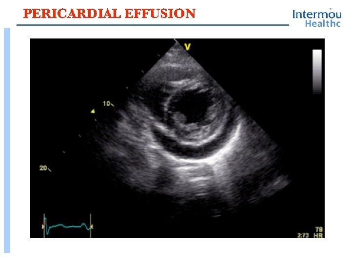
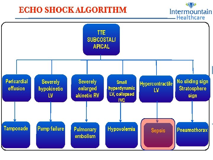
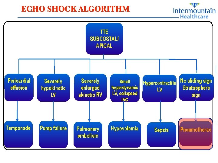
Echocardiographic Approach to Shock Hypovolemic Shock Acute Heart


 Diastolic dysfunction? High LAP Normal")
 / LVEDA normal range of")
![Step 2: Estimate Systolic Function LVESA [FAC] FAC= (EDA – ESA) / EDA a.](https://slidetodoc.com/presentation_image_h2/67dcca0daeeb2e01b4b557cca9655c1c/image-26.jpg "Step 2: Estimate Systolic Function LVESA [FAC] FAC= (EDA – ESA) / EDA a.")
, whereas")
 method of assessing ventricular function LV contraction is visualised in the")


- Slides: 39
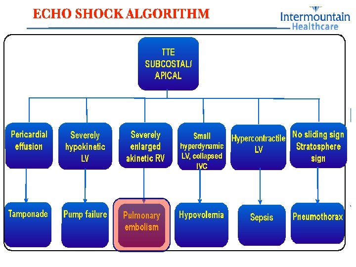
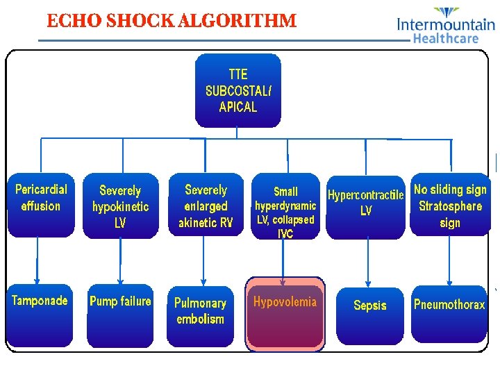
Echocardiographic Approach to Shock Hypovolemic Shock & Acute Heart Failure Rasoul Azarfarin Prof. of Anesthesiology MD, FACC Rajaie Cardiovascular Medical & Rearch Center Jan 18 2018
Definition of Shock • Inadequate oxygen delivery to meet metabolic demands • Results in global tissue hypoperfusion and metabolic acidosis • Shock can occur with a normal blood pressure and hypotension can occur without shock
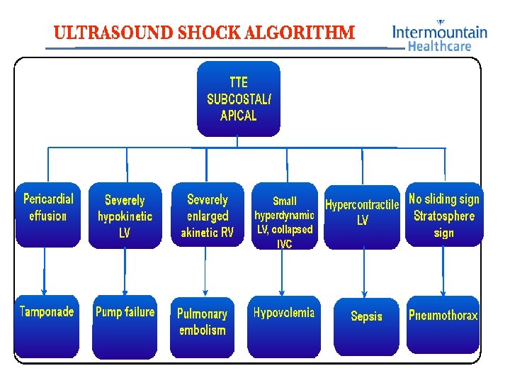
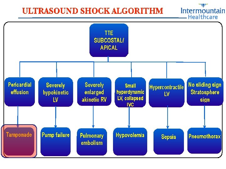
Types of Shock • Hypovolemic • Septic • Cardiogenic • Anaphylactic • Neurogenic • Obstructive
Cardiogenic Shock • Defined as: • SBP < 90 mm. Hg • CI < 2. 2 L/m/m 2 • PCWP > 18 mm. Hg • Signs: • • • Cool, mottled skin Tachypnea Hypotension Altered mental status Narrowed pulse pressure Rales, murmur
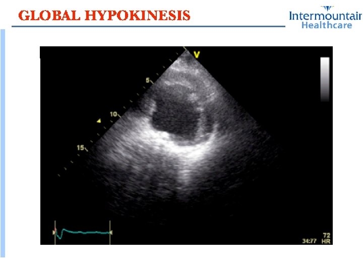
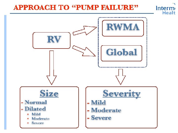
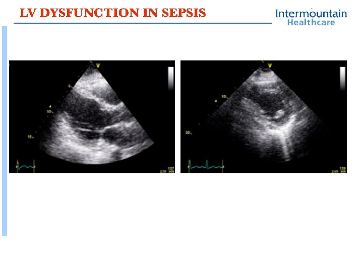
Global LV Dysfunction
Global LV Dysfunction
Global LV Dysfunction
Step 3: Estimate LAP (High > 15 mm. Hg) Diastolic dysfunction? High LAP Normal LAP Low LAP In general terms, the trend in PCWP is in the same direction as the change in inter-atrial septal pattern.
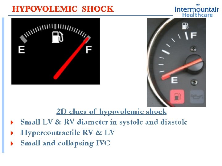
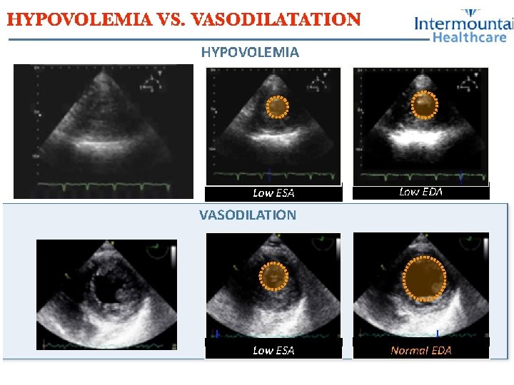
Step 1: Estimate Left Ventricular End Diastolic Volume (Preload) / LVEDA normal range of LVEDA= 8 -14 cm 2 ØAn LVEDA < 8 cm 2 is consistent with hypovolaemia, and ØLVEDA > 14 cm 2, consistent with a dilated left ventricle
Step 2: Estimate Systolic Function LVESA [FAC] FAC= (EDA – ESA) / EDA a. Increased (FAC > 65%) b. Normal (FAC 50 -65%) c. Reduced (<50%).
With low LV afterload cardiac index is usually high (>3. 5 L/min/m 2), whereas with hypovolemia cardiac index is usually low (<2. 5 L/min/m 2)
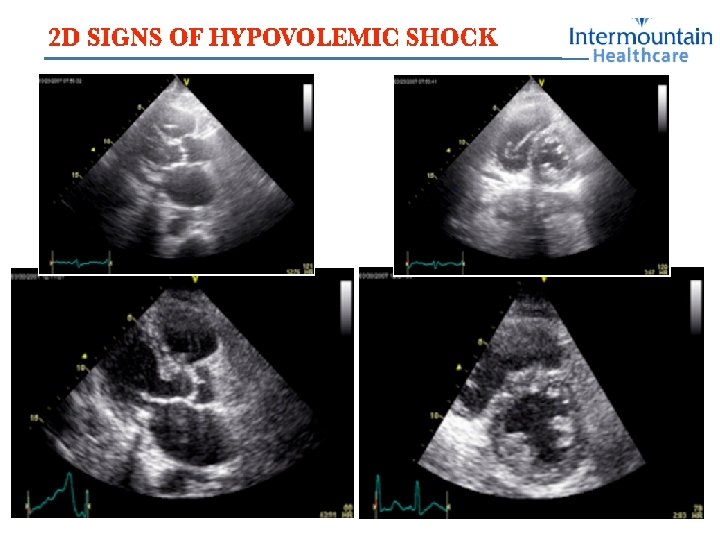
Semiquantative (Visual: eyeballing) method of assessing ventricular function LV contraction is visualised in the entire standard transthoracic 2 -D imaging planes: ·PLAX & PSAX view ·AP 4 Ch & AP 2 Ch view ·Apical long axis (Aplax) view ·subcostal long (Sublax) and short axis (Subsax) views The grading of the global systolic contraction can be described as: ·normal ·reduced ·severely reduced
RV Rupture+ PE
RV Rupture+ PE
RV Rupture+ PE