Echinococcus granulosus The dog tapeworm Dr Bindhusaran M
 Assistant Professor Dept.")
Echinococcus granulosus The dog tapeworm Dr. Bindhusaran M. D. (Hom. ) Assistant Professor Dept. of Pathology & Microbiology

Habitat and morphology • Intermediate host: Man - harbors larval stage • D. H: dog and other canine animals Morphology Adult worm: 3 -6 mm length Head : 4 suckers, rostellum bears hooks Neck: short and thick Strobila: 3 segments

• Egg • Ovoid and resembles eggs of other taenia • Consits of hexacanth embryo with hooks • Infective to cattle and man Larval form: • Found in the hydatid cyst • Bears scolex which posses suckers and hooks

Life Cycle • The worm passes its life cycle in two hosts. • 1. Definitive Hosts: Dog, wolf, fox and jackal. The adult worm lives in the small intestine of these animals. The dog is the optimum definitive host. • 2. Intermediate Hosts. Sheep, pig, cattle, horse, goat and man. • The larval stage is passed in these animals and man giving rise to hydatid cyst. • The sheep appears to be the optimum intermediate host.

Life cycle • The eggs are discharged with the faeces of the definitive hosts (dog and allied animals). • These are swallowed by the intermediate hosts, sheep and other domestic animals while grazing in the field, and also by man (particularly children) due to intimate handling of infected dogs. • In the duodenum, the hexacanth embryos are hatched out. And bore their way through the intestinal wall and enter the radicles of the portal vein.

Life cycle • The embryos are carried to the liver to be arrested in the sinusoidal capillaries (the liver acts as the first filter). • Some of the embryos may pass through the hepatic capillaries, enter the pulmonary circulation and filter out in the lungs (lungs act as the second filter). • A few of the embryos may pass the pulmonary capillaries, enter the general blood stream and lodge in the various organs. • Practically, all the organs of the domestic animals may be invaded but they are chiefly found in the liver and lungs. Wherever the embryo settles, it forms a hydatid cyst, the young larva being transformed into a hollow bladder
. From the inner side of the cyst, brood")
Life cycle (hydatis, drop of water). From the inner side of the cyst, brood capsules with a number of scolices are developed. A hydatid cyst developing from a single egg (oncosphere) may contain thousands of scolices. A fully developed scolex is an end-product and its presence inside the hydatid cyst is a sign of “a complete biological development”. These fertile hydatids, when ingested by the dog, are capable of growing into adult worms in about 6 to 7 weeks’ time in the intestine. Thus, the cycle is repeated.

Life cycle • As the dogs have no access to the hydatid cyst developed in the viscera of man, the life cycle of the parasite comes to a dead-end. The natural cycle is thus maintained by dog and sheep. • Life span of the adult worm in the canine host is short (about 6 months).

Pathogenicity. • The adult worms of E. granulosus in dogs do not cause much inconvenience. • The larval worm of E. granulosus in man causes unilocular hydatid disease.

Pathogenesity Infecting Agent Eggs, in dog's faeces. Portal of Entry- Alimentary tract. Sites of localization Viscera (liver, lungs and other organs). EVOLUTION OF HYDATID CYST Cyst wall cosists of 2 layers: (1) Outer Cuticular Layer (Ectocyst). I t is a laminated hyaline membrane having a thickness up to • 1 mm. To the naked eye, the ectocyst has the appearance of white of a hard_boiled egg. it is elastic • (2) Inner or Germinal Layer (Endocyst) : It is cellular and consists of a number of nuclei embedded in a protoplasmic mass • It is very thin and measureas about 22 to 25 micrometre in thickness. It is the vital layer of the cyst and gives rise to brood capsules with scolices (b ) secretets the specific hydatid fluid, and (c) forms the outer layer. • • •
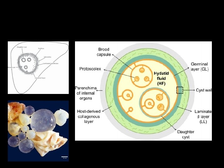
 Clear colourless fluid (may be pale yellow in colour).")
Composition of hydatid fluid (i) Clear colourless fluid (may be pale yellow in colour). (ii) Specific gravity low, 1. 005 to 1. 010. (iii) Reaction slightly acid, p. H 6. 7. (iv) Contains sodium chloride, sodium sulphate, sodium phosphate and sodium and calcium salts of succinic acid _ (V) Antigenic, being used for immunological tests. (vi) Highly toxic, when absorbed gives rise to anaphylactic symptoms. (vii) Hydatid sand A granular deposit found to settle at the bottom. It consists of liberated brood capsules free scolices and loose hooklets.

Development of brood capsule and scolices • Brood capsule sprout form germinal layer • 5 -20 scolices develops in these capsules • Rate of growth: by 1 year it is 4 cm in diameter • Distribution of the cyst: first the liver and any other organ can be involved

Lab diagnosis • Casoni’s reaction It Immediate hypersensitivity skin test to diagnose hyadatid cyst Intradermal injection 0. 2 ml of sterile hydatid fluid is injected with in 30 minutes produce a large wheal ( 5 cm In diameter) it fades in 1 hour.

Molecular methods such as DNA probe and PCR have been developer value because of their technical complexity. • 4. Exploratory Cyst Puncture. Though an accurate diagnosis may be made by withdrawing a few millilitres of the hydatid fluid and examining it under the microscope for scolices or hooklets, yet it is often attended with serious results and is therefore not advised.

• 5. Radiological. This is often helpful in the diagnosis of hydatid cysts of lungs and liver. Shows a characteristic circular shadow with a sharp outline • In cases where the long bones are involved a mottled appearance is seen in the skiagram. • Ultrasonography of whole abdomen is useful in locating the site of hydatid cyst of the abdominal organs. • CT scan is more helpful than MRI scan in the diagnosis of diseases of different organs.

CLASS TREMATODA • Leaf shaped unsegmented worms called flukes • Posses 2 suckers oral sucker and a ventral sucker • Sexes are not separate • Body cavity is absent • They are oviparous and liberate eggs • Eggs are operculated and can develop only in water

Larval satges Miracidium Sporocyst Redia Cercaria Metacercaria

Classification of trematodes according to habitat • Intestinal trematodes • Hepatic trematodes • Lung trematodes • Blood trematodes( blood fluke)
- Slides: 19