
ECG etc Miscellaneous ECGs Rey Vivo MD Assistant

- Slides: 26
ECG etc… (Miscellaneous ECGs) Rey Vivo, MD Assistant Professor of Medicine Texas Tech University Health Sciences Center
Objectives • Describe the following miscellaneous ECGs: – – – – Lead reversal Dextrocardia Hypothermia Pericarditis Cardiac Tamponade Wolff-Parkinson-White Pulmonary embolism • Practice, practice!!!
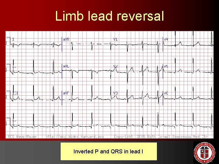
Limb lead reversal Inverted P and QRS in lead I
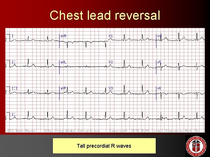
Chest lead reversal Tall precordial R waves
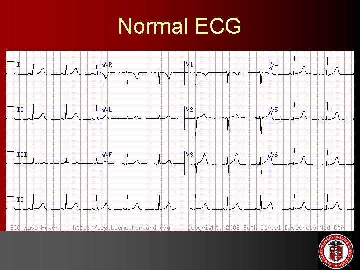
Normal ECG
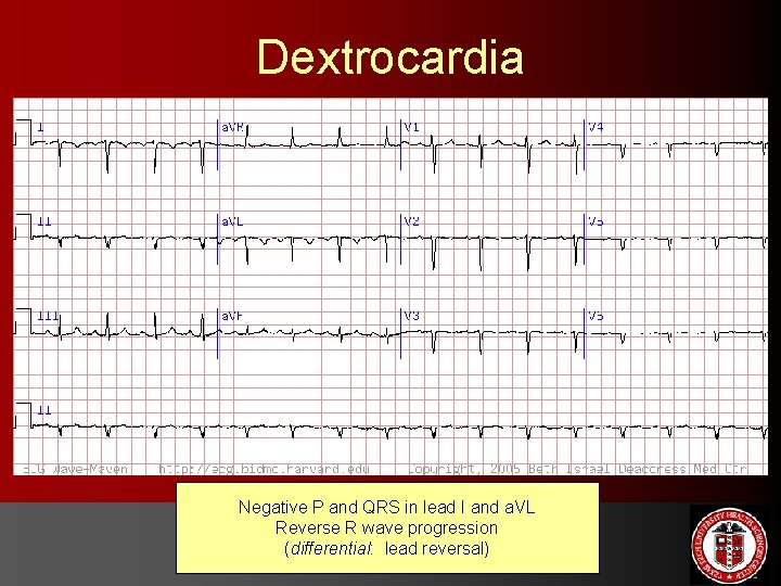
Dextrocardia Negative P and QRS in lead I and a. VL Reverse R wave progression (differential: lead reversal)
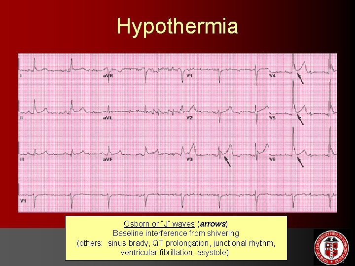
Hypothermia Osborn or “J” waves (arrows) Baseline interference from shivering (others: sinus brady, QT prolongation, junctional rhythm, ventricular fibrillation, asystole)
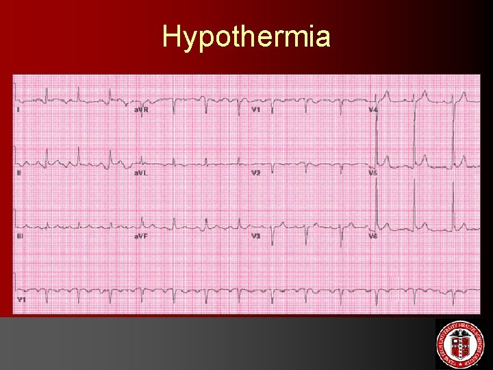
Hypothermia
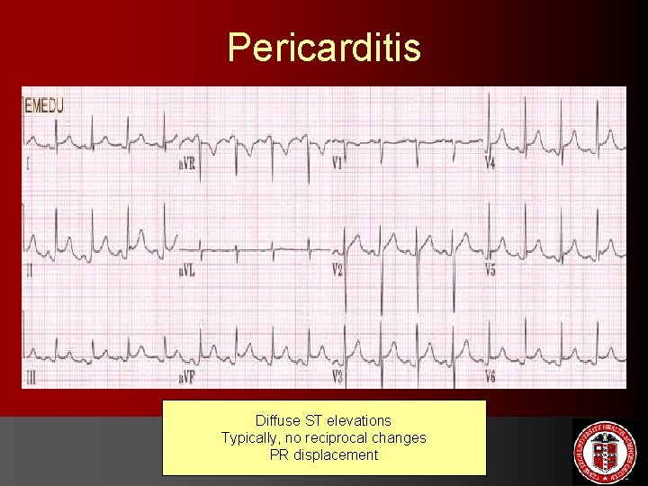
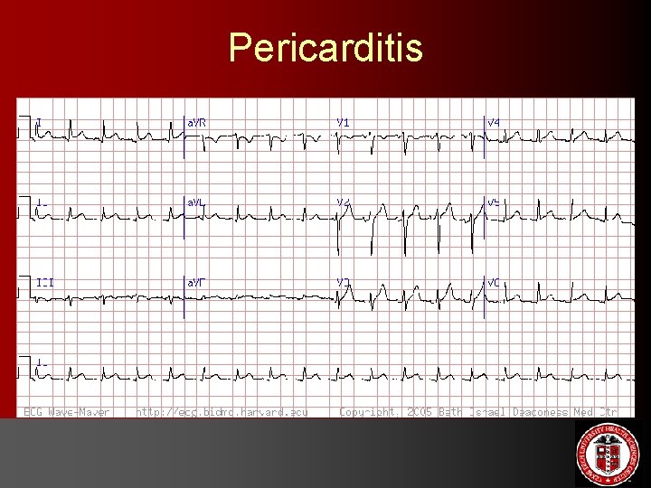
Pericarditis Diffuse ST elevations Typically, no reciprocal changes PR displacement
Pericarditis
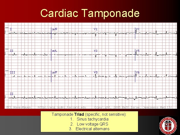
Cardiac Tamponade Triad (specific, not sensitive): 1. Sinus tachycardia 2. Low voltage QRS 3. Electrical alternans
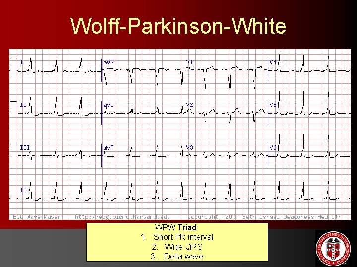
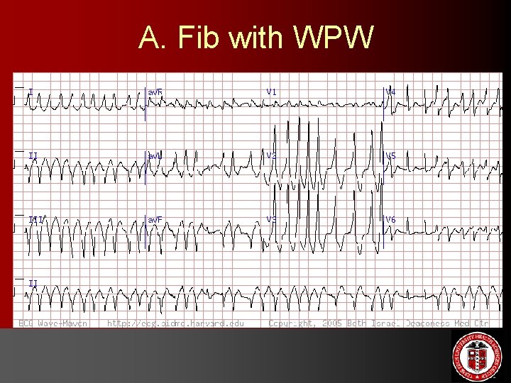
Wolff-Parkinson-White WPW Triad: 1. Short PR interval 2. Wide QRS 3. Delta wave
A. Fib with WPW
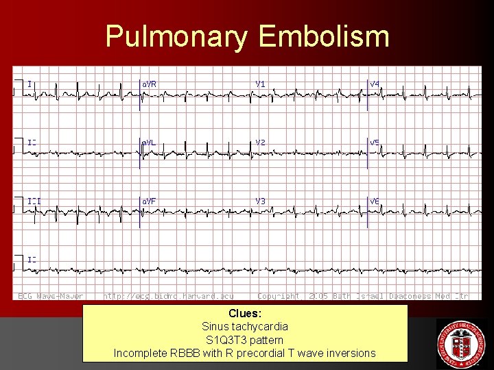
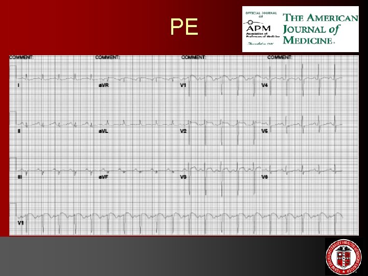
Pulmonary Embolism Clues: Sinus tachycardia S 1 Q 3 T 3 pattern Incomplete RBBB with R precordial T wave inversions
PE
Lets’ practice!
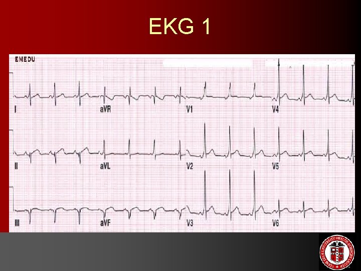
EKG 1
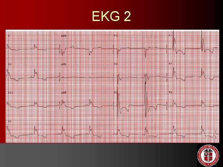
EKG 2
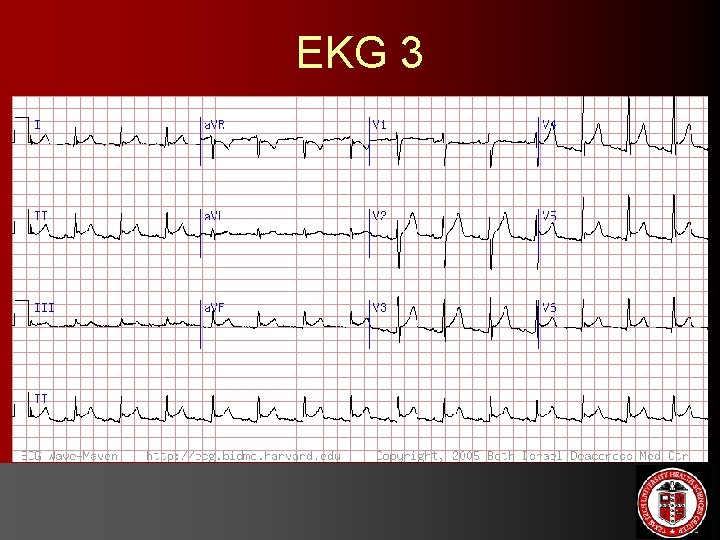
EKG 3
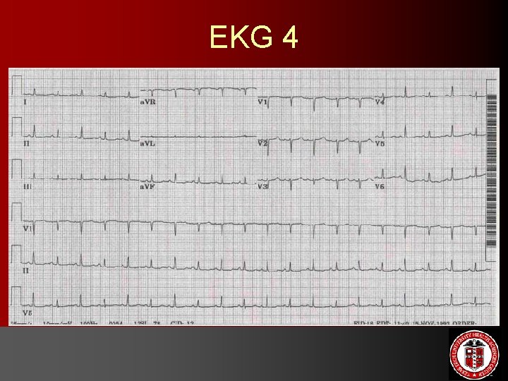
EKG 4
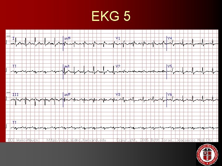
EKG 5
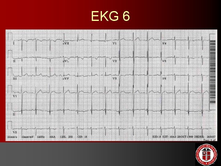
EKG 6
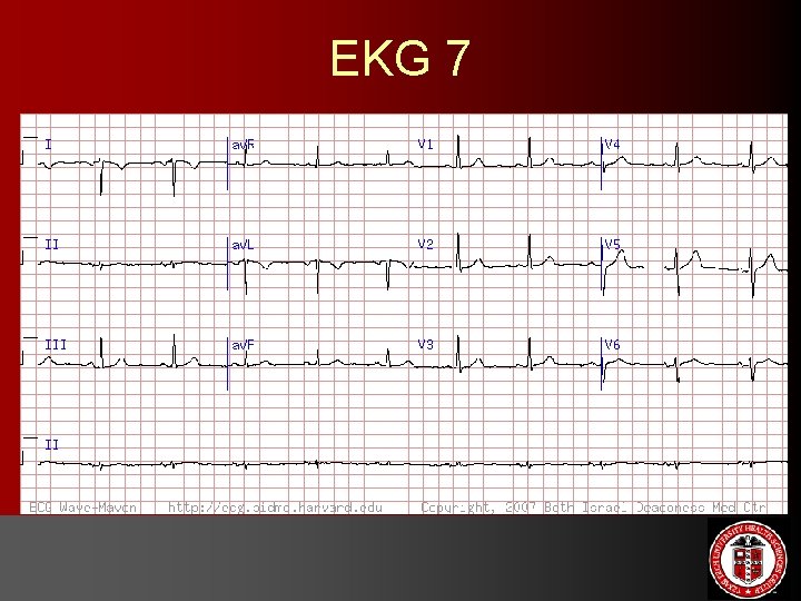
EKG 7
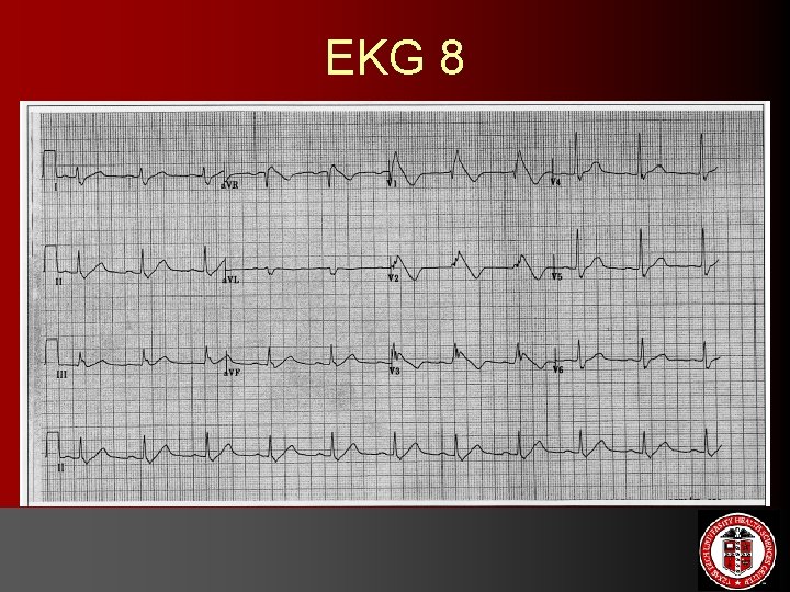
EKG 8
Brugada syndrome • • • Defined in 1992 by brothers Pedro and Josep Brugada in J Am Coll Cardiol Rare condition associated with sudden cardiac death (SCD) or ventricular fibrillation/polymorphic VT in structurally normal hearts Mean age of presentation: 35 -40 years; male predominance (notably in SE Asia where it is endemic) Familial with autosomal dominant inheritance Clinical presentation: syncope or SCD is most common initial event; typically occurs in sleep or in early morning ECG criteria: ST segment elevation in V 1 -V 3 with characteristic RBBB pattern Defect in sodium channel gene (SCN 5 A) on chromosome 3 Lifetime risk of SCD Consider ICDs; refer for genetic counseling
The End