
EBM Journal reading Longterm Pattern of Progression of
EBM Journal reading Long-term Pattern of Progression of Myopic Maculopathy A Natural History Study Ophthalmology 2010; 117: 1595– 1611.
Purpose Investigate the long-term progression pattern of myopic maculopathy Determine the visual prognosis of each progression stage Retrospective, observational case series.
Introduction Pathologic myopia is a leading cause of visual impairment The fourth to ninth most frequent cause of blindness worldwide The axial elongation of the eye and the development of a posterior staphyloma result in a thinning of the retina and choroid, which then lead to the development of different types of myopic maculopathy.
Introduction Myopic maculopathy on visual impairment is important because the maculopathy is often bilateral, irreversible, and frequently affects individuals during their most productive years

Introduction In this study, myopic maculopathy defined as 1. tessellated fundus 2. diffuse chorioretinal atrophy 3. lacquer cracks 4. patchy chorioretinal atrophy 5. CNV 6. macular atrophy

Tessellated fundus 20 -year-old woman, -14. 0 diopters, axial length: 26. 7 mm
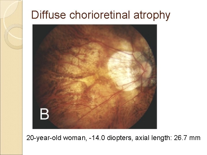
Diffuse chorioretinal atrophy 20 -year-old woman, -14. 0 diopters, axial length: 26. 7 mm
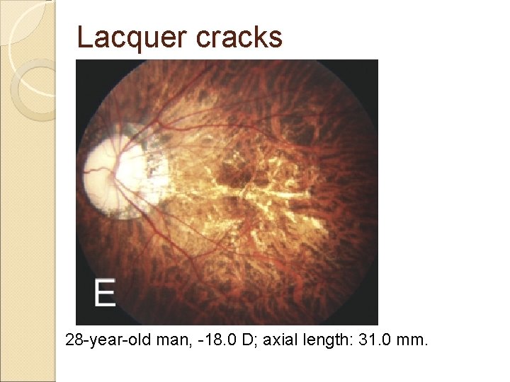
Lacquer cracks 28 -year-old man, -18. 0 D; axial length: 31. 0 mm.
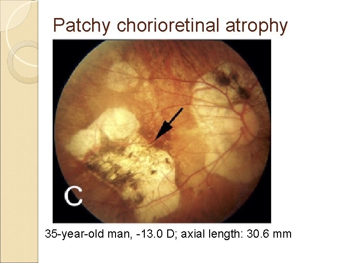
Patchy chorioretinal atrophy 35 -year-old man, -13. 0 D; axial length: 30. 6 mm
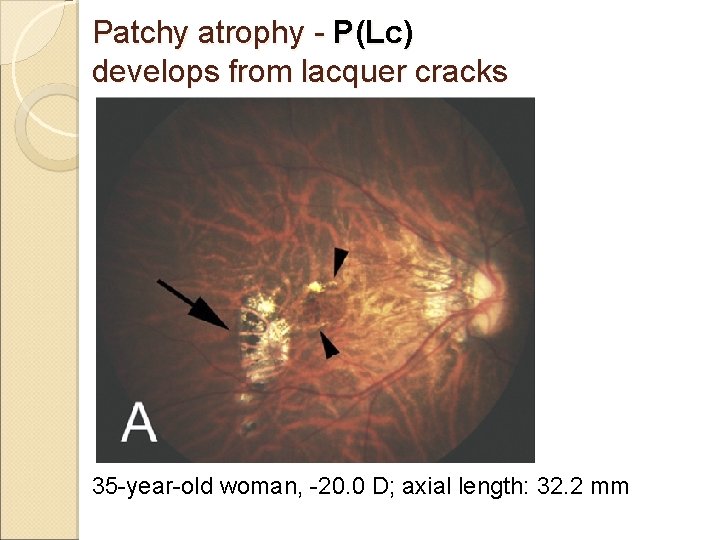
Patchy atrophy - P(Lc) develops from lacquer cracks 35 -year-old woman, -20. 0 D; axial length: 32. 2 mm
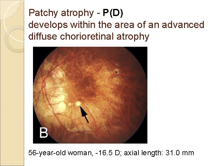
Patchy atrophy - P(D) develops within the area of an advanced diffuse chorioretinal atrophy 56 -year-old woman, -16. 5 D; axial length: 31. 0 mm
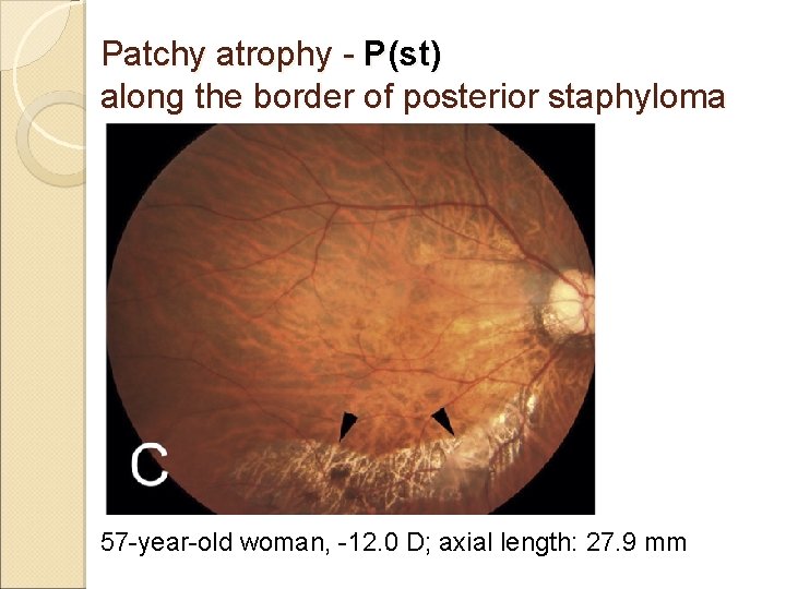
Patchy atrophy - P(st) along the border of posterior staphyloma 57 -year-old woman, -12. 0 D; axial length: 27. 9 mm

CNV choroidal neovascular membrane 51 -year-old woman, -13. 0 D; axial length: 30. 1 mm
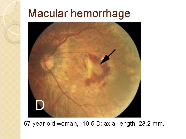
Macular hemorrhage 67 -year-old woman, -10. 5 D; axial length: 28. 2 mm.
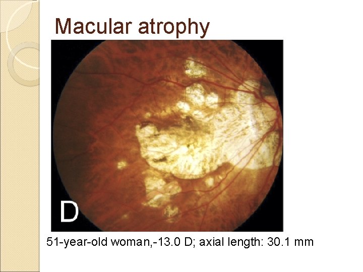
Macular atrophy 51 -year-old woman, -13. 0 D; axial length: 30. 1 mm
Patients and Methods 806 eyes of 429 patients with high myopia (refractive error more than -8. 00 diopters or axial length > 26. 5 mm) who were followed for 5– 32 years
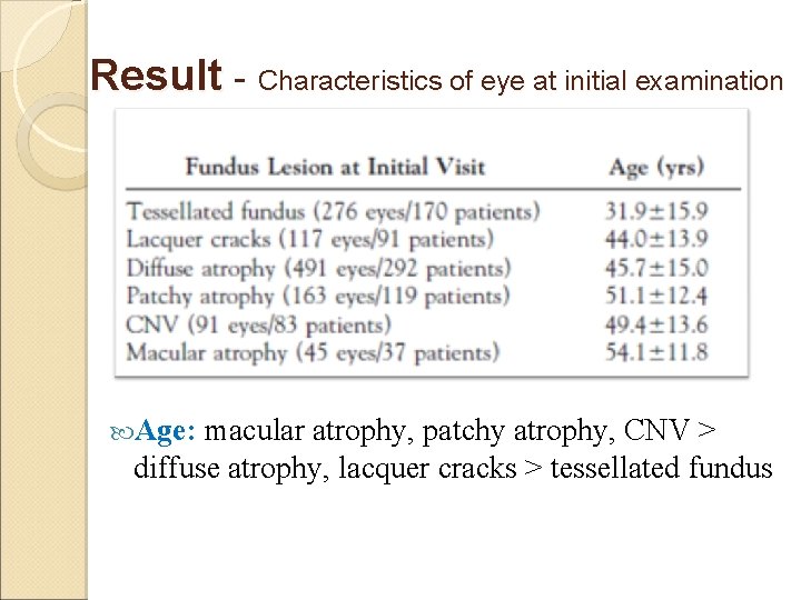
Result - Characteristics of eye at initial examination Age: macular atrophy, patchy atrophy, CNV > diffuse atrophy, lacquer cracks > tessellated fundus
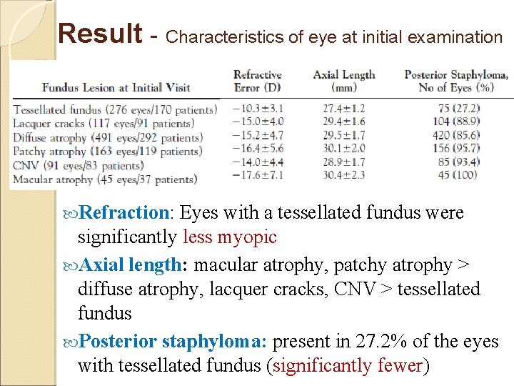
Result - Characteristics of eye at initial examination Refraction: Eyes with a tessellated fundus were significantly less myopic Axial length: macular atrophy, patchy atrophy > diffuse atrophy, lacquer cracks, CNV > tessellated fundus Posterior staphyloma: present in 27. 2% of the eyes with tessellated fundus (significantly fewer)
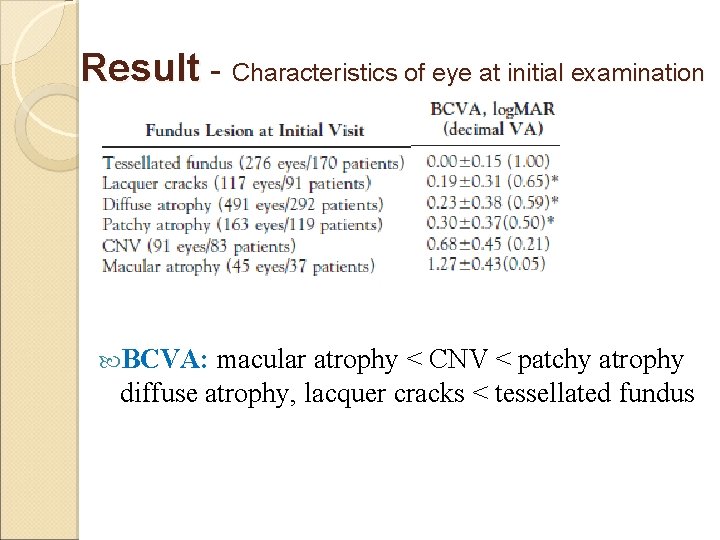
Result - Characteristics of eye at initial examination BCVA: macular atrophy < CNV < patchy atrophy diffuse atrophy, lacquer cracks < tessellated fundus
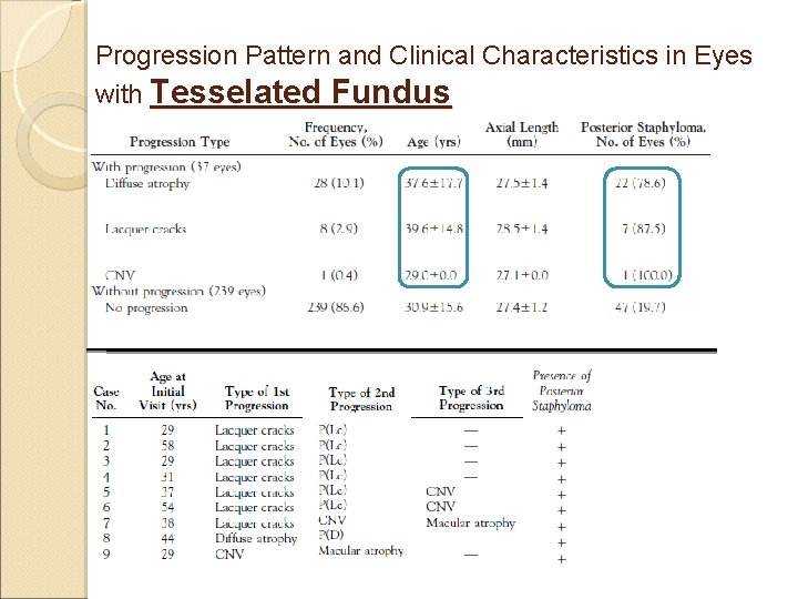
Progression Pattern and Clinical Characteristics in Eyes with Tesselated Fundus
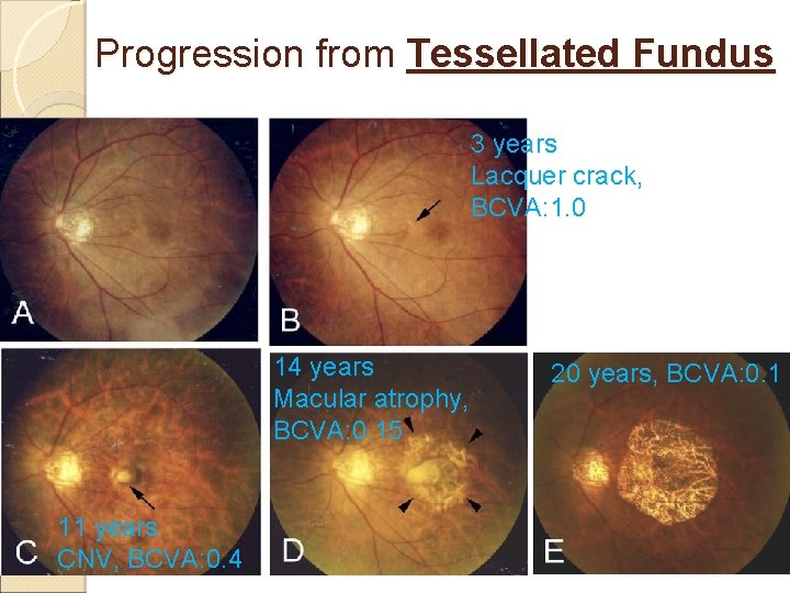
Progression from Tessellated Fundus 3 years Lacquer crack, BCVA: 1. 0 14 years Macular atrophy, BCVA: 0. 15 11 years CNV, BCVA: 0. 4 20 years, BCVA: 0. 1
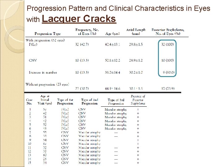
Progression Pattern and Clinical Characteristics in Eyes with Lacquer Cracks
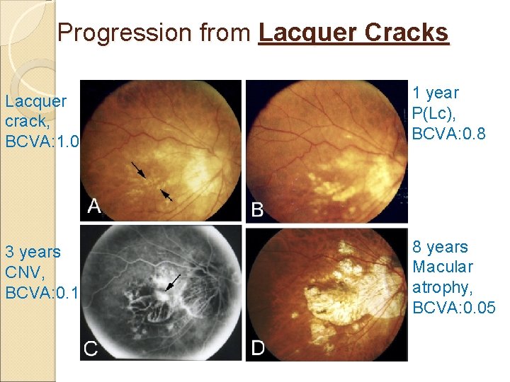
Progression from Lacquer Cracks Lacquer crack, BCVA: 1. 0 1 year P(Lc), BCVA: 0. 8 3 years CNV, BCVA: 0. 1 8 years Macular atrophy, BCVA: 0. 05
Progression pattern in the eyes with lacquer cracks was the development of a CNV at the foveal edge in eyes with the P(Lc) type of patchy atrophy and a final progression to macular atrophy. We should carefully monitor and watch for the development of a CNV when lacquer cracks increase their width and progress to P(Lc).
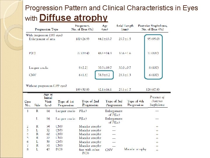
Progression Pattern and Clinical Characteristics in Eyes with Diffuse atrophy
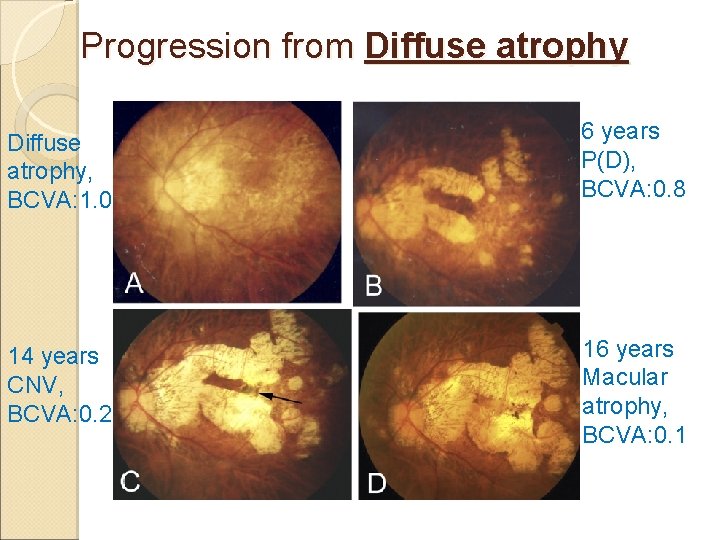
Progression from Diffuse atrophy, BCVA: 1. 0 6 years P(D), BCVA: 0. 8 14 years CNV, BCVA: 0. 2 16 years Macular atrophy, BCVA: 0. 1
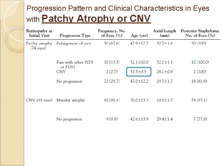
Progression Pattern and Clinical Characteristics in Eyes with Patchy Atrophy or CNV
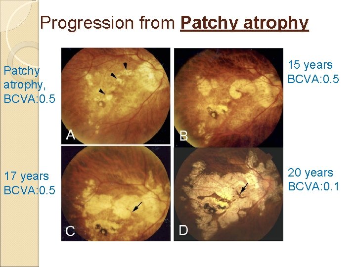
Progression from Patchy atrophy, BCVA: 0. 5 15 years BCVA: 0. 5 17 years BCVA: 0. 5 20 years BCVA: 0. 1
Result - Progression Pattern of Myopic Maculopathy 327 of the 806 eyes (40. 6%) showed a progression of the myopic maculopathy Tessellated fundus: 37 of 276 eyes (13. 4%) Lacquer cracks: 52 of 75 eyes (69. 3%) Diffuse atrophy: 183 of 372 eyes (49. 2%) Patchy chorioretinal atrophy: 52 of 74 eyes (70. 3%) CNV: 82 of 91 eyes (90. 1%)
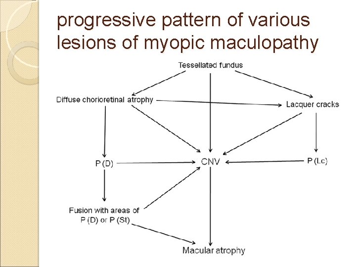
progressive pattern of various lesions of myopic maculopathy
Tessellated fundus may be the first fundus lesion observed in eyes with pathologic myopia Posterior staphyloma was critically important for the progression of myopic maculopathy. The fusion of patchy atrophy, the development of CNV, and macular atrophy all led to significant visual decreases

Conclusion Predicting the progression and the vision in highly myopic patients Knowledge of natural course of myopic maculopathies will allow us to determine the effectiveness of the treatments.

Thanks for your Listening!
- Slides: 33