
EARLY PREGNANCY COMPLICATIONS DO DR OLU AP Abortion
EARLY PREGNANCY COMPLICATIONS DOÇ. DR. OLUŞ APİ
Abortion Loss of a pregnancy during the first 20 weeks of pregnancy, at a time that the fetus cannot survive. Such a loss may be involuntary (a "spontaneous" abortion), or it may be voluntary ("induced" or "elective" abortion). Miscarriage is the term used for spontaneous abortion, an unexpected 1 st trimester pregnancy loss.
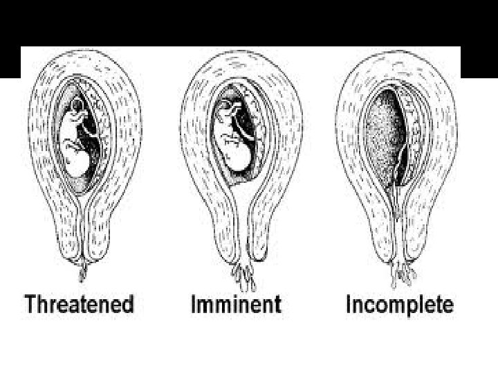
Categories of Abortions These include: 1. Threatened 2. Inevitable 3. Incomplete 4. Complete 5. Septic
Facts about abortion Such losses are common, occurring in about one out of every 6 pregnancies. These losses are unpredictable and unpreventable. About 2/3 are caused by chromosome abnormalities. About 30% are caused by placental malformations and are similarly not treatable. The remaining miscarriages are caused by miscellaneous factors but are not usually associated with: Ø Ø Minor trauma Intercourse Medication Too much activity
Following a miscarriage, the chance of having another miscarriage with the next pregnancy is about 1 in 6.
Spontaneous abortion (also known as miscarriage) is the expulsion of an embryo or fetus due to accidental trauma or natural causes before approximately the 22 nd week of gestation; the definition by gestational age varies by country. [ Most miscarriages are due to incorrect replication of chromosomes; they can also be caused by environmental factors
Induced A pregnancy can be intentionally aborted in many ways. The manner selected depends chiefly upon the gestational age of the embryo or fetus, which increases in size as the pregnancy progresses. Specific procedures may also be selected due to legality, regional availability, and doctor-patient preference. Reasons for procuring induced abortions are typically characterized as eitherapeutic or elective.
Induced abortion 1. 2. 3. Therapeutic abortion when it is performed to: save the life of the pregnant woman preserve the woman's physical or mental health terminate pregnancy that would result in a child born with a congenital disorder that would be fatal or associated with significant morbidity or selectively reduce the number of fetuses to lessen health risks associated with multiple pregnancy.
Induced abortion An elective abortion: When it is performed at the request of the woman "for reasons other than maternal health or fetal disease.
Threatened Abortion A threatened abortion means the woman has experienced symptoms of bleeding or cramping. At least one-third of all pregnant women will experience these symptoms. Half will abort spontaneously. The other half , bleeding and crampingwill disappear and the remainder of the pregnancy will be normal. These women who go on to deliver their babies at full term can be reassured that the bleeding in the first trimester will have no effect on the baby and that you expect a full-term, normal, healthy baby.
Threatened abortion (Features) 1. History Mild vaginal bleeding. No abdominal pain or mild abdominal pain 2. Examination Good general condition. The cervix is closed The uterus is usually the correct size for date 3. U/S which is essential for the diagnosis Showed the presence of fetal heart activity
Threatened abortion (Management) 1. Reassurance If fetal heart activity is present, > 90% of cases will be progressed satisfactorily 2. Advice: Decrease physical activity (bed rest is of no therapeutic value) avoid intercourse 3. Hormones i. e. Progesterone & h. CG Which are used in the first trimester to support pregnancy, (but they are of no proven value) 4. Anti- D: An adequate dose of anti-D should be given to all Rh –ve, non-immunised patients, whose husbands are Rh +ve 5. ANC as high risk patients Because those patients are liable to late pregnancy complications such as APH and preterm labour.
Inevitable abortion A condition in which: Vaginal bleeding has been profuse The cervix has become dilated Abortion will invetably occur.
Inevitable and incomplete abortions (Features) 1. History Ø Heavy vaginal bleeding. with no passage of products conception (inevitable) with the passage of products of conception (incomplete abortion) Ø Severe lower abdominal pain which follows the bleeding
Inevitable and incomplete abortions (Features) 2. Examinations Poor general condition. Ø The cervix is dilating and products of conception may be passing trough the os Ø The uterus may be the correct size for date (inevitable abortion) or small for date (incomplete abortion) 3. U/S Fetal heart activity may or may not present in inevitable abortion or retained products of conception ( RPOC ) in incomplete abortion Ø
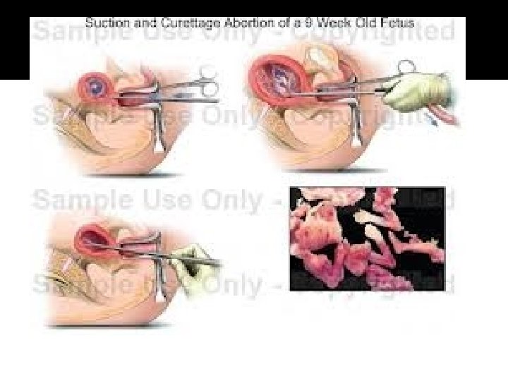
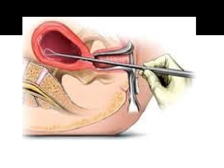
Inevitable and incomplete abortions (management) 1. 2. CBC , blood grouping , XM 2 units of blood Resuscitation large IV line, fluids & blood transfusion 3. Oxytoxic drugs Ergometrine 0. 5 mg IM + Oxytocin infusion (20 -40 units in 500 cc saline) 4. 5. Evacuation & curettage. Post-abortion management.
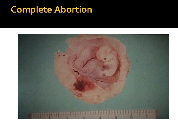
Complete Abortion
Complete abortion (Features) 1. History Ø Ø 2. Examination Ø 3. Heavy vaginal bleeding which has been stopped. lower abdominal pain which follows the bleeding which has been stopped. The cervix is closed U/S Ø showed empty uterine cavity or PROP
Complete abortion (Management) - Evacuation & curettage in the presence of RPOC. 2. Post-abortion management. 1.
Missed abortion Retention of products for several weeks No increase in fundal height Absence of FHT Regressions of signs of pregnancy Loss of wight
Missed abortion (Features) 1. Most of missed abortions are diagnosed accidentally during routine U/S in early pregnancy. In some cases there may be a history of : Ø Ø Ø 2. Episodes of mild vaginal bleeding Regression of early symptoms of pregnancy. Stop of fetal movements after 20 weeks gestation. Examination Ø The uterus may be small for date
Missed abortion (Features) 3. U/S (which is essential for diagnosis ) diagnosed if two ultrasound ( T/V or T/A) at least 7 days apart showed an embryo of > 7 weeks gestation ( CRL > 6 mm in diameter and gestational sac > 20 mm in diameter ) with no evidence of heart activity.
Missed abortion (Management) CBC , blood grouping Platelets count, to exclude the risk of DIC NB : DIC does not occur before 5 weeks of missed abortion or IUFD and if occurred will be of mild grade 1. 2.
Missed abortion (Management) 3. Options of treatment Ø Conservative treatment: if left alone spontaneous expulsion will occur Ø Surgical evacuation of the uterus; by D & C: Indicated in 1 st trimester missed abortion Ø Medical termination of pregnancy: by Misoprostol (PGE 1) Cytotec: Indicated in 1 st & 2 nd trimesters missed abortions. Ø Cytotec vaginal ( is the best) or oral tab. 200 μg, 2 tab/ 3 hrs/ up to 5 doses daily, which can be repeated next day if there is no response in the first day Ø Subsequent surgical evacuation is needed in cases of RPOC Ø The main side effects of cytotec are nausea, vomiting and fever. 4. Post-abortion management.

Anembryonic pregnancy (Blighted ovum) It is due to an early death and resorption of the embryo with the persistence of the placental tissue Ø It is diagnosed if two ultrasound ( T/V or T/A) at least 7 days apart showed after 7 weeks of gestation i. e. gestational sac > 20 mm , an empty gestational sac with no fetal echoes seen. Ø It is treated in a similar way to missed abortion. Ø
Septic abortion Spontaneous or induced termination of a pregnancy in which the mother's life may be threatened because of the invasion of germs into the endometrium, myometrium, and beyond. The woman requires immediate and intensive care Massive antibiotic therapy Evacuation of the uterus Emergency hysterectomy to prevent death from overwhelming infection and septic shock.
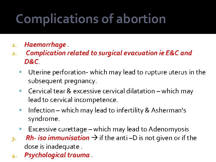
Complications of abortion 1. 2. 3. 4. Haemorrhage. Complication related to surgical evacuation ie E&C and D&C. Uterine perforation- which may lead to rupture uterus in the subsequent pregnancy. Cervical tear & excessive cervical dilatation – which may lead to cervical incompetence. Infection – which may lead to infertility & Asherman's syndrome. Excessive curettage – which may lead to Adenomyosis Rh- iso immunisation if the anti –D is not given or if the dose is inadequate. Psychological trauma.
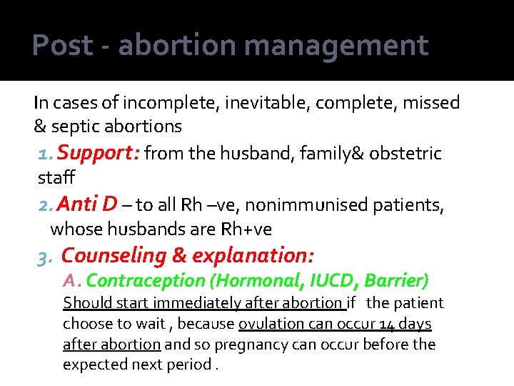
Post - abortion management In cases of incomplete, inevitable, complete, missed & septic abortions 1. Support: from the husband, family& obstetric staff 2. Anti D – to all Rh –ve, nonimmunised patients, whose husbands are Rh+ve 3. Counseling & explanation: A. Contraception (Hormonal, IUCD, Barrier) Should start immediately after abortion if the patient choose to wait , because ovulation can occur 14 days after abortion and so pregnancy can occur before the expected next period.
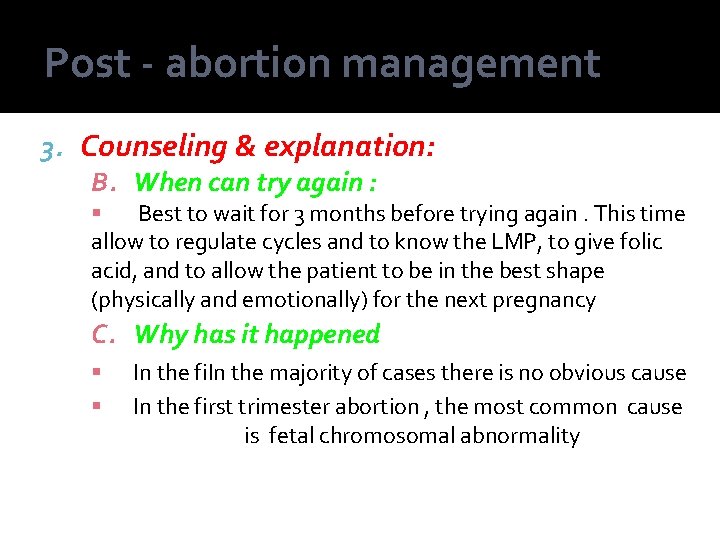
Post - abortion management 3. Counseling & explanation: B. When can try again : Best to wait for 3 months before trying again. This time allow to regulate cycles and to know the LMP, to give folic acid, and to allow the patient to be in the best shape (physically and emotionally) for the next pregnancy C. Why has it happened In the fi. In the majority of cases there is no obvious cause In the first trimester abortion , the most common cause is fetal chromosomal abnormality
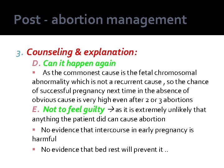
Post - abortion management 3. Counseling & explanation: D. Can it happen again As the commonest cause is the fetal chromosomal abnormality which is not a recurrent cause , so the chance of successful pregnancy next time in the absence of obvious cause is very high even after 2 or 3 abortions E. Not to feel guilty as it is extremely unlikely that anything the patient did can cause abortion No evidence that intercourse in early pregnancy is harmful No evidence that bed rest will prevent it. .
Recurrent abortion Definition : Ø Is defined as 3 or more consecutive spontaneous abortions Ø It may presented clinically as any of other types of abortions. Types : Ø Primary : All pregnancies have ended in loss Ø Secondary : One pregnancy or more has proceeded to viability(>24 weeks gestation) Incidence : with all others ending in loss Ø occurs in about 1% of women of reproductive age.
Recurrent abortion Causes Idiopathic recurrent abortion, in about 50%, in which no cause can be found. The known causes include the followings : 1. Chromosomal disorders: Ø Fetal chromosomal abnormalities & structural abnormalities Ø Parental balanced translocation 2. Anatomical disorders: Ø Ø Cervical incompetence: →congenital and aquired Uterine causes: → submucous fibroids, uterine anomalies & Asherman’s syndrome
Recurrent abortion Causes Medical disorders: 3. Endocrine disorders : diabetes , thyroid disorders , PCOS & corpus luteum insufficiency. Ø Immunological disorders : Anticardiolipin syndrome & SLE. Ø Thrombophilia: congenital deficiency of Protein C&S and antithrombin III, & presence of factor V leiden. Ø Infections Ø To. RCH - CMV may be a cause of recurrent abortion, but To. RH are not causes of recurrent abortion. Genital tract infection e. g Bacterial vaginosis Ø Rh – isoimmunization
Recurrent abortion Diagnosis : History : Ø Previous abortions : gestational age and place of abortions & fetal abnormalities. Ø Medical history : DM , thyroid disorders, PCOS, autoimmune diseases & thrombophilia. 2. Examination : 1. Ø Ø General : weight , thyroid & hair distribution Pelvic: cervix ( length & dilatation ) and uterine size.
Recurrent abortion Diagnosis : 3. investigations : A. Investigations for medical disorders: Blood grouping & indirect Coomb’s test in Rh –ve women Endocrinal screening: Blood sugar , TFT & LH /FSH ratio Immunological screening: Anti anticardiolipine antibodies & lupus inhibitor. Ø Thrombophilia screening: Protein C & S, antithrombin III levels, factor V leiden, APTT and PT. Ø Infection screening Ø Ø Ø High vaginal & cervical swabs To. RCH profile ( which scientifically is not necessary )
Recurrent abortion Diagnosis : 3. investigations : B. Investigations for anatomical disorders: TV/US: fibroids, cervical incompetence & PCOS. Hystroscopy or HSG, fibroids, cervical incompetence, uterine anomalies & Asherman's syndrome Ø Ø C. Investigations for chromosomal disorders: Ø Ø Parental karyotyping: Parental balanced translocation. Fetal karyotyping: Fetal chromosomal anomalies.
Recurrent abortion Management: 3. in idiopathic recurrent abortion. With support and good antenatal care , the chance of successful spontaneous pregnancy is about 60 -70% Ø Support : from husband, family & obstetric staff. Ø Advice : stop smoking & alcohol intake, decrease physical activity Ø Tender loving care Ø Drug therapy • Progesterone & h. CG: start from the luteal phase & up to 12 weeks. • Low dose aspirin ( 75 mg/day ) start from the diagnosis of pregnancy & up to 37 weeks • LMWH (20 -40 mg/day) start from the diagnosis of fetal heart activity & up to 37 ws
Recurrent abortion Management: 3. In the presence of a cause treatment is directed to control the cause Ø Endocrine disorders • Control DM and thyroid disorders before pregnancy • Ovulation induction drugs , ovarian drilling or IVF in PCOS. • Progesterone or h. CG in corpus luteum insufficiency. Ø : In anti-cardiolipin syndrome: • Low dose aspirin ( 75 mg/day ) & prednisilone ( 20 -30 mg / day), starting when pregnancy is diagnosed till 37 weeks. • These drugs are not teratogenic.
Recurrent abortion Management: In thrombophilia: • Low dose aspirin ( 75 mg/day) starting when pregnancy is diagnosed and low molecular weight heparin ie LMWH ( 20 -40 mg/day) starting when fetal heart activity diagnosed & to continue both till 37 weeks. Ø In uterine disorders Ø • Cervical cerclage in cervical incompetence, best time at the 14 weeks of pregnancy. • Myomectomy in submucus fibroid, excision of uterine septum in septate & subseptate uterus & adhesolysis in Asherman's syndrome.
Recurrent abortion Management: Ø Ø Ø In infection: : treatment of the genital tract infection. In Rh isoimmunization: Repeated intrauterine transfusion In parental balanced translocation • Explain the risk of fetal chromosomal disorders ( about 30% ) • Encourage to try again or adoption.
Gestational trophoplastic disease GTD Definition : It is a clinical term used to indicate three closely related conditions characterized by active abnormal proliferation of trophoblastic cells : : vhydatidiform mole , vinvasive hydatidiform v. Choriocarcinoma mole and
Gestational trophoplastic disease GTD These neoplasm's retain certain characteristic of the normal placenta such as invasive tendencies and the ability to make h. CG hormone
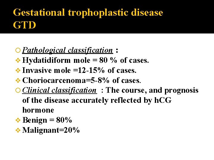
Gestational trophoplastic disease GTD classification : v Hydatidiform mole = 80 % of cases. v Invasive mole =12 -15% of cases. v Choriocarcenoma=5 -8% of cases. Clinical classification : The course, and prognosis of the disease accurately reflected by h. CG hormone v Benign = 80% v Malignant=20% Pathological
Hydatidiform mole The hydatidiform mole incidence ranges from 1 in 522 pregnancies in Japan To 1 in 1500 pregnancies in USA, and Sweden, this variation is not understood, but ethnic factors have been suggested. The incidence is higher in the poorest socioeconomic classes than the semiprivate(4 times), and (8 times) than the privet , and these mostly related to diet especially protein deficiency.
Hydatidiform mole The maternal age over 40 years found to have a 5. 2 -fold increased risk of trophoblastic disease in compression to the mothers below the age of 35 years.

Hydatidiform mole can be subdivided into: complete and vpartial mole v
Hydatidiform mole Based 1. Genetic on: and 2. Histopathologic al features
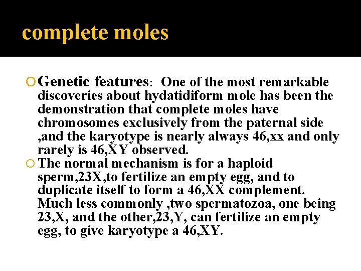
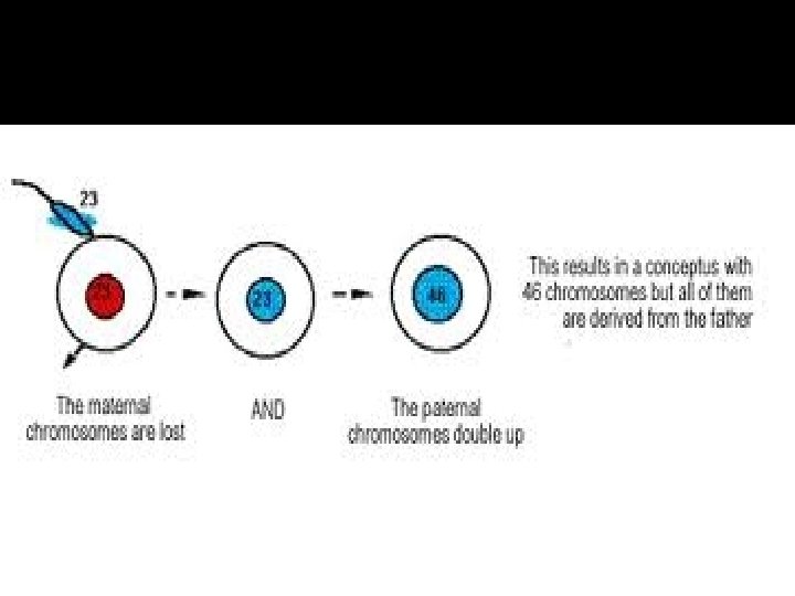
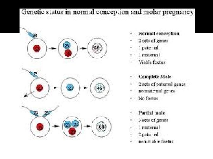
complete moles Genetic features: One of the most remarkable discoveries about hydatidiform mole has been the demonstration that complete moles have chromosomes exclusively from the paternal side , and the karyotype is nearly always 46, xx and only rarely is 46, XY observed. The normal mechanism is for a haploid sperm, 23 X, to fertilize an empty egg, and to duplicate itself to form a 46, XX complement. Much less commonly , two spermatozoa, one being 23, X, and the other, 23, Y, can fertilize an empty egg, to give karyotype a 46, XY.
Partial moles Are triploid in origin with two sets of paternal haploid genes and one set of maternal haploid genes
Partial moles They occur, in almost all cases, following dispermic fertilization of an ovum. There is usually evidence of a fetus or fetal red blood cells
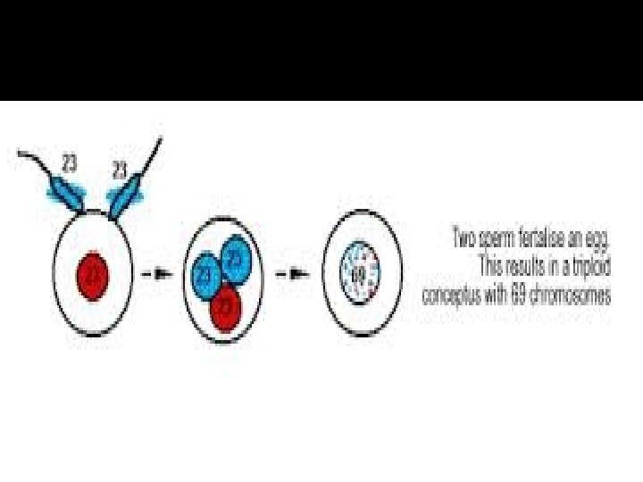
partial moles In the partial moles , the normal finding is a triploid karyotype , 69 chromosomes instead of normal 46. The most common mechanism appears to be fertilization of normal egg by two sperm, giving a complement of 69, XXY.

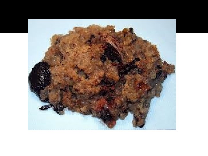
Pathological features v. Complete q there are mole : numerous edematous vesicles, which looks like a bunch of small clear grapes, q; usually no fetus, or membranes. ,
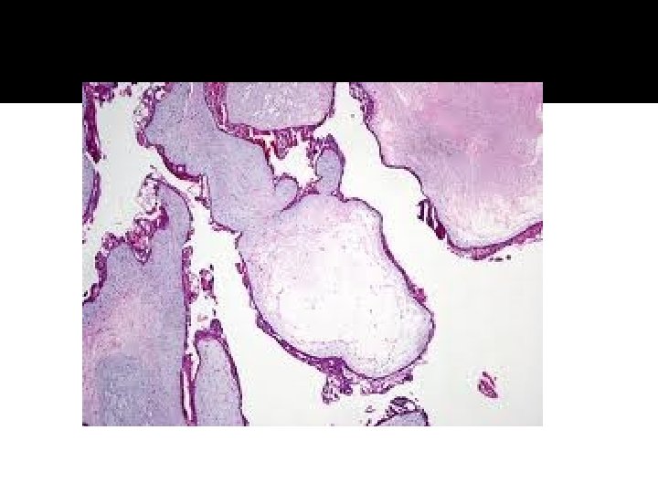
Pathological features microscopically: there are: Ø large oedematous enlarged villi , Ø a vascular , Ø with variable degree of trophoblastic hyperplasia. Ø carries greater risk of malignancy and requires longer follow up than the partial mole.
Pathological features v Partial mole: Ø Shows a less clear -cut picture , with the formation of vesicles usually focal , Ø fetus and membranes may present, Ø the vesicles have degree of vascularity.
Pathological features In general , the more active trophoblastic appearance the greater the risk of malignancy. So the subsequent management depends more on the h. CG results than the histological reports.
Clinical features v. Bleeding : Bleeding in early pregnancy after variable period of amenorrhea is the most common clinical sign of the mole (occurs in 90% of cases), with the passage of the vesicles.
Pathological features v. Hyperemesis gravidarum : Occurs into 25% of cases of moles , and appears more common when the uterus is much enlarged and h. CG levels are very high.
Pathological features v Uterine enlargement: The uterus is commonly “large for date”in 50% of case of moles , ü although, in a small proportion of cases the uterus corresponding to the gestational age or smaller than date. ü The uterus having a doughy consistency. ü The fetal parts are not palpable, and fetal heart is absent.
Pathological features v. Large theca lutein cysts of the ovary are present in “ 20%”of moles , these may be exaggerated the clinical picture of large for date uterus. These cysts are manifestation of excessive h. CG.
Pathological features Pre-eclampsia : Occur in association with the moles with range widely from 12 -54%, these due to differing times of diagnosis, the longer pregnancy progresses , the greater chance to developing pre-eclampsia. If the signs of pre-eclampsia appears early in pregnancy , the possibility of hydatidiform mole should be looked for with out delay.
Pathological features v Hyperthyrodism: Develop in small proportion of women , and this may be due to thyrotrophic effects of the human chorionic thyrotrophin , which may lead to goitre, fine tremor , supra-ventricular tachycardia, and weight loss. v DIC can develop in long-standing hydatidiform moles , when there is embolization of trophoblastic tissue to the lung, leads to thromboplastic substances which stimulate fibrin, and platelet deposition.
Diagnosis of hydatidiform moles ØHistory and examination : From the history of amenorrhea , passage of vesicles vaginally with bleeding ; the size and consistency of the uterus.
Pathological features ØThe increasing use of ultrasound in early pregnancy has probably led to the earlier diagnosis of molar pregnancy
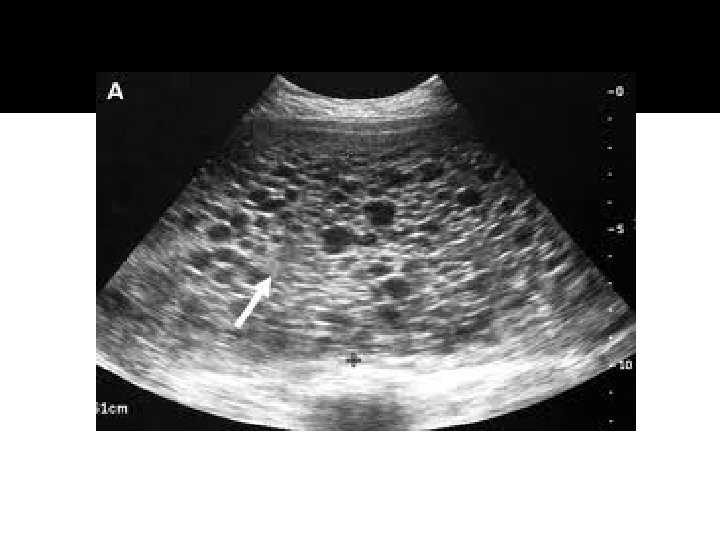
Pathological features ØBy the U/S examination can be diagnosed from very early pregnancy , characterized by “Snow-storm" appearance.
Pathological features ØBy very high levels of serum h. CG than the normal singleton pregnancy , which is diagnostic and prognostic to the course of the disease , with very short dappling time.
Prognosis The risks of hydatidiform mole are: Immediate hemorrhage , sepsis, or preeclampsia; the treatment of these conditions has vastly improved recently. Molar metastases : Of a non proliferative ”benign” type can occur.
Prognosis Choriocarcinoma: The most important danger association with the hydatidiform mole is the development of malignant GTD(Invasive mole or choriocarcinoma) in about 10% of cases,

ECTOPIC PREGNANCY
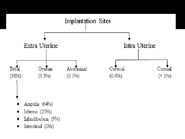
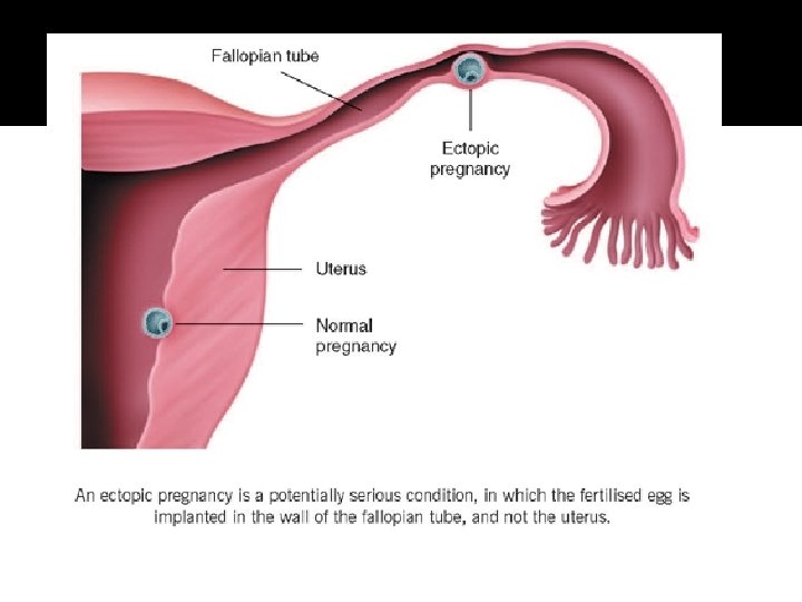
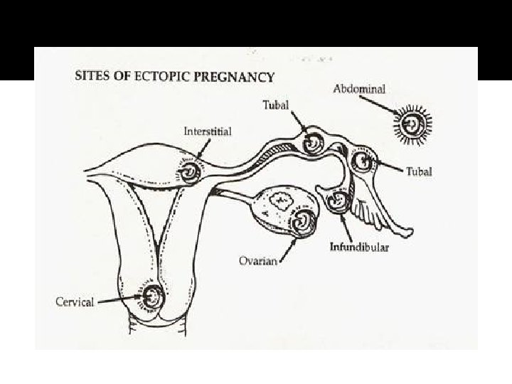
Definition: Implantation outside uterine cavity Most common site is within fallopian tube 98%, in the distal ampulla than in the proximal isthmus, followed by corneal 2% and abdominal 1. 4%, ovarian 0. 15% and cervical os 0. 15% Incidence: I in 100 of all pregnancies and to 1 in 30 in high risk population arising in the west in parallel with number of cases of chlamydia infection
Risk Factor for Ectopic Pregnancy Previous PID – chlamydia infection Previous ectopic pregnancy Tubal ligation Previous tubal surgery Intrauterine device Prolonged infertility Diethylstilbestrol (DES) exposure in-utero Multiple sexual partners
THE OUTCOME OF ECTOPIC PREGNANCY The muscle wall of the tube has not the capacity of uterine muscles for hypertrophy and distention and tubal pregnancy nearly always end in rupture and the death of the ovum. Tubal abortion – usually in ampullary about 8 weeks – forming pelvic haematocele Rupture into the peritoneal cavity ▪ Occur mainly from the narrow isthmus before 8 weeks or later from the interstitial portion of the tube. Haemorrhage is likely to be severe. ▪ Sometimes rupture is extraperitoneal between the leaves of the broad ligament – Broad ligament haematoma. Haemorrhage is likely to be controlled
Tubal pregnancy – effect on uterus The uterus enlarge in first 3 months as if the implantation were normal, reach the size of a gravid uterus of the same maturity. Uterine decidua grows abundantly and when the embryo dies bleeding occurs as the decidua degenerates due to effect of oestrogen withdrawal.
Clinical Finding: Variable - Early diagnosis - location of the implantation - Whether rupture has occurred Classic symptom trait with unruptured ectopic pregnancy: Amenorrhoea, abdominal pain, abnromal vagina bleeding Classic signs – adnexal or cervical motion tenderness. With ruptured ectopic pregnancy, finding parallel with the degree of internal bleeding and hypovolemia – abdominal guarding and rigidity, shoulder pain and fainting attacks and shock.
Symptoms and Signs: Pain – constant - Cramp-like - It may be referred to the shoulder if blood tracks to the diaphragm and stimulate the phernic nerve and it may be severe as to cause fainting. - The pain caused by the distension of the gravid tube by its effort to contract and expel the ovum and by irritation of the peritoneum, by leakage of blood. - Vaginal bleeding – occur usually after death of the ovum and is an effect of oestrogen withdrawal. It is dark, scanty and its irregularity may lead the patient to confuse it with the menstrual flow and give misleading history. 25% of cases presents without any vaginal bleeding
Cont. - Internal blood loss – severe and rapid. The usual sign of collapse and chock and it is less common than the condition presenting by slow trickle of blood into the pelvic cavity. - Peritoneal irritation – muscle guarding - frequency of micturation - fever - misleading of appendicitis - Pelvic examination – extreme tenderness - cystic mass may be felt - Abdominal - tenderness in one or other fossa. - General tenderness and resistance to palpation over whole abdomen.
Differential diagnosis: 1. Salpingitis 2. Abortion 3. Appendecitis 4. Torsion of pedicle of ovarian cyst 5. Rupture of corpus luteum or follicular cyst 6. Perforation of peptic ulcer.
n Diagnosis: - Careful history about LMP its timing and appearance. - Always think of tubal pregnancy women with lower abdomen pain in whom there is possibility of pregnancy should be regarded as having an ectopic until proved otherwise. - Pregnancy test nearly always be found by the time of clinical presentation. - Ultrasound to exclude intrauterine pregnancy - Laparoscopy: for identifying an unruptured tubal pregnancy which is producing equivocal symptoms and for exclude salpingitis and bleeding from small ovarian cyst. - For operative treatment using minimally invasive methods.
n Treatment: n If haemorrhage and shock present n n n Restore blood volume by the transfusion of red cells or volume expander Proceed with Laparotomy The earlier diagnosis of tubal pregnancy has allowed a more conservative approach to management where the tube is less damage. n n Pregnancy removed from the tube by laparoscopy (salpingostomy) hopefully retaining tubal function. Trophoblast destroyed by chemotherapeutic agent such as methotrexate
DIAGNOSIS: BHCG level TVU Medical Managment Methotrexate 1 mg/kg body weight Indicationss: Haemodynamically stable, no active bleeding, No haemoperitneum, minimal bleeding and no pain No contra indication to methotrexate Able to return for follow up for several weeks Non laparoscopic diagnosis of ectopic pregnancy General anaesthesia poses a significant risk Unruptured adenexal mass < 4 cm in size by scan No cardiac activity by scan

If sure of date of LMP and /or Regular cycle, i. e. >6 wks. gestation, Arrange TV ultrasound If unsure of date of LMP and /or irregular cycle, Measure serum h. CG If h. CG <100 (? early Intrauterine/ ? Ectopic pregnancy If Hcg >1000, use protocol for suspected Ectopic pregnancy 3. Empty uterus + free fluid in POD + adnexal + FH serum h. CG > 1000 Meet criteria for Methorexate treatment Use methotrexate protocol Does not meet criteria for methotrexate treatment Laproscopic /salpingotomy/ Salpingectomy ? Proceed to laparotomy OR Laparotomy if haemodynamically unstable
- Slides: 92