
Dysrhythmias Chapter 35 Copyright 2017 Elsevier Inc All
- Slides: 128
Dysrhythmias Chapter 35 Copyright © 2017, Elsevier Inc. All Rights Reserved.
Properties of Cardiac Cells • • Automaticity Excitability Conductivity Contractility Copyright © 2017, Elsevier Inc. All Rights Reserved.
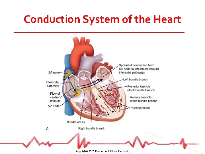
Conduction System of the Heart Copyright © 2017, Elsevier Inc. All Rights Reserved.
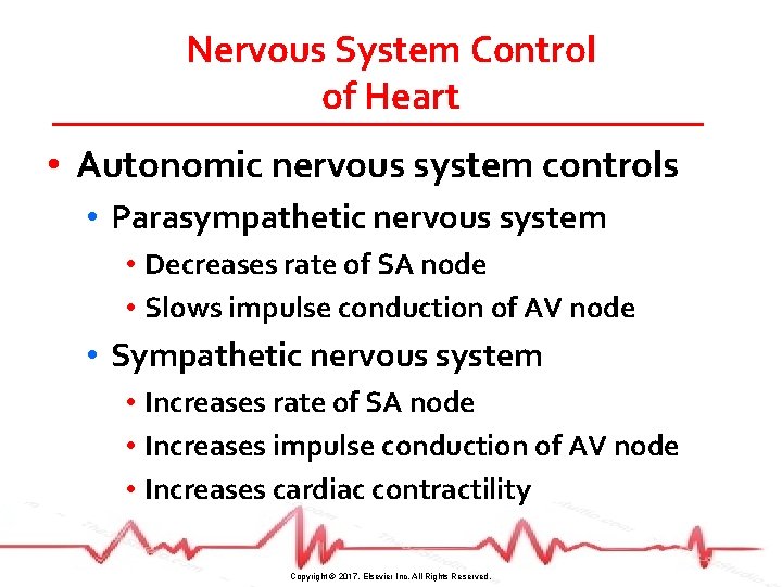
Nervous System Control of Heart • Autonomic nervous system controls • Parasympathetic nervous system • Decreases rate of SA node • Slows impulse conduction of AV node • Sympathetic nervous system • Increases rate of SA node • Increases impulse conduction of AV node • Increases cardiac contractility Copyright © 2017, Elsevier Inc. All Rights Reserved.
Dysrhythmias • Disorder of impulse formation, conduction of impulses, or both • SA node normal pacemaker of heart (60– 100 beats/minute) • Secondary pacemakers • AV node (40– 60 beats/minute) • His-Purkinje fibers (20– 40 beats/minute) Copyright © 2017, Elsevier Inc. All Rights Reserved.
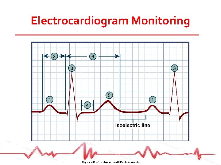
Electrocardiogram Monitoring • Graphic tracing of electrical impulses produced by heart • Waveforms of ECG represent activity of charged ions across membranes of myocardial cells Copyright © 2017, Elsevier Inc. All Rights Reserved.
Electrocardiogram Monitoring Copyright © 2017, Elsevier Inc. All Rights Reserved.
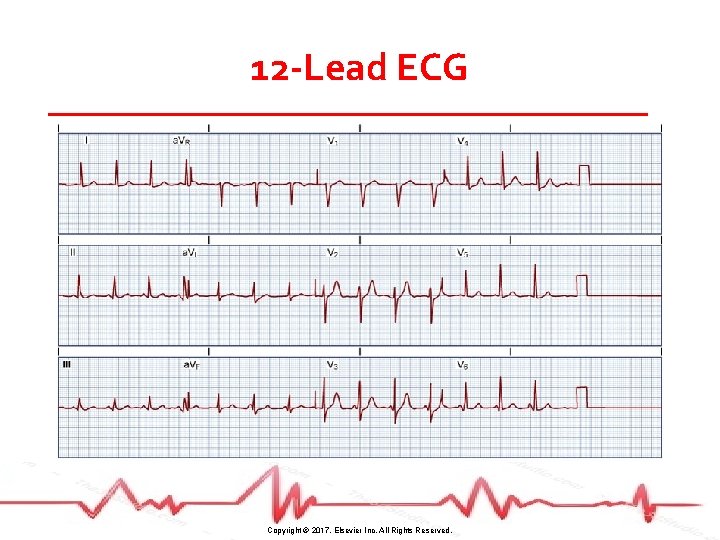
12 -Lead ECG Copyright © 2017, Elsevier Inc. All Rights Reserved.
Case Study (©i. Stockphoto/Thinkstock) • S. D. is a 45 -year-old woman who comes to the ED complaining of sudden onset of palpitations and shortness of breath. • Standard protocol requires you to obtain a 12 -lead ECG and attach S. D. to the cardiac monitor for continuous monitoring. Copyright © 2017, Elsevier Inc. All Rights Reserved.
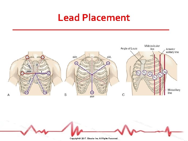
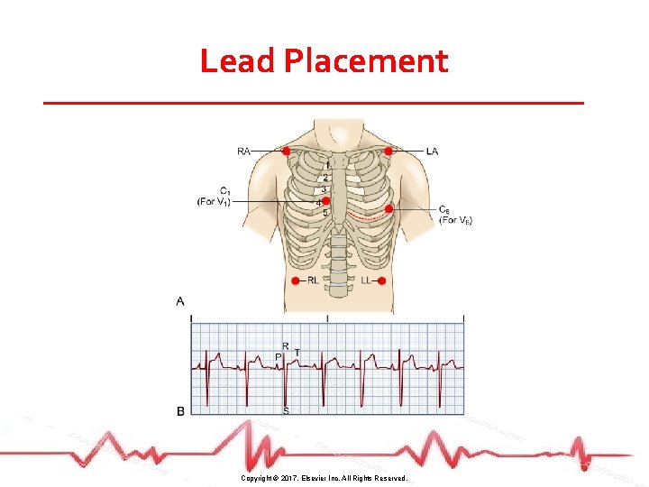
Case Study (©i. Stockphoto/Thinkstock) • Describe the appropriate location to apply the leads for both the 5 -lead cardiac monitor as well as the 12 -lead ECG. Copyright © 2017, Elsevier Inc. All Rights Reserved.
Lead Placement Copyright © 2017, Elsevier Inc. All Rights Reserved.
Lead Placement Copyright © 2017, Elsevier Inc. All Rights Reserved.
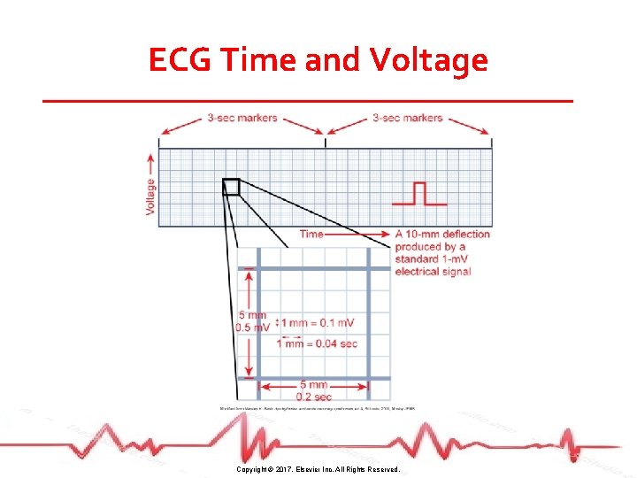
ECG Time and Voltage Copyright © 2017, Elsevier Inc. All Rights Reserved.
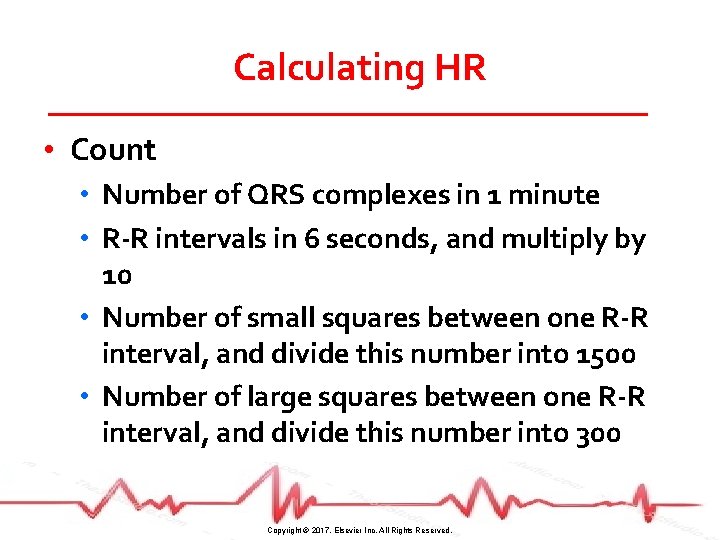
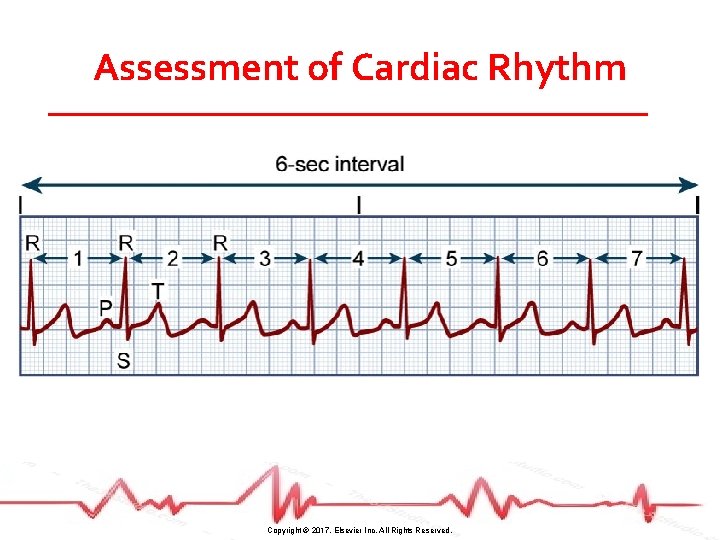
Calculating HR • Count • Number of QRS complexes in 1 minute • R-R intervals in 6 seconds, and multiply by 10 • Number of small squares between one R-R interval, and divide this number into 1500 • Number of large squares between one R-R interval, and divide this number into 300 Copyright © 2017, Elsevier Inc. All Rights Reserved.
Assessment of Cardiac Rhythm Copyright © 2017, Elsevier Inc. All Rights Reserved.
Patient Preparation • • Clip excessive hair on chest wall Rub skin with dry gauze May need to use alcohol for oily skin Apply electrode pad Copyright © 2017, Elsevier Inc. All Rights Reserved.
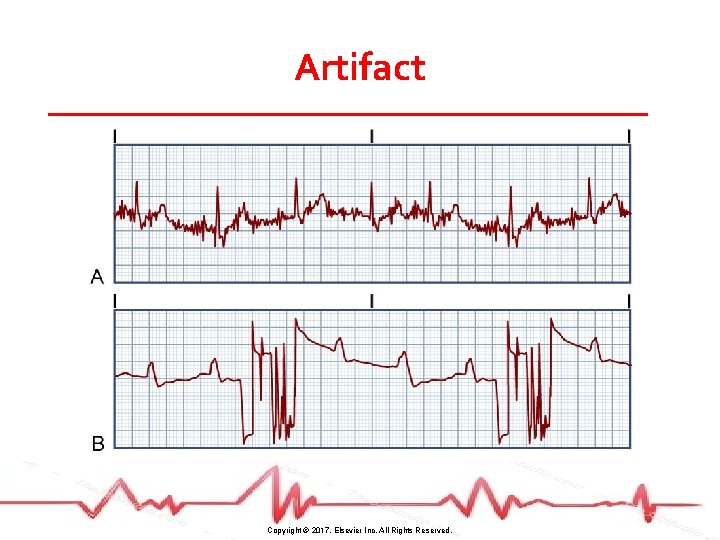
Artifact Copyright © 2017, Elsevier Inc. All Rights Reserved.
Telemetry Monitoring • Observation of HR and rhythm at a distant site • Two types • Centralized monitoring system • Advanced alarm system alerts when it detects dysrhythmias, ischemia, or infarction Copyright © 2017, Elsevier Inc. All Rights Reserved.
Assessment of Heart Rhythm • Interpret the rhythm AND assess the clinical status of the patient • Is the patient hemodynamically stable? • Determine cause of dysrhythmia • Assess and treat the patient, not the monitor! Copyright © 2017, Elsevier Inc. All Rights Reserved.
Case Study (©i. Stockphoto/Thinkstock) • The UAP obtains the 12 -lead ECG recording for S. D. while you print out a rhythm strip from the cardiac monitor. • Describe the method you would use to assess S. D. ’s rhythm strip. Copyright © 2017, Elsevier Inc. All Rights Reserved.
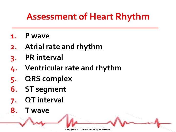
Assessment of Heart Rhythm 1. 2. 3. 4. 5. 6. 7. 8. P wave Atrial rate and rhythm PR interval Ventricular rate and rhythm QRS complex ST segment QT interval T wave Copyright © 2017, Elsevier Inc. All Rights Reserved.
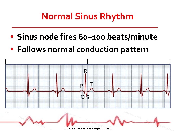
Normal Sinus Rhythm • Sinus node fires 60– 100 beats/minute • Follows normal conduction pattern Copyright © 2017, Elsevier Inc. All Rights Reserved.
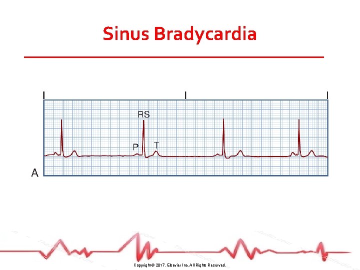
Sinus Bradycardia Copyright © 2017, Elsevier Inc. All Rights Reserved.
Sinus Bradycardia • Normal rhythm in aerobically trained athletes and during sleep • Can occur in response to parasympathetic nerve stimulation and certain drugs • Also associated with some disease states Copyright © 2017, Elsevier Inc. All Rights Reserved.
Sinus Bradycardia • Manifestations • • Hypotension Pale, cool skin Weakness Angina Dizziness or syncope Confusion or disorientation Shortness of breath Copyright © 2017, Elsevier Inc. All Rights Reserved.
Sinus Bradycardia • Treatment • Atropine • Pacemaker • Stop offending drugs Copyright © 2017, Elsevier Inc. All Rights Reserved.
Audience Response Question A patient’s cardiac rhythm is sinus bradycardia with a heart rate of 34 beats/minute. If the bradycardia is symptomatic, the nurse would expect the patient to exhibit a. Palpitations. b. Hypertension. c. Warm, flushed skin. d. Shortness of breath. Copyright © 2017, Elsevier Inc. All Rights Reserved.
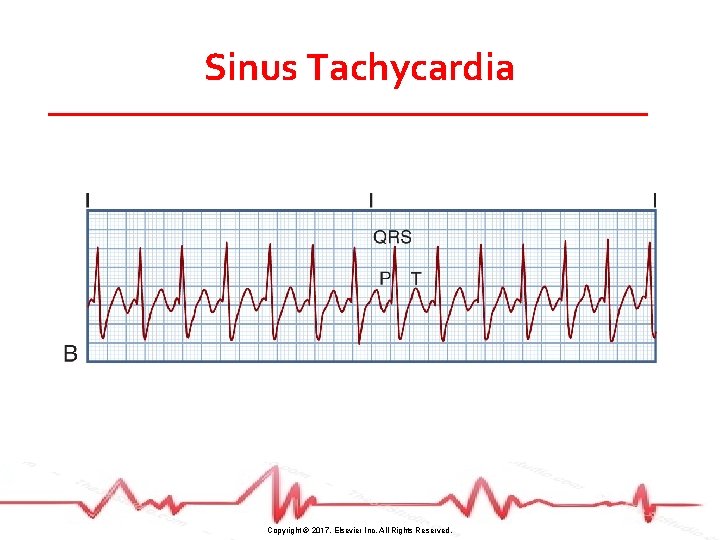
Sinus Tachycardia Copyright © 2017, Elsevier Inc. All Rights Reserved.
Sinus Tachycardia • Caused by vagal inhibition or sympathetic stimulation • Associated with physiologic and psychologic stressors • Drugs can increase rate Copyright © 2017, Elsevier Inc. All Rights Reserved.
Sinus Tachycardia • Manifestations • • Dizziness Dyspnea Hypotension Angina in patients with CAD Copyright © 2017, Elsevier Inc. All Rights Reserved.
Sinus Tachycardia • Treatment • Guided by cause (e. g. , treat pain) • Vagal maneuver • β-blockers Copyright © 2017, Elsevier Inc. All Rights Reserved.
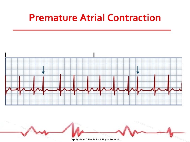
Premature Atrial Contraction Copyright © 2017, Elsevier Inc. All Rights Reserved.
Premature Atrial Contraction • Contraction originating from ectopic focus in atrium in location other than SA node • Travels across atria by abnormal pathway, creating distorted P wave • May be stopped, delayed, or conducted normally at the AV node Copyright © 2017, Elsevier Inc. All Rights Reserved.
Premature Atrial Contraction • Causes • • Stress Fatigue Caffeine Tobacco Alcohol Hypoxia Electrolyte imbalance Disease states Copyright © 2017, Elsevier Inc. All Rights Reserved.
Premature Atrial Contraction • Manifestations • Palpitations • Heart “skips a beat” • Treatment • Monitor for more serious dysrhythmias • Withhold sources of stimulation • β-blockers Copyright © 2017, Elsevier Inc. All Rights Reserved.
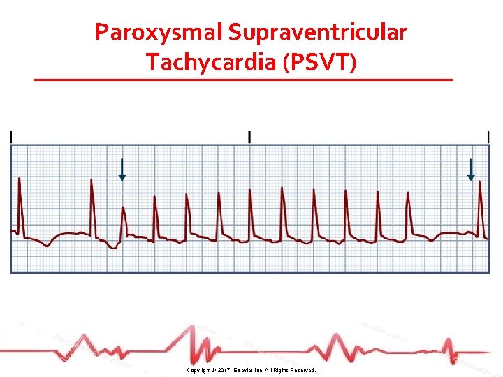
Paroxysmal Supraventricular Tachycardia (PSVT) Copyright © 2017, Elsevier Inc. All Rights Reserved.
Paroxysmal Supraventricular Tachycardia (PSVT) • Reentrant phenomenon: PAC triggers a run of repeated premature beats • Paroxysmal refers to an abrupt onset and termination • Associated with overexertion, stress, deep inspiration, stimulants, disease, digitalis toxicity Copyright © 2017, Elsevier Inc. All Rights Reserved.
Paroxysmal Supraventricular Tachycardia (PSVT) • Manifestations • HR is 150– 220 beats/minute (add for clarification) • HR > 180 leads to decreased cardiac output and stroke volume • Hypotension • Dyspnea • Angina Copyright © 2017, Elsevier Inc. All Rights Reserved.
Paroxysmal Supraventricular Tachycardia (PSVT) • Treatment • • • Vagal stimulation IV adenosine IV β-blockers Calcium channel blockers Amiodarone DC cardioversion Copyright © 2017, Elsevier Inc. All Rights Reserved.
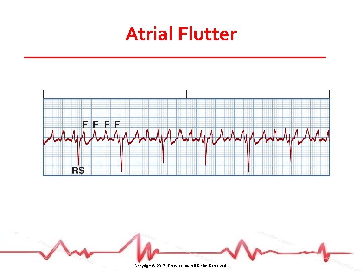
Atrial Flutter Copyright © 2017, Elsevier Inc. All Rights Reserved.
Atrial Flutter • Typically associated with disease • Symptoms result from high ventricular rate and loss of atrial “kick” → decreased CO → heart failure • Increases risk of stroke Copyright © 2017, Elsevier Inc. All Rights Reserved.
Atrial Flutter • Treatment • Pharmacologic agent • Electrical cardioversion • Radiofrequency ablation Copyright © 2017, Elsevier Inc. All Rights Reserved.
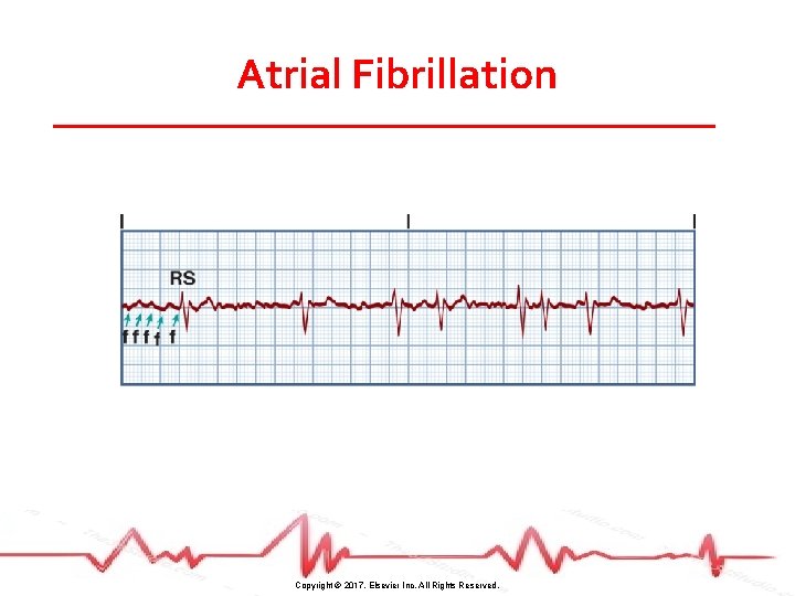
Case Study (©i. Stockphoto/Thinkstock) • S. D. ’s ECG reveals atrial fibrillation with a rapid ventricular response (HR = 168). • Describe what S. D. ’s rhythm would look like. • What might be the cause of this dysrhythmia for S. D. ? Copyright © 2017, Elsevier Inc. All Rights Reserved.
Atrial Fibrillation Copyright © 2017, Elsevier Inc. All Rights Reserved.
Atrial Fibrillation Paroxysmal or persistent Most common dysrhythmia Prevalence increases with age Usually occurs in patients with underlying heart disease • Can also occur with other disease states • • Copyright © 2017, Elsevier Inc. All Rights Reserved.
Case Study (©i. Stockphoto/Thinkstock) • S. D. ’s blood pressure is 94/58 with HR of 168. • What treatment might you expect the health care provider to initially order for S. D. ’s atrial fibrillation? Copyright © 2017, Elsevier Inc. All Rights Reserved.
Atrial Fibrillation • As with atrial flutter – causes a decrease in CO and an increased risk of stroke Copyright © 2017, Elsevier Inc. All Rights Reserved.
Atrial Fibrillation • Treatment • Drugs to control ventricular rate and/or convert to sinus rhythm (amiodarone and ibutilide most common) • Electrical cardioversion • Anticoagulation • Radiofrequency ablation • Maze procedure with cryoablation Copyright © 2017, Elsevier Inc. All Rights Reserved.
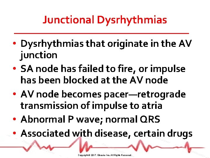
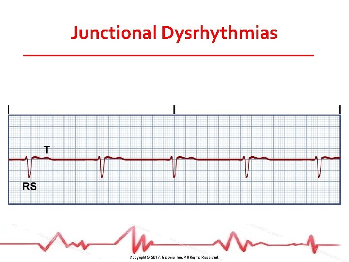
Junctional Dysrhythmias • Dysrhythmias that originate in the AV junction • SA node has failed to fire, or impulse has been blocked at the AV node • AV node becomes pacer—retrograde transmission of impulse to atria • Abnormal P wave; normal QRS • Associated with disease, certain drugs Copyright © 2017, Elsevier Inc. All Rights Reserved.
Junctional Dysrhythmias Copyright © 2017, Elsevier Inc. All Rights Reserved.
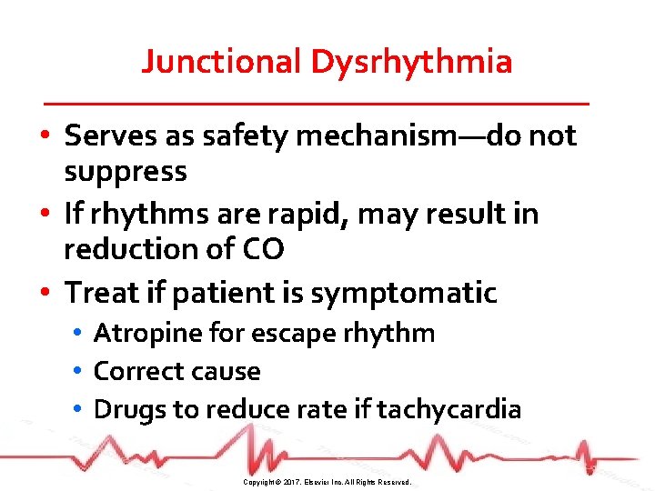
Junctional Dysrhythmia • Serves as safety mechanism—do not suppress • If rhythms are rapid, may result in reduction of CO • Treat if patient is symptomatic • Atropine for escape rhythm • Correct cause • Drugs to reduce rate if tachycardia Copyright © 2017, Elsevier Inc. All Rights Reserved.
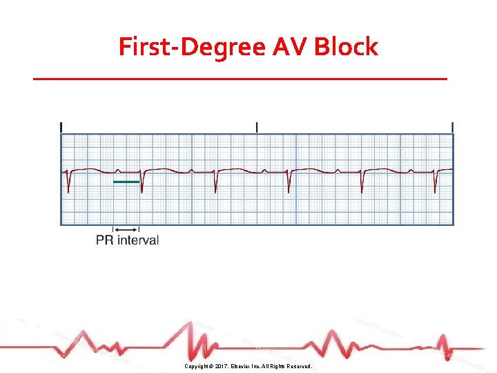
First-Degree AV Block Copyright © 2017, Elsevier Inc. All Rights Reserved.
First-Degree AV Block • Associated with disease states and certain drugs • Typically not serious • Patients asymptomatic • No treatment • Monitor for changes in heart rhythm Copyright © 2017, Elsevier Inc. All Rights Reserved.
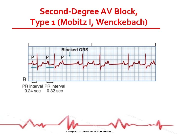
Second-Degree AV Block, Type 1 (Mobitz I, Wenckebach) Copyright © 2017, Elsevier Inc. All Rights Reserved.
Second-Degree AV Block, Type 1 (Mobitz I, Wenckebach) • • May result from drugs or CAD Typically associated with ischemia Usually transient and well tolerated Treat if symptomatic • Atropine • Pacemaker • If asymptomatic, monitor closely Copyright © 2017, Elsevier Inc. All Rights Reserved.
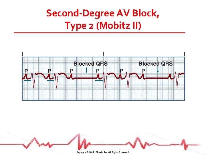
Second-Degree AV Block, Type 2 (Mobitz II) Copyright © 2017, Elsevier Inc. All Rights Reserved.
Second-Degree AV Block, Type 2 (Mobitz II) • Associated with heart disease and drug toxicity • Often progressive and results in decreased CO • Treat with pacemaker Copyright © 2017, Elsevier Inc. All Rights Reserved.
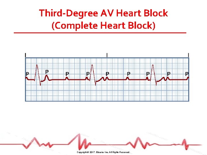
Third-Degree AV Heart Block (Complete Heart Block) Copyright © 2017, Elsevier Inc. All Rights Reserved.
Third-Degree AV Heart Block (Complete Heart Block) • Associated with severe heart disease, some systemic diseases, certain drugs • Usually results in decreased CO • Can lead to syncope, HF, shock • Treat with pacemaker • Drugs to increase heart rate if needed while awaiting pacing Copyright © 2017, Elsevier Inc. All Rights Reserved.
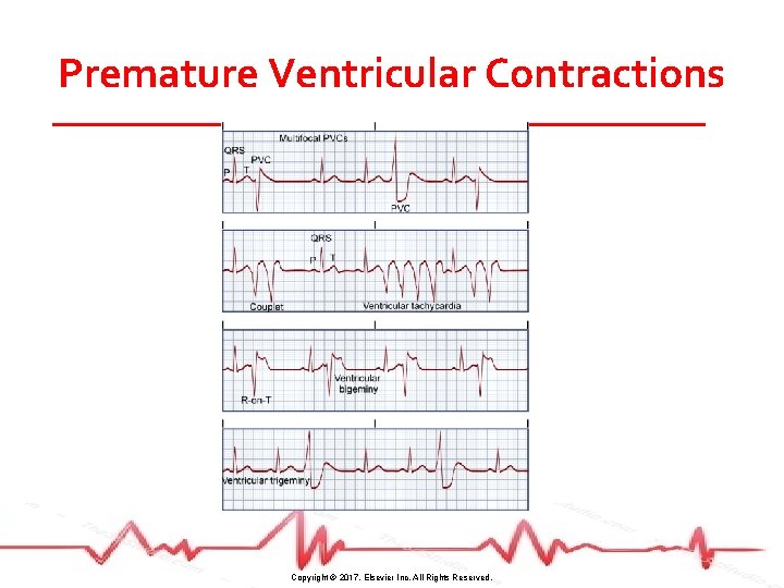
Premature Ventricular Contractions Copyright © 2017, Elsevier Inc. All Rights Reserved.
Premature Ventricular Contractions • Associated with stimulants, electrolyte imbalances, hypoxia, heart disease • Not harmful with normal heart but CO reduction, angina, and HF in diseased heart • Assess apical-radial pulse deficit Copyright © 2017, Elsevier Inc. All Rights Reserved.
Premature Ventricular Contractions • Treatment • Correct cause • Antidysrhythmics Copyright © 2017, Elsevier Inc. All Rights Reserved.
Audience Response Question A patient has a diagnosis of acute myocardial infarction, and his cardiac rhythm is sinus bradycardia with 6 to 8 premature ventricular contractions (PVCs) per minute. The pattern that the nurse recognizes as the most characteristic of PVCs is a. An irregular rhythm. b. An inverted T wave. c. A wide, distorted QRS complex. d. An increasingly long PR interval. Copyright © 2017, Elsevier Inc. All Rights Reserved.
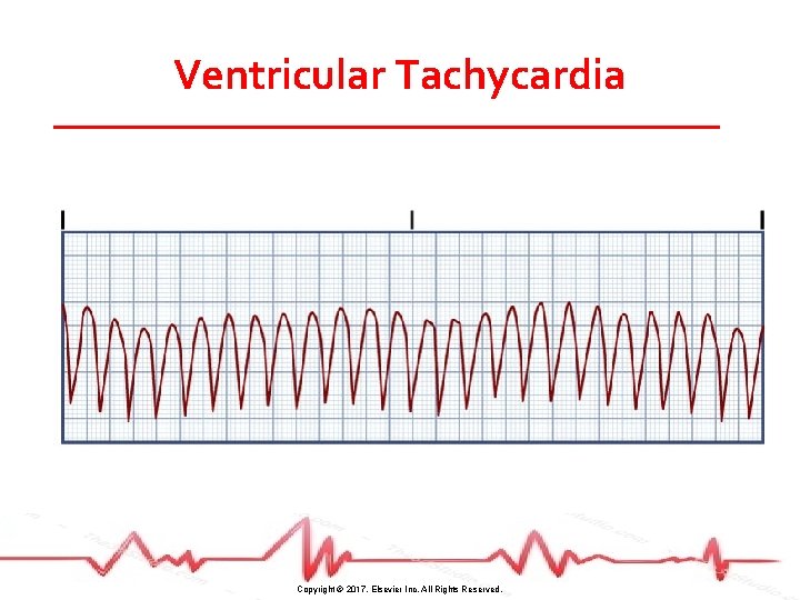
Ventricular Tachycardia Copyright © 2017, Elsevier Inc. All Rights Reserved.
Ventricular Tachycardia • Ectopic foci take over as pacemaker • Monomorphic, polymorphic, sustained, and nonsustained • Considered life-threatening because of decreased CO and the possibility of deterioration to ventricular fibrillation Copyright © 2017, Elsevier Inc. All Rights Reserved.
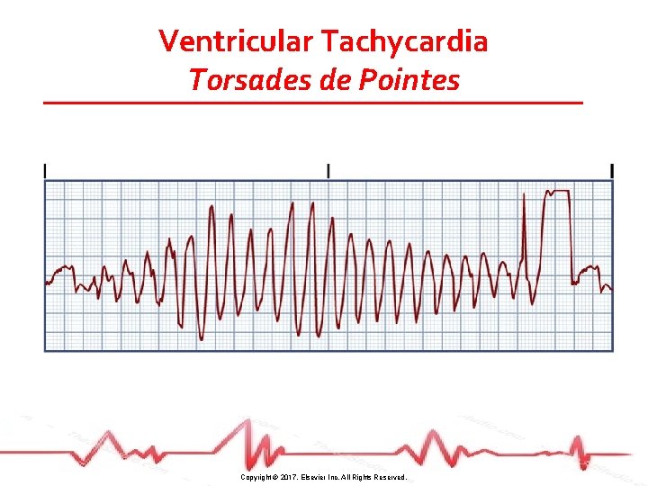
Ventricular Tachycardia Torsades de Pointes Copyright © 2017, Elsevier Inc. All Rights Reserved.
Ventricular Tachycardia • Associated with heart disease, electrolyte imbalances, drugs, CNS disorder • Can be stable (patient has a pulse) or unstable (pulseless) • Sustained VT causes severe decrease in CO • Hypotension, pulmonary edema, decreased cerebral blood flow, cardiopulmonary arrest Copyright © 2017, Elsevier Inc. All Rights Reserved.
Ventricular Tachycardia • Precipitating causes must be identified and treated (e. g. , hypoxia) • VT with pulse (stable) treated with antidysrhythmics or cardioversion • Pulseless VT treated with CPR and rapid defibrillation Copyright © 2017, Elsevier Inc. All Rights Reserved.
Accelerated Idioventricular Rhythm (AIVR) • Develops when the intrinsic pacemaker rate (SA node or AV node) becomes less than that of ventricular ectopic pacemaker • Rate is between 40 and 100 beats/minute • Atropine if patient symptomatic • Temporary pacing • Do not suppress rhythm Copyright © 2017, Elsevier Inc. All Rights Reserved.
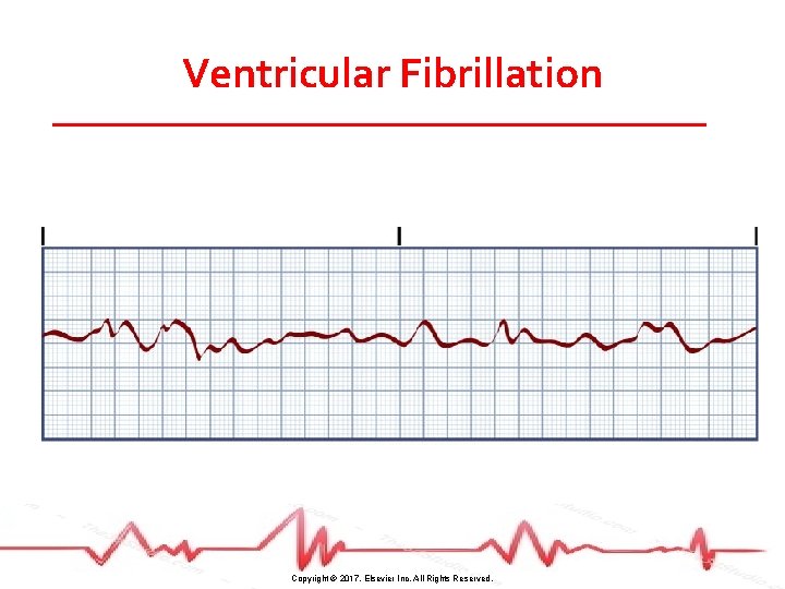
Ventricular Fibrillation Copyright © 2017, Elsevier Inc. All Rights Reserved.
Ventricular Fibrillation • Associated with MI, ischemia, disease states, procedures • Unresponsive, pulseless, and apneic • If not treated rapidly, death will result • Treat with immediate CPR and ACLS • Defibrillation • Drug therapy (epinephrine, vasopressin) Copyright © 2017, Elsevier Inc. All Rights Reserved.
Audience Response Question A patient in the coronary care unit develops ventricular fibrillation. The first action the nurse should take is to a. Perform defibrillation. b. Initiate cardiopulmonary resuscitation. c. Prepare for synchronized cardioversion. d. Administer IV antidysrhythmic drugs per protocol. Copyright © 2017, Elsevier Inc. All Rights Reserved.
Asystole • Represents total absence of ventricular electrical activity • No ventricular contraction • Patient unresponsive, pulseless, apneic • Must assess in more than one lead Copyright © 2017, Elsevier Inc. All Rights Reserved.
Asystole • Usually result of advanced cardiac disease, severe conduction disturbance, or end-stage HF • Treat with immediate CPR and ACLS measures • Epinephrine and/or vasopressin • Intubation • Poor prognosis Copyright © 2017, Elsevier Inc. All Rights Reserved.
Pulseless Electrical Activity • Electrical activity can be observed on the ECG, but no mechanical activity of the heart is evident, and the patient has no pulse • Prognosis is poor unless underlying cause quickly identified and treated Copyright © 2017, Elsevier Inc. All Rights Reserved.
Pulseless Electrical Activity Hs and Ts Pneumonic • Hypovolemia • Hypoxia • Hydrogen ion (acidosis) • Hyper-/hypokalemia • Hypoglycemia • Hypothermia • Toxins • Tamponade (cardiac) • Thrombosis (MI and pulmonary) • Tension pneumothorax • Trauma Copyright © 2017, Elsevier Inc. All Rights Reserved.
Pulseless Electrical Activity • Treatment • CPR followed by intubation and IV epinephrine • Treatment is directed toward correction of the underlying cause Copyright © 2017, Elsevier Inc. All Rights Reserved.
Sudden Cardiac Death (SCD) • Death from a cardiac cause • Majority of SCDs result from ventricular dysrhythmias • Ventricular tachycardia • Ventricular fibrillation Copyright © 2017, Elsevier Inc. All Rights Reserved.
Prodysrhythmia • Life-threatening dysrhythmias caused by antidysrhythmia drugs • Severe LV dysfunction increases risk • Digoxin and class IA, IC, and III antidysrhythmia drugs • Most susceptible first few days of drug therapy Copyright © 2017, Elsevier Inc. All Rights Reserved.
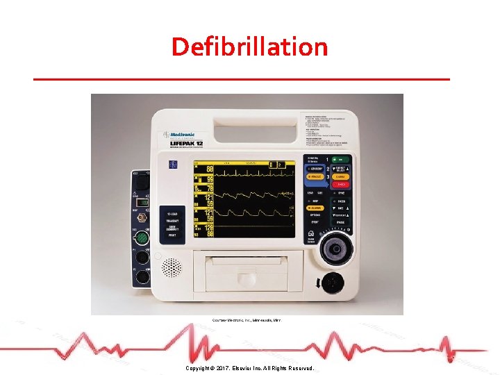
Defibrillation • Treatment of choice for VF and pulseless VT • Most effective when completed within 2 minutes of dysrhythmia onset • Passage of DC electrical shock through the heart to depolarize myocardial cells • Allows SA node to resume pacemaker role Copyright © 2017, Elsevier Inc. All Rights Reserved.
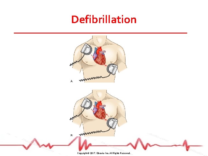
Defibrillation • Monophasic defibrillators deliver energy in one direction • Biphasic defibrillators deliver energy in two directions • Use lower energies • Fewer postshock ECG dysrhythmias Copyright © 2017, Elsevier Inc. All Rights Reserved.
Defibrillation Copyright © 2017, Elsevier Inc. All Rights Reserved.
Defibrillation • Output is measured in joules or watts per second • Recommended energy for initial shocks in defibrillation • Biphasic: 120 to 200 joules • Monophasic: 360 joules • Immediate CPR after first shock Copyright © 2017, Elsevier Inc. All Rights Reserved.
Defibrillation Copyright © 2017, Elsevier Inc. All Rights Reserved.
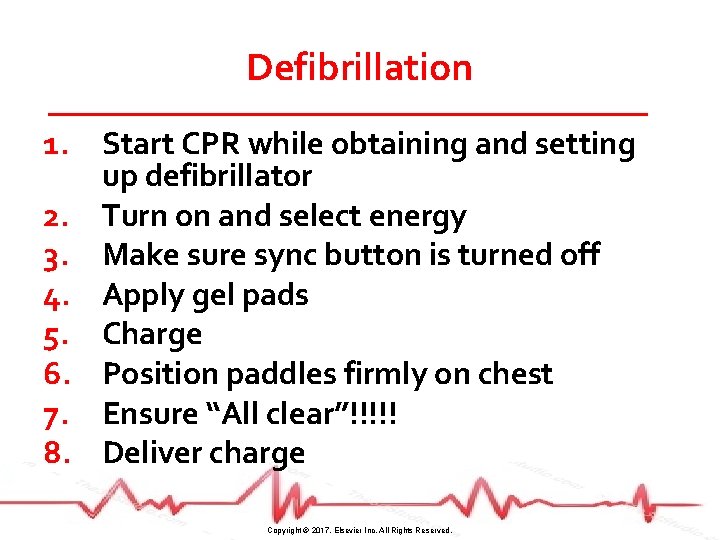
Defibrillation 1. Start CPR while obtaining and setting up defibrillator 2. Turn on and select energy 3. Make sure sync button is turned off 4. Apply gel pads 5. Charge 6. Position paddles firmly on chest 7. Ensure “All clear”!!!!! 8. Deliver charge Copyright © 2017, Elsevier Inc. All Rights Reserved.
Case Study (©i. Stockphoto/Thinkstock) • S. D. was admitted to the telemetry unit and an IV amiodarone drip was started. • The purpose of the drug was to convert her atrial fibrillation to normal sinus rhythm. Copyright © 2017, Elsevier Inc. All Rights Reserved.
Case Study (©i. Stockphoto/Thinkstock) • Although her heart rate has decreased to 108 beats/minute, she remains in atrial fibrillation 24 hours later. • A cardiologist was consulted and electrical cardioversion is planned. Copyright © 2017, Elsevier Inc. All Rights Reserved.
Case Study (©i. Stockphoto/Thinkstock) • What would you teach S. D. about the scheduled procedure? • What are three differences between defibrillation and cardioversion? Copyright © 2017, Elsevier Inc. All Rights Reserved.
Synchronized Cardioversion • Choice of therapy for ventricular ( VT with a pulse) or supraventricular tachydysrhythmias • Synchronized circuit delivers a countershock on the R wave of the QRS complex of the ECG Copyright © 2017, Elsevier Inc. All Rights Reserved.
Synchronized Cardioversion • Procedure similar to defibrillation except sync button turned ON • If patient stable, sedate prior • Initial energy lower • 70– 75 joules (biphasic) • 100 joules (monophasic) • If patient becomes pulseless, turn sync button off and defibrillate Copyright © 2017, Elsevier Inc. All Rights Reserved.
Implantable Cardioverter-Defibrillator (ICD) • Appropriate for patients who • Have survived SCD • Have spontaneous sustained VT • Have syncope with inducible ventricular tachycardia/fibrillation during EPS • Are at high risk for future life-threatening dysrhythmias • Decreases mortality Copyright © 2017, Elsevier Inc. All Rights Reserved.
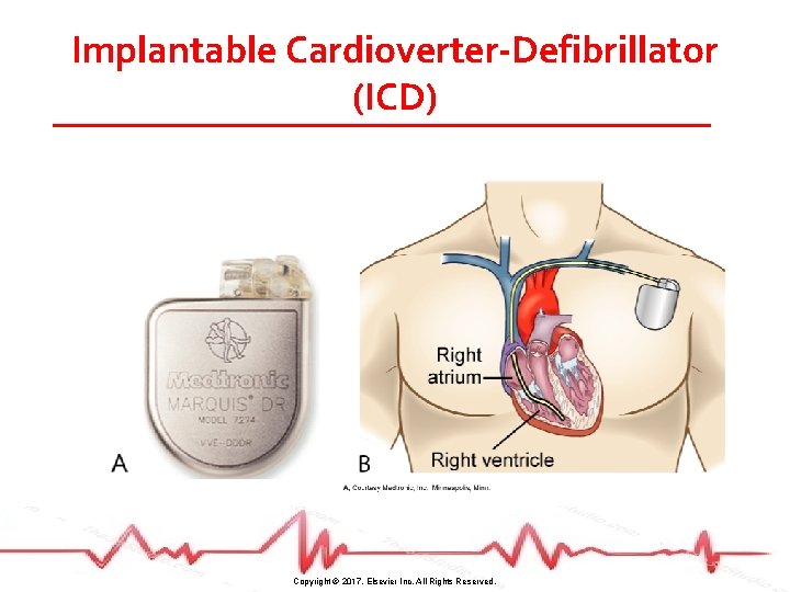
Implantable Cardioverter-Defibrillator (ICD) • Consists of a lead system placed via subclavian vein to the endocardium • Battery-powered pulse generator is implanted subcutaneously • Sensing system monitors HR and rhythm – delivering 25 joules or less to heart when detects lethal dysrhythmia Copyright © 2017, Elsevier Inc. All Rights Reserved.
Implantable Cardioverter-Defibrillator (ICD) Copyright © 2017, Elsevier Inc. All Rights Reserved.
Implantable Cardioverter-Defibrillator (ICD) • Includes antitachycardia and antibradycardia pacemakers • Overdrive pacing for tachycardias • Backup pacing for bradycardias • Preprocedure and postprocedure care same as pacemaker • S-ICD Copyright © 2017, Elsevier Inc. All Rights Reserved.
Implantable Cardioverter-Defibrillator (ICD) • Variety of emotions are possible • • Fear of body image change Fear of recurrent dysrhythmias Expectation of pain with ICD discharge Anxiety about going home • Participation in an ICD support group should be encouraged Copyright © 2017, Elsevier Inc. All Rights Reserved.
Implantable Cardioverter-Defibrillator (ICD) Patient and Caregiver Teaching 1. 2. 3. 4. 5. 6. 7. Follow-up appointments Incision care Arm restrictions Sexual activity Driving Avoid direct blows Avoid large magnets, MRI Copyright © 2017, Elsevier Inc. All Rights Reserved.
Implantable Cardioverter-Defibrillator (ICD) Patient and Caregiver Teaching 8. 9. 10. 11. 12. 13. Air travel not restricted Avoid antitheft devices What to do if ICD fires Medic Alert ID ICD identification card Caregivers to learn CPR Copyright © 2017, Elsevier Inc. All Rights Reserved.
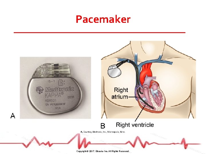
Pacemakers • Used to pace the heart when the normal conduction pathway is damaged • Pacing circuit consists of • Programmable pulse generator (power source) • One or more conducting (pacing) leads to myocardium Copyright © 2017, Elsevier Inc. All Rights Reserved.
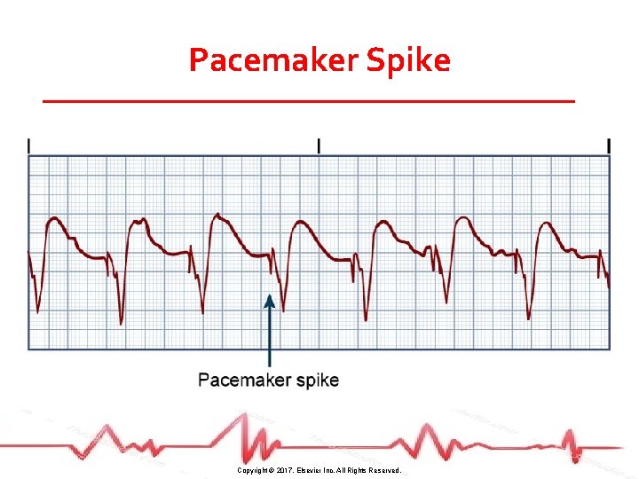
Pacemaker Spike Copyright © 2017, Elsevier Inc. All Rights Reserved.
Pacemakers • Pace atrium and/or one or both of ventricles • Most pace on demand, firing only when HR drops below preset rate • Sensing device inhibits pacemaker when HR adequate • Pacing device triggers when no QRS complexes within set time frame Copyright © 2017, Elsevier Inc. All Rights Reserved.
Pacemakers • Antitachycardia pacing: delivery of a stimulus to the ventricle to terminate tachydysrhythmias • Overdrive pacing: pacing the atrium at rates of 200– 500 impulses/minute to terminate atrial tachycardias Copyright © 2017, Elsevier Inc. All Rights Reserved.
Pacemaker Copyright © 2017, Elsevier Inc. All Rights Reserved.
Pacemakers • Cardiac resynchronization therapy (CRT) • Resynchronizes the heart cycle by pacing both ventricles • Biventricular pacing • Used to treat patients with heart failure • Can be combined with ICD for maximum therapy Copyright © 2017, Elsevier Inc. All Rights Reserved.
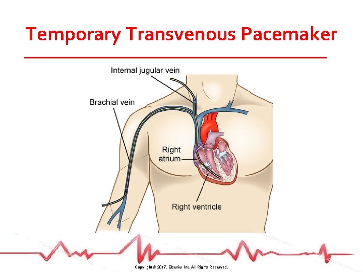
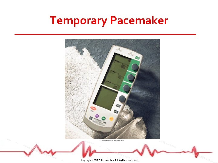
Temporary Pacemakers • Power source outside the body • Transvenous • Epicardial • Transcutaneous Copyright © 2017, Elsevier Inc. All Rights Reserved.
Temporary Transvenous Pacemaker Copyright © 2017, Elsevier Inc. All Rights Reserved.
Epicardial Pacing • Leads placed on epicardium during heart surgery • Passed through chest wall and attached to external power source as needed Copyright © 2017, Elsevier Inc. All Rights Reserved.
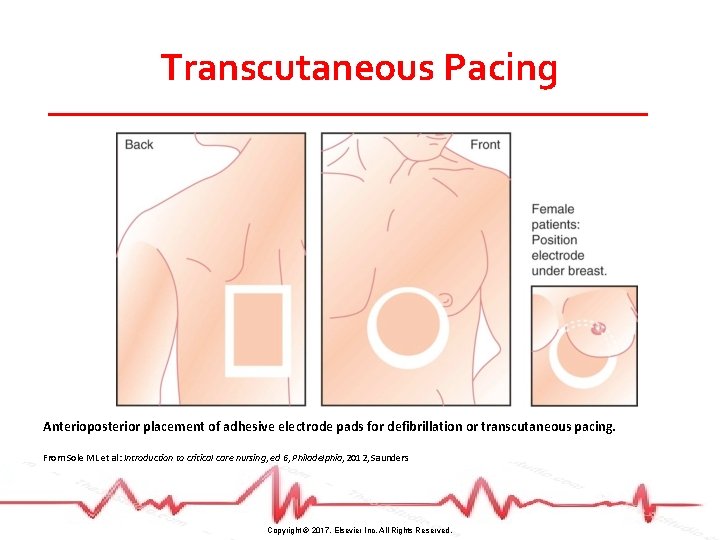
Transcutaneous Pacing • For emergency pacing needs • Noninvasive • Bridge until transvenous pacer can be inserted • Use lowest current that will “capture” • Patient may need analgesia/sedation Copyright © 2017, Elsevier Inc. All Rights Reserved.
Transcutaneous Pacing Anterioposterior placement of adhesive electrode pads for defibrillation or transcutaneous pacing. From Sole ML et al: Introduction to critical care nursing, ed 6, Philadelphia, 2012, Saunders Copyright © 2017, Elsevier Inc. All Rights Reserved.
Temporary Pacemaker Copyright © 2017, Elsevier Inc. All Rights Reserved.
Pacemakers • ECG monitoring for malfunction • Failure to sense • Causes inappropriate firing • Failure to capture • Lack of pacing when needed leads to bradycardia or asystole Copyright © 2017, Elsevier Inc. All Rights Reserved.
Pacemakers • Monitor for other complications • • • Infection Hematoma formation Pneumothorax Atrial or ventricular septum perforation Lead misplacement Copyright © 2017, Elsevier Inc. All Rights Reserved.
Pacemakers • Postprocedure care • OOB once stable • Limit arm and shoulder activity • Monitor insertion site for bleeding and infection • Patient teaching important Copyright © 2017, Elsevier Inc. All Rights Reserved.
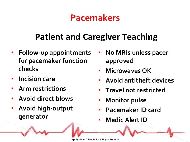
Pacemakers Patient and Caregiver Teaching • Follow-up appointments for pacemaker function checks • Incision care • Arm restrictions • Avoid direct blows • Avoid high-output generator • No MRIs unless pacer approved • Microwaves OK • Avoid antitheft devices • Travel not restricted • Monitor pulse • Pacemaker ID card • Medic Alert ID Copyright © 2017, Elsevier Inc. All Rights Reserved.
Radiofrequency Catheter Ablation Therapy • Electrode-tipped ablation catheter “burns” accessory pathways or ectopic sites in the atria, AV node, and ventricles • Nonpharmacologic treatment of choice for several atrial dysrhythmias • Postcare similar to cardiac catheterization Copyright © 2017, Elsevier Inc. All Rights Reserved.
Case Study (©i. Stockphoto/Thinkstock) • S. D. ’s electrical cardioversion was successful. • S. D. ’s sinus rhythm has remained stable for 24 hours and she is ready to go home. Copyright © 2017, Elsevier Inc. All Rights Reserved.
Case Study (©i. Stockphoto/Thinkstock) • As you enter her room to provide discharge teaching, she tells you she is experiencing some chest “heaviness and tightness right under her breast bone. ” Copyright © 2017, Elsevier Inc. All Rights Reserved.
Case Study (©i. Stockphoto/Thinkstock) • You ask the UAP to obtain a 12 -lead ECG while you notify S. D. ’s health care provider. • What specific ECG change will you be looking for to determine if S. D. ’s chest pain is related to cardiac ischemia? Injury? Infarction? Copyright © 2017, Elsevier Inc. All Rights Reserved.
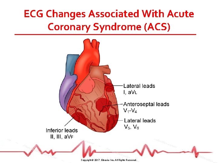
ECG Changes Associated With Acute Coronary Syndrome (ACS) Copyright © 2017, Elsevier Inc. All Rights Reserved.
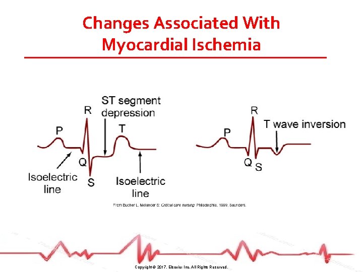
ECG Changes Associated With Acute Coronary Syndrome (ACS) • Ischemia • ST-segment depression and/or T wave inversion • ST-segment depression is significant if it is at least 1 mm (one small box) below the isoelectric line • Changes reverse when adequate blood flow is restored to myocardium Copyright © 2017, Elsevier Inc. All Rights Reserved.
Changes Associated With Myocardial Ischemia Copyright © 2017, Elsevier Inc. All Rights Reserved.
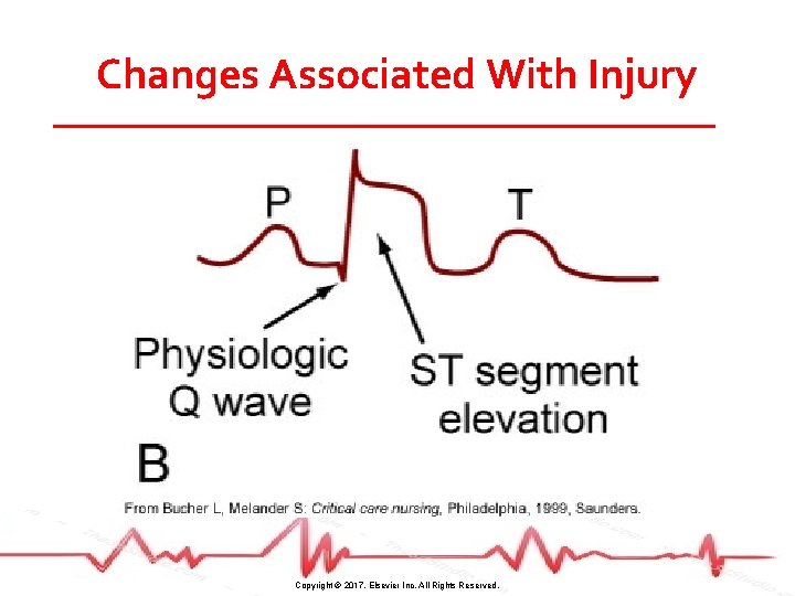
ECG Changes Associated With Acute Coronary Syndrome (ACS) • Injury • ST-segment elevation occurs • Significant if >1 mm above the isoelectric line • If treatment is prompt and effective, may avoid or limit infarction • Absence of serum cardiac markers confirms no infarction Copyright © 2017, Elsevier Inc. All Rights Reserved.
Changes Associated With Injury Copyright © 2017, Elsevier Inc. All Rights Reserved.
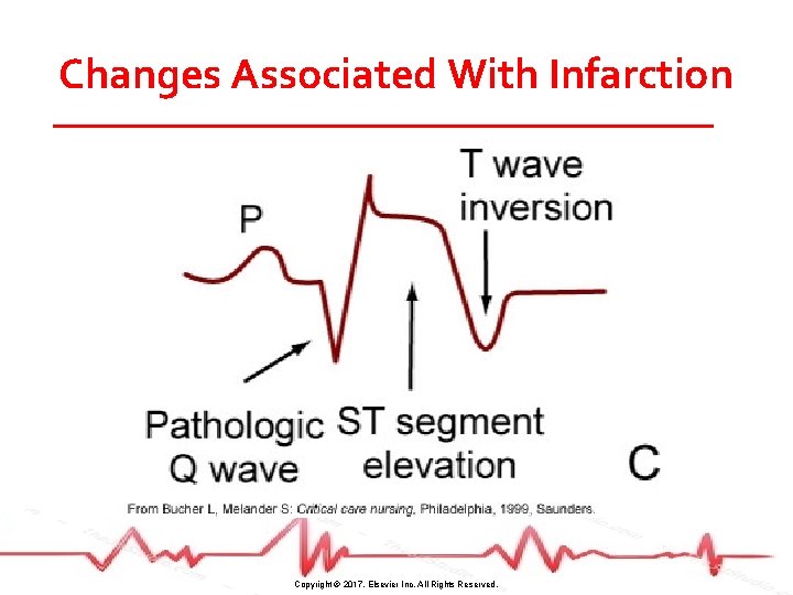
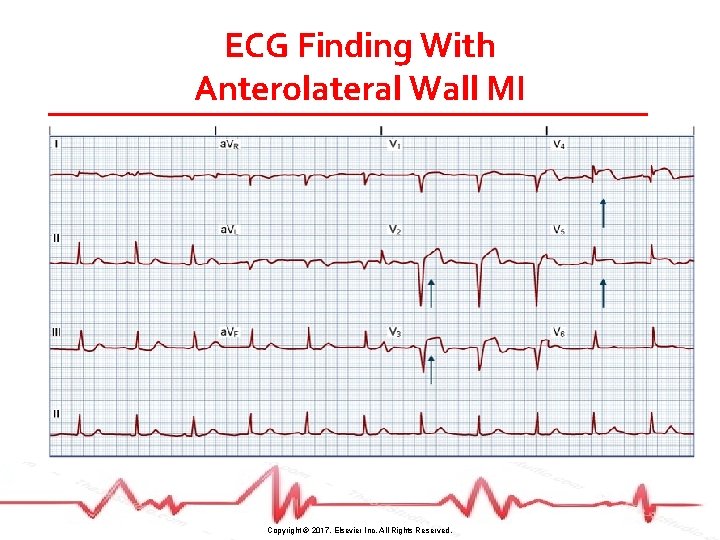
ECG Changes Associated With Acute Coronary Syndrome (ACS) • Infarction • Physiologic Q wave is the first negative deflection following the P wave • Small and narrow (<0. 04 second in duration) • Pathologic Q wave is deep and >0. 03 second in duration Copyright © 2017, Elsevier Inc. All Rights Reserved.
Changes Associated With Infarction Copyright © 2017, Elsevier Inc. All Rights Reserved.
ECG Finding With Anterolateral Wall MI Copyright © 2017, Elsevier Inc. All Rights Reserved.
Syncope • Brief lapse in consciousness accompanied by a loss in postural tone (fainting) • Noncardiovascular causes • • • Stress Hypoglycemia Dehydration Stroke Seizure Copyright © 2017, Elsevier Inc. All Rights Reserved.
Syncope • Cardiovascular causes • Cardioneurogenic or “vasovagal” syncope • Carotid sinus sensitivity • Dysrhythmias (tachycardias, bradycardias) • Prosthetic valve malfunction • Pulmonary emboli • HF Copyright © 2017, Elsevier Inc. All Rights Reserved.
Syncope • Diagnostic studies • • Echocardiography Stress test EPS Head-up, tilt test • To assess for cardioneurogenic syncope • Abnormal response to position change causes paradoxic vasodilation and bradycardia (vasovagal response) Copyright © 2017, Elsevier Inc. All Rights Reserved.