Drug Interactions antagonist one drug diminishes the effect


 ◦ After action in postsynaptic cleft,")


- Slides: 108
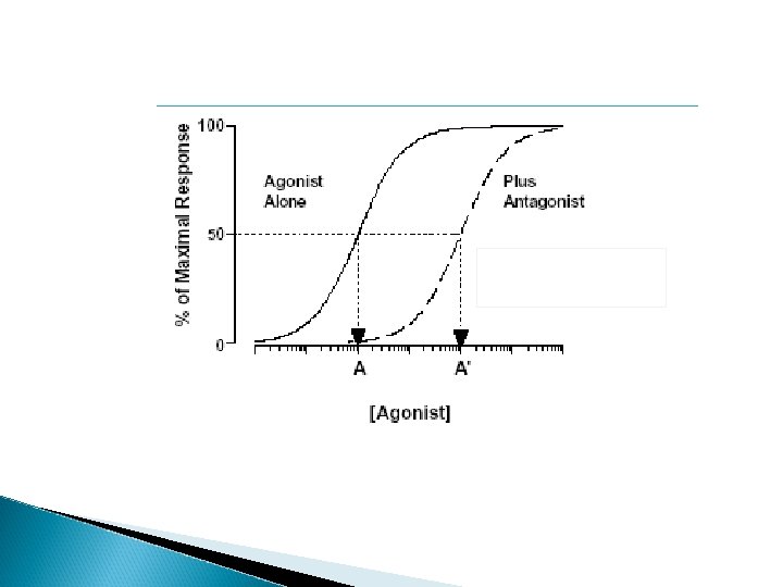
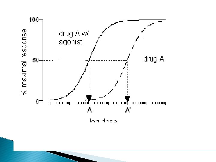
Drug Interactions antagonist - one drug diminishes the effect of another ◦ Shifts the DRC to the right agonist – one drug is additive to the effect of another
Toxicity due to drugs expected results – due to the principal actions of the drugs less expected – no drug is completely selective
Drug Tolerance definition? types of tolerance ◦ metabolic tolerance – enzyme induction ◦ pharmacodynamic tolerance –
chemical see-saw drug brain response
The brain wants to rebalance the activity
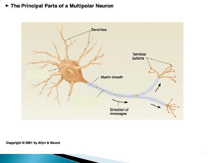
CHAPTER 3 The Neuron, Synaptic Transmission, Neurotransmitters and the CNS
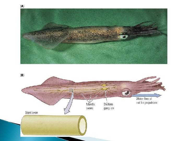
How do neurons communicate?
a b c
How do neurons communicate? Need to think about this question 2 ways
How do neurons communicate? 1. within neurons – 2. between neurons-
Neuron receiving info Information traveling down neuron
How do neurons communicate within neurons – electrically between neurons – chemically ◦ Synapse – space between neurons
Neurons can exist in one of 3 states the “resting” state the “active” state ◦ neuron is firing ◦ action potential the “refractory” state
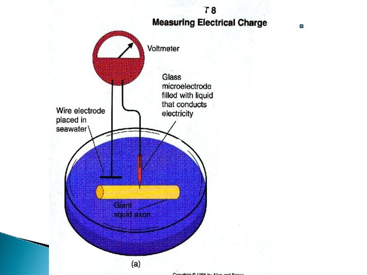
At rest: inside of the axon has a slightly negative charge relative to outside the axon ◦ called the membrane potential ◦ usually around -70 m. V
At rest: inside of the axon has a slightly negative charge relative to outside the axon ◦ called the membrane potential why?
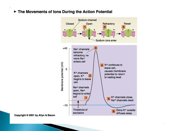
action potential or spike
Neuron stimulated (either electrically or by receiving a “message” see depolarization (change from negative inside neuron to more positive)
action potential or spike
Neuron stimulated (either electrically or by receiving a “message” see depolarization (change from negative inside neuron to more positive) ◦ “threshold” – if a great enough depolarization occurs, an action potential will occur
Neuron stimulated (either electrically or by receiving a “message” see depolarization (change from negative inside neuron to more positive) ◦ “threshold” – if a great enough depolarization occurs, an action potential will occur ◦ action potential – very quick – milliseconds Other terms – spike, firing, generating an AP
action potential or spike
Hyperpolarization return to negative this is the refractory or recovery period
action potential or spike
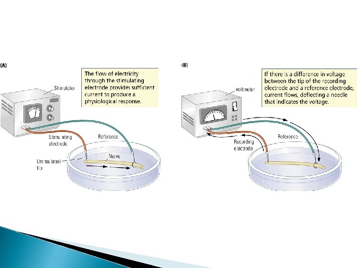
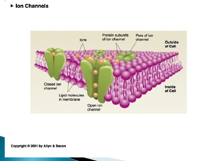
What causes these changes in electrical potential and the action potential? All axons and cells have a membrane thin lipid (fat) bilayer The membranes have channels (to allow ions in or out) Ions – molecules with a charge These channels can be open or shut
What causes these changes in electrical potential? Ions flowing across the membrane causes the changes in the potential Ions are molecules that contain a positive or negative charge anion – negative charge cation – positive charge
Some important ions for neuronal communication Na+ sodium ◦ HIGHER CONCENTRATION OUTSIDE THE AXON Cl- chloride K+ potassium ◦ HIGHER CONCENTRATION OUTSIDE AXON ◦ higher concentration inside the axon Aanions -large (-) molecules with a negative charge (stuck inside the axon)
Some forces that play a role in maintaining membrane potential concentration gradient – ◦ ions diffuse from higher concentration to lower concentration
example of concentration forces
What would each ion do if the ion channel opened based on the concentration gradient? Na+ K+ Cl-
What would each ion do if the ion channel opened based on the concentration gradient? Na+ K+ Cl- Na+ would enter axon
Concentration Gradient Na+ would enter axon K+ K+ would leave axon Cl-
Concentration Gradient Na+ would enter axon K+ K+ would leave axon Cl- would enter axon
Some forces that play a role in maintaining membrane potential concentration gradient – ◦ ions diffuse from higher concentration to lower concentration electrical gradient - ◦ opposite charges attract so ions are attracted to an environment that has a charge that is opposite of the charge they carry!
example of electrostatic forces
What would each ion do if the ion channel opened based on electrostatic forces ? Na+ K+ Cl-
Electrical Gradient Na+ K+ Cl- go in
Electrical Gradient Na+ go in K+ stay in Cl-
Electrical Gradient Na+ go in K+ stay in Cl- stay out
Concentration Gradient Electrical Gradient Na+ go in K+ go out stay in Cl- go in stay out
What drives the action potential? opening of Na+ channels and influx of Na+ ions
What happens if sodium channels are blocked? lidocaine, novocaine, cocaine TTX – tetrototoxin Sagitoxin◦ red tides
Concentration Gradient Na+ go in K+ go out Cl- go in Electrical Gradient as cell is depolarized (+ intracellular)
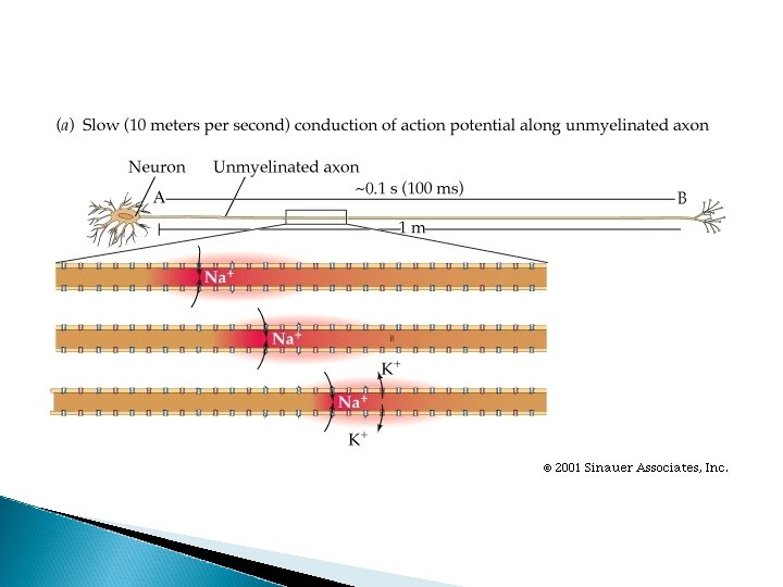
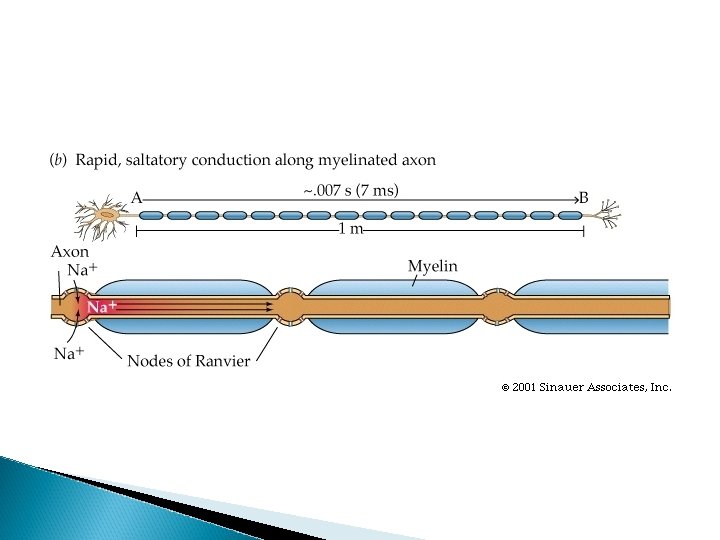
nodes of ranvier
nodes of ranvier
What about communication between neurons?
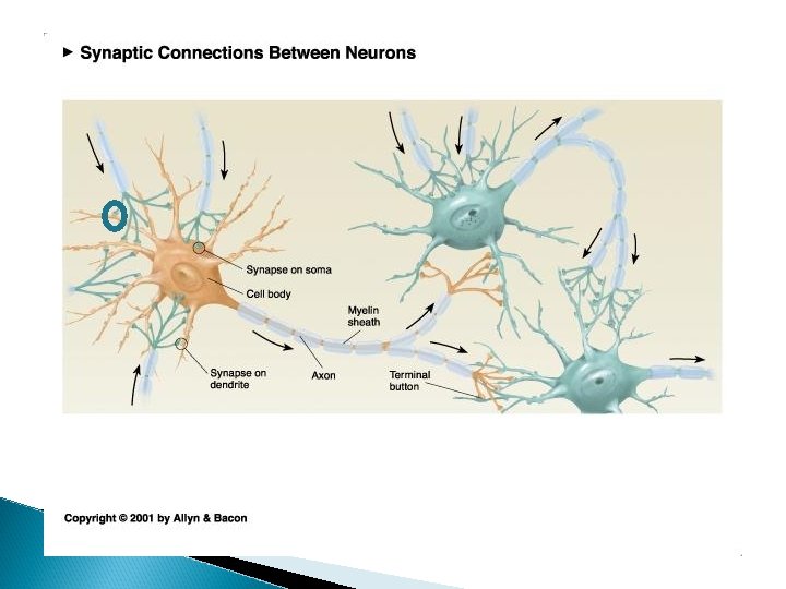
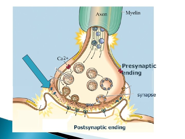
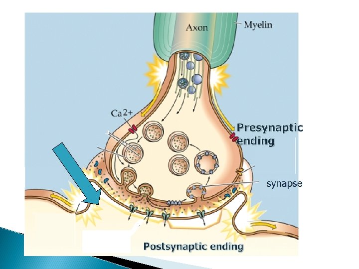
Some terms……. presynaptic ending – ◦ portion of the axon conveying information to the next neuron
Some terms……. presynaptic ending – ◦ the portion of the axon that is conveying information to the next neuron synapse or synaptic cleft ◦ the space between neurons where communication occurs
Some terms……. presynaptic ending – ◦ the portion of the axon that is conveying information to the next neuron synapse or synaptic cleft ◦ the space between neurons where communication occurs postsynaptic membrane ◦ the portion of the neuron (usually dendrite) that receives information
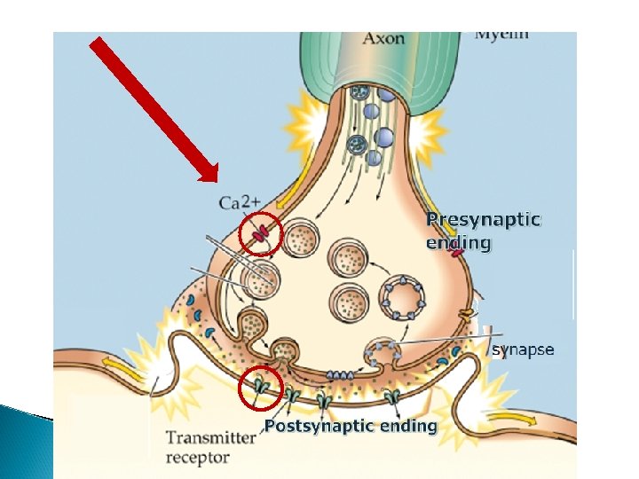
Some terms……. presynaptic ending – ◦ the portion of the axon that is conveying information to the next neuron synapse or synaptic cleft ◦ the space between neurons where communication occurs postsynaptic membrane ◦ the portion of the neuron (usually dendrite) that receives information pre and postsynaptic receptors ◦ proteins in both the presynaptic and postsynaptic ending that allow for information to be transferred
synaptic vesicles --small enclosed membranes that contain neurotransmitter found in presynaptic ending neurotransmitter – substance in vesicles that are released in synapse and convey info to the next neuron
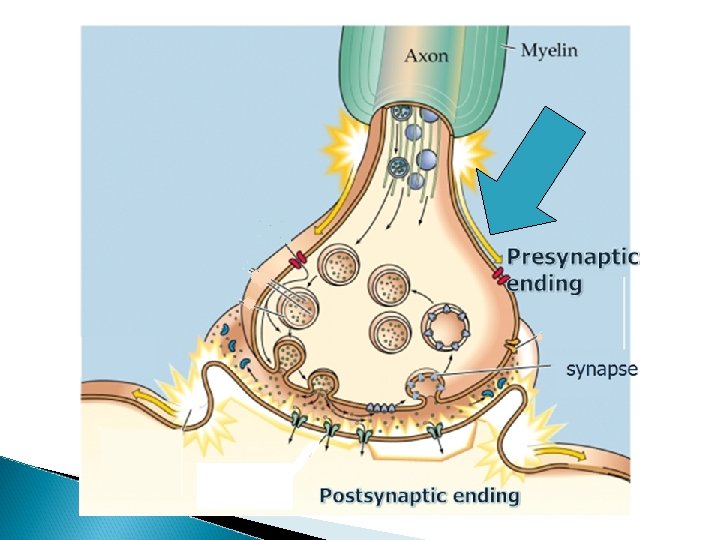
Presynaptic ending synapse Postsynaptic ending
What happens at level of synapse? AP reaches presynaptic ending. Ca+2 channels in presynaptic ending open and Ca+2 enters
Why are Ca+2 ions important? Ca+2 entry into the presynaptic ending critical for neurotransmitter release
Figure 3. 5 A. Photomicrograph of a synapse in action, taken with the electron microscope. B. Schematic of the process Julien: A Primer of Drug Action, Eleventh Edition Copyright © 2008 by Worth Publishers
postsynaptic receptors protein embedded in membrane mechanism for neurotransmitter to influence postsynaptic activity by binding to receptor
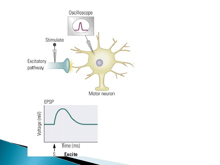
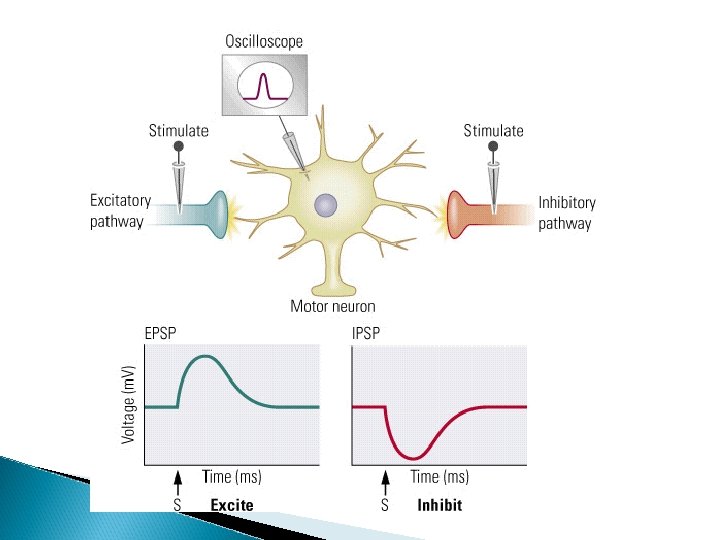
Summary NT binds to postsynaptic receptors and causes small local changes in electrical potential (depolarizations or hyperpolarizations)◦ Called graded potentials
What happens to convey info from one neuron to the next ◦ Graded Potentials- increase or decrease the likelihood of the neuron receiving info to generate an action potential
Graded potentials graded potentials that increase the likelihood of an action potential are called EPSPs (excitatory postsynaptic potentials)
Graded potentials graded potentials that increase the likelihood of an action potential are called EPSPs (excitatory postsynaptic potentials) graded potentials that decrease the likelihood of an action potential are called IPSPs (inhibitory postsynaptic potentials)
How does the neurotransmitter cause EPSPs and IPSPs? NT binding to postsynaptic receptors cause local ion channels to open
How does the neurotransmitter cause EPSPs and IPSPs? – chemically dependent ion channels in contrast with electrically dependent ion channels
How does the neurotransmitter cause EPSPs and IPSPs?
How does the neurotransmitter cause EPSPs and IPSPs? postsynaptic receptors open ion channels – ◦ ion channels in postsynaptic membrane (that we need to worry about) include Na+, Cl- and K+
Two kinds of Graded Potentials EPSPs – excitatory postsynaptic potentials - increase the likelihood of an AP - opening of
EPSPs – excitatory postsynaptic potentials opening of local Na+ channels IPSPs – inhibitory postsynaptic potentials
◦ IPSPs – inhibitory postsynaptic potentials • decreases the liklihood of an action potential opening of
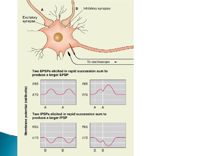
What happens to convey info from one neuron to the next? ◦ graded potentials are summed at axon hillock
Axon hillock
What happens to convey info from one neuron to the next? ◦ EPSPs and IPSPs are summed a axon hillock……. . AND
What happens to convey info from one neuron to the next Graded potentials are localized – has impact in limited region; AP travels down the axon
Neurotransmitters and Receptors General Principles Synthesis 1. Formation of transmitters 2. Precursors are the main ingredient. Brought to the neuron by the bloodstream. Taken up by cell body and/or terminal. Often come from substances in the diet. 3. Enzymes put the ingredients together.
Neurotransmitters and Receptors Transmitters Stored in Vesicles 1. Concentration 2. Protection
Neurotransmitters and Receptors Release = exocytosis ◦ Vesicles fuse with presynaptic membrane and release transmitters into the synapse. Binding = attachment of transmitter to receptor
Neurotransmitters and Receptors There are different varieties of receptors. ◦ Some respond fast ◦ Called Ionotropic ◦ Direct reaction to the transmitter
Neurotransmitters and Receptors Different varieties of receptors: ◦ Other types of receptors respond more slowly. ◦ Indirectly ◦ Called Metabotropic, or G protein-coupled ◦ Initiates a second signal (messenger) inside the neuron.
Neurotransmitters and Receptors Inactivation: Termination of Synaptic Transmission 1. Metabolism 2. Re‑uptake 3. Re-uptake by glial cell (glutamate only)
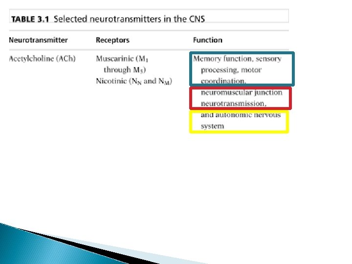
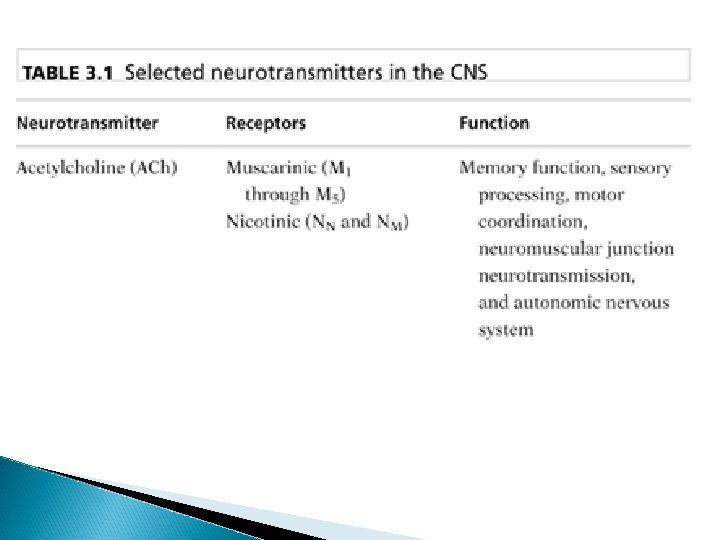
Neurotransmitters Acetylcholine Catecholamines ◦ norepinephrine ◦ dopamine Indoleamines ◦ serotonin amino acids ◦ gaba ◦ glutamate peptides ◦ opiates biogenic amines ◦ histamine
Neurotransmitters and Receptors Acetylcholine—first to be recognized, because of peripheral actions • Synthesis – Acetyl-Co. A (in mitochondria) + choline (from diet)
Published in 1939
Neurotransmitters and Receptors Inactivation: ◦ Acetylcholinesterase (ACh. E) ◦ After action in postsynaptic cleft, ACh. E degrades ACh to choline and acetate, which are taken back up into the neuron.
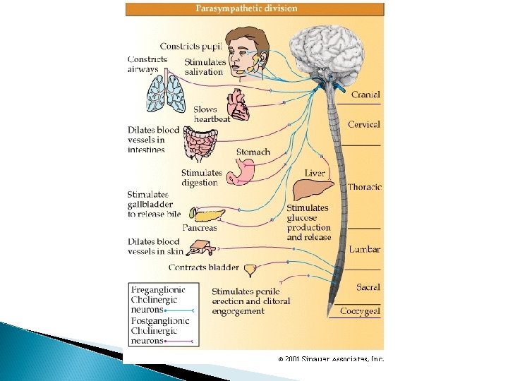
Neurotransmitters and Receptors Where is ACh produced? Septal nucleus and nucleus basalis ◦ Projects to forebrain. Midbrain ◦ Projects to reticular formation, pons, cerebellum, and cranial nerve nuclei. Ach NE Ach
Cholinergic system
Neurotransmitters and Receptors ◦ Nicotinic ◦ Muscarinic ACh. E Inhibitors ◦ Irreversible Often toxic Include pesticides and nerves gases ◦ Reversible Cognitive enhancers Treating Alzheimer’s