DRACUNCULUS MEDINENSIS GUINEA WORM Dracunculiasis documented as far
")


- Slides: 25
DRACUNCULUS MEDINENSIS (GUINEA WORM )
Dracunculiasis documented as far back as 1550 BC
Dracunculus medinensis causes dracunculiasis or Guinea worm disease Longest nematodes infecting humans Longest male: 2. 5 cmx 0. 4 mm. Longest female: 60 cm-1 meterx 1. 5 mm.
LARVAE. Size-500 -700 x 17 -20 µm Anterior end is rounded Tail is pointed and long Larvae is taken by cyclops
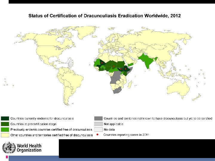
EPIDEMIOLOGY Areas where step wells or shallow ponds, lake water is used. Found on sub saharan africa, Egypt. Arabian peninsula, Iran, Afganistan, Pakstan, Western india and S. india INCIDENCE- about 10. million people affected
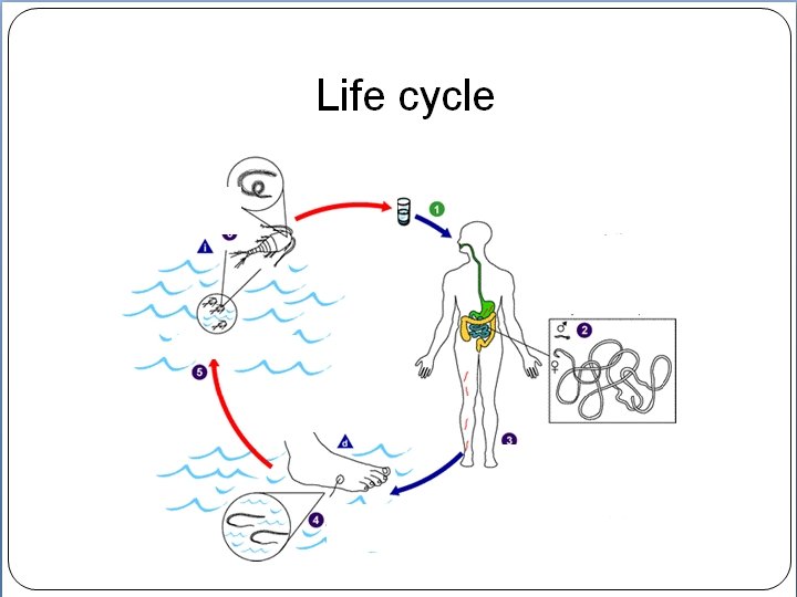
LIFE CYCLE. Definitive host- Man harbor the adult in the sub cutaneous tissue. Intermediate host- Water crustaciancyclops, larvae undergoes developmental changes Each cyclops can ingest 15— 20 larvae.
Larvae develop for two weeks inside copepods
Ingestion of contaminated water leads to human D. medinensis transmission
Pathogenisis. Incubation period-About a year. Symptoms do not occur until gravid female migrate to skin 1 -SYSTEMIC SYMPTOMS before formation of blister patient may show nausea, diarrhoea, urticarial rash. 2 -BLISTER FORMATION 1 - year after infection, a reddish papule appear where the head of the worm lies. Toxic fluid by the worm cause a blister, repture result in a shallow ulcer. the worm protrudes through a hole in the ulcer whenever the part com in contact with water
D. Medinensis migrate to lower limbs and induce blisters
LAB. DIAGNOSIS 1. Detection of adult worm during larval discharge. 2. Detection of larvae- the blister is flooded with water, induces the release of larvae. 3. X-ray- may show live or calcified worm in the connective tissue
TREATMENT. 1 -Removal of the worm by slowly being wound around a stick. 2 - Surgical removal. 3 -Drugs, niridazole, thiabendazole, metronidazole.
PREVENTION 1. PROVISION OF SAFE DRINKING WATER 2. PUBLIC AWARENCE.
Diagnosis made by observing worm head protruding from blister
No vaccines or medicine can treat or prevent Guinea Worm Disease
Only way to remove D. medinensis is by extracting entire worm inch by inch
Community education and awareness is a critical step towards prevention
Prevention through filtering water sources
Eradication efforts have reduced cases by 99%!
The end of dracunculiasis as we know it?
… Not so fast