Dr Vaishali Sanjay Mandhana Asso Prof Anatomy MBBS
 MBBS, DCP, MD.")
Dr. Vaishali Sanjay Mandhana Asso. Prof. (Anatomy) MBBS, DCP, MD.

Introduction Respiration-2 Phases ¡ Inspiration-Active Process ¡ l ¡ 1 sec. in quiet breathing Expiration-Mostly Passive (In quiet breathing) l For 3 sec. RR ¡ 16 -20 / min. -in adults. ¡ 25 -30 / min. – in children. ¡ 30 -40/ min. - in infants. ¡
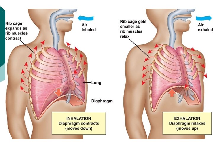

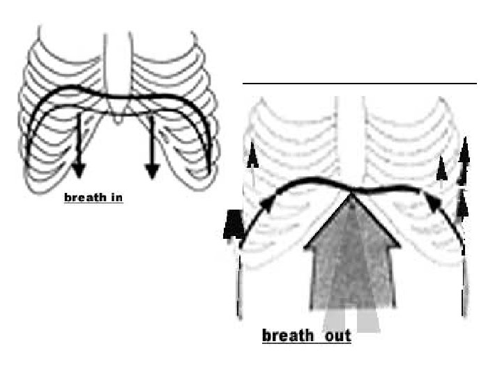
¡ For Inspiration we want to increase 1. AP diameter of the thoracic cage. ¡ 2. Transverse diameter of thoracic cage. ¡ The above 2 are increased by rib movements. ¡ 3. Vertical diameter of thoracic cage ¡ l It is increased by contraction of diaphragm.

Principles of Movements The Respiratory movements occure because of some peculiarities of ribs. ¡ Factors increasing AP Dia. ¡ 1. Ribs act as a lever. l Its fulcrum is just lateral to the tubercle. l Hence 2 unequal segments l Slight movements at posterior end causes magnified movement at anterior end. l

¡ ¡ 2. Anterior end at lower level than Posterior end. Hence during elevation of the rib anterior end also moves forward. Hence AP diameter is increased due to up and down movement of Ribs(more due to 2 -6 th rib) This is pump handle movement.

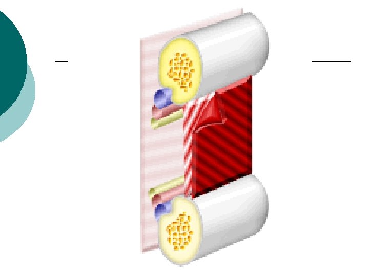
¡ Factors increasing Transverse Dia 3. Shaft lies at lower level than both the ends. l So during elevation of the Ribs, shaft moves outward , more prominently in 7 -10 th ribs (210 th ribs) l This is bucket handle movement. l

¡ 4. Thoracic cage as a cone. Hence during elevation of ribs, lower larger rib occupies the position of upper smaller rib. ¡ This increases the transverse diameter. ¡

Movements of the Ribs ¡ Pump handle movement. ¡ Bucket handle movement. ¡ Line of the axis of movement passes through costotransverse joint, costovertebral joint and contralateral costochondral joint.


Movements of the Ribs ¡ Pump handle movement ¡ Increases ¡ Occurs A. P. diameter primarily at 2 nd - 6 th rib. ¡ Movement occurs at costovertebral joint.
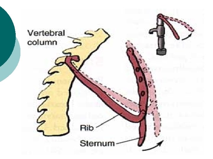

Pump Handle Movement

Movement of the Ribs ¡ Bucket handle movement ¡ Increases diameter. ¡ Occurs rib. transverse primarily in 7 th-10 th ¡ Movement occurs at costotransverse joint.


Bucket Handle Movement

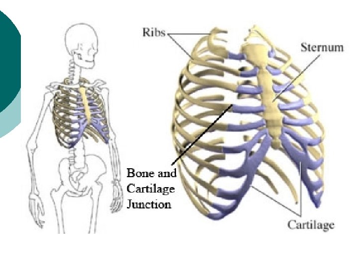
WHY STERNAL ANGLE IS PRODUCED? ¡ ¡ ¡ Movement of the sternum Direction of 2 nd to 6 th ribs & cartilage is downward n medially. Their elevation moves the body of sternum forward. 7 th to 10 th ribs directed downward n forward but their cartilages are directed upward n medially. Their elevation moves the body of sternum backward

Hence body of sternum shows Forward movement—with 2 nd to 6 th ribs and…… Backward movement—with 7 th to 10 th cartilages. ¡ This forward and backward movement produces sternal angle.

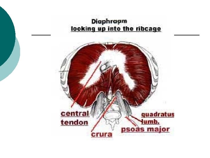
Muscles of Inspiration ¡ Diaphragm l l ¡ Intercostal Muscles l l l ¡ Elevates 1 st rib (Deep Insp. ) Pectoral Mus. & Serratus Anterior Mus. l ¡ Straightening the thoracic part of v. column. Scalani & Sternocledomastoid Mus. l ¡ 1 cm. circumference – 200 ml. air. Sucked Lower mus. -in quiet inspiration-EMG. Upper mus. -in deep inspiration-EMG. Erector spinae l ¡ 2/3 in quiet breathing. Only Muscle in neonats and infants Act in forced inspiration. Quadratus Lumborum. l Fixes the last rib.


External and Internal Intercostal Muscles

Muscles of Expiration ¡ ¡ ¡ Elastic Recoil of the lung. Muscles of Anterior Abdominal Wall. Latissmus Dorsi mus.

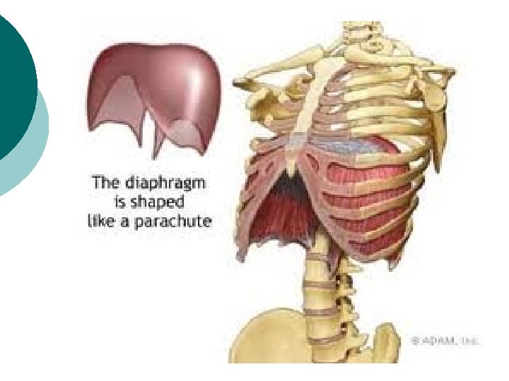
Role of Diaphragm ¡ Contraction of Diaphragm –piston movement. l l ¡ Range of Movement l l l ¡ Lower Ribs fixed and Vault Desends. Ant. Abd. Wall bulges. Central Tendon becomes fixed. Further contraction elevates lower ribs 1. 5 cms. in quiet breathing. . Surface area of Diaphragm = 270 sq. cm 1. 5 cm descent = increases 400 cc volume 4 th to 5 th costal cartilage after forced expiration. 6 -10 cm descend in forceful inspiration i. e. Goes upto 11 th to 12 th vertebra. Position of Diaphragm l l Highest in supine position. Then in erect position. Lowest in sitting position. Higher on that side on which the body lies.

Summery ¡ Quiet Inspiration l l l ¡ Deep Inspiration l l l ¡ AP Diameter-2 -6 ribs. (1 st rib fixed) Transverse Diameter-7 -10 ribs. Vertical Diameter-Diaphragm Above movements Increased. Scalani & Sternocledomastoid(for 1 st rib) Erector spinae(reduces concavity of spine Forced Inspiration l l l Above movements exagerated Trapezius Levator scapulae Rhomboidus elevate & fix the scapulae. Seratus ant. & pectoralis minor act on ribs Erector spinae action exagerated.

Summery… ¡ Quiet Expiration l ¡ Elastic Recoil Deep/Forceful Expiration l l Abdominal Muscles Latissmus Dorsi

Applied Anatomy Dyspnoea-Difficulty in breathing. ¡ Respiratory Diseases ¡ l l Restrictive-Asthma, Status Asthmaticus Obstructive-COPD. Phrenic Nerve injury¡ Paralysis of Hemi diaphragm. ¡ Plural effusiondyspnoea, collapse of lungs. ¡ Pnumothorax-open/tension ¡
 Funnel chest Deformity. (Pectus Excavetum)")
Applied Anatomy ¡ ¡ Pigeon Chest Deformity. (Pectus Carinatum) Funnel chest Deformity. (Pectus Excavetum) Barrel Chest Deformity. Flail Chest Deformity-Several Rib # paradoxical movement of chest wall.

Pigeon Chest Deformity Funnel chest Deformity

Barrel Chest Deformity


Joints of Thoracic Cage

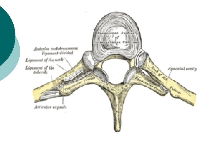
Joints of Thoracic cage ¡ Manubrio-sternal Joint. l l ¡ Xiphi-sternal Joint. l ¡ Secondary cartilagenous jt. Angular and AP movements. 2 nd -6 th Ribs moves it forward. 7 th – 10 th Ribs move it backward. Primary cartilagenous Jt. (no movements. ) Costovertebral Joint. l l Rib with own vertebra & higher vertebra. Plain synovial joint. Lig. -Intra articular ¡ Capsular ¡ Triradiate(forms hypo chordal bow) Movement-Pump handle movement. (2 nd-6 th Rib).

Joints of Thoracic cage… ¡ Costotransverse Joint. l l l ¡ Costochondral Joint. l ¡ Primary cartilagenous-No movement. Chondro-sternal Joint. l l ¡ Synovial jt. Lig. -Superior costotransverse -Inferior costotransverse -Lateral costotransverse Movement-Bucket handle(7 th-10 th rib) 1 st-Primary cartilagenous 2 nd-7 th Synovial jt. Inter-Chondral Joint. l Synovial jt.

Costochondral Joint & Chondro-sternal Joint.

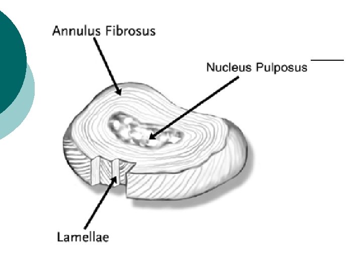
Joints of Thoracic cage… ¡ Inter-Vertebral Joint. l l ¡ Intervertebral Disc l l ¡ ¡ ¡ 2 joints with articular facets-Zygapophysial jt. Body of vertebras-Secondary cartilegenous jt. Annulus fibrosus Nucleus Pulposus(Ramnant of notocord) Ligaments. . -Ant. Longitudinal lig. -Post. Longitudinal lig. -Intertransverse lig. -Interspinous lig. -Supraspinous lig. -Ligamenta flava Movements-Flexion –Cervical and Lumbar V. -Extension –Cervical and Lumbar V. -Lat. Flexion-Rotation Thoracic V.

Joints of thorax ¡ Sternoclavicular joint l ¡ Saddle typesynovial, compound, complex jt. Lig. -capsule l l l Articular disc. Interclavicular lig. Costoclavicular lig

Inter-Vertebral Joint.

Inter-Vertebral Joint.

- Slides: 48