
Dr Tsukasa Shibue MIT Tsukasa Shibue MD Ph
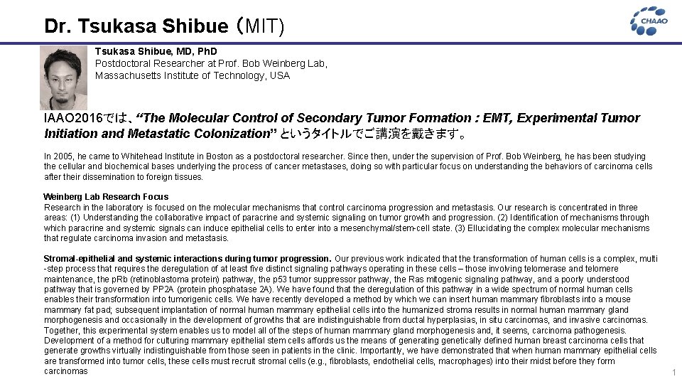
Dr. Tsukasa Shibue (MIT) Tsukasa Shibue, MD, Ph. D Postdoctoral Researcher at Prof. Bob Weinberg Lab, Massachusetts Institute of Technology, USA IAAO 2016では、“The Molecular Control of Secondary Tumor Formation : EMT, Experimental Tumor Initiation and Metastatic Colonization” というタイトルでご講演を戴きます。 In 2005, he came to Whitehead Institute in Boston as a postdoctoral researcher. Since then, under the supervision of Prof. Bob Weinberg, he has been studying the cellular and biochemical bases underlying the process of cancer metastases, doing so with particular focus on understanding the behaviors of carcinoma cells after their dissemination to foreign tissues. Weinberg Lab Research Focus Research in the laboratory is focused on the molecular mechanisms that control carcinoma progression and metastasis. Our research is concentrated in three areas: (1) Understanding the collaborative impact of paracrine and systemic signaling on tumor growth and progression. (2) Identification of mechanisms through which paracrine and systemic signals can induce epithelial cells to enter into a mesenchymal/stem-cell state. (3) Ellucidating the complex molecular mechanisms that regulate carcinoma invasion and metastasis. Stromal-epithelial and systemic interactions during tumor progression. Our previous work indicated that the transformation of human cells is a complex, multi -step process that requires the deregulation of at least five distinct signaling pathways operating in these cells – those involving telomerase and telomere maintenance, the p. Rb (retinoblastoma protein) pathway, the p 53 tumor suppressor pathway, the Ras mitogenic signaling pathway, and a poorly understood pathway that is governed by PP 2 A (protein phosphatase 2 A). We have found that the deregulation of this pathway in a wide spectrum of normal human cells enables their transformation into tumorigenic cells. We have recently developed a method by which we can insert human mammary fibroblasts into a mouse mammary fat pad; subsequent implantation of normal human mammary epithelial cells into the humanized stroma results in normal human mammary gland morphogenesis and occasionally in the development of growths that are indistinguishable from ductal hyperplasias, in situ carcinomas, and invasive carcinomas. Together, this experimental system enables us to model all of the steps of human mammary gland morphogenesis and, it seems, carcinoma pathogenesis. Development of a method for culturing mammary epithelial stem cells affords us the means of generating genetically defined human breast carcinoma cells that generate growths virtually indistinguishable from those seen in patients in the clinic. Importantly, we have demonstrated that when human mammary epithelial cells are transformed into tumor cells, these cells must recruit stromal cells (e. g. , fibroblasts, endothelial cells, macrophages) into their midst before they form carcinomas 1

Dr. Tsukasa Shibue Our research has revealed that several mesenchymal cell types that form the tumor-associated stroma are important in supporting carcinoma cell growth within the tumor. Thus, mesenchymal stem cells and myofibroblasts in the tumor-associated stroma can release paracrine signals that impart invasive and metastatic powers to nearby carcinoma cells. At the same time, primary carcinomas release endocrinal signals that impinge on the bone marrow and spleen and induce the formation of several types of inflammatory cells that may then be recruited by tumors , via the general hematogenous circulation, to help the tumors to rapidly assemble a highly functional, tumor-supporting stroma. We are currently investigating the mechanism(s) by which this recruitment occurs. In addition, we have determined that the stromal fibroblasts that are present in human mammary carcinomas are biologically abnormal and are in an activated state reminiscent of that of myofibroblasts. Together, these interactions indicate that cancer is a systemic disease long before systemic dissemination of cancer cells has begun. Paracrine and systemic signals that induce epithelial cells to enter into the mesenchymal/stem-cell state. The epithelial-mesenchymal transition (EMT) represents a cell-biological program that enables both normal and neoplastic epithelial cells to acquire mesenchymal cell attributes, such as motility, invasiveness, and a resistance to apoptosis. At the same time, our work has revealed that epithelial cells that are forced through an EMT acquire many of the attributes of normal and neoplastic stem cells. This holds implications for the pathogenesis of metastasis, since carcinoma cells that have acquired mesenchymal attributes that enable them to physically disseminate also acquire the self-renewal trait that allows them to seed macroscopic metastases. To date our research has revealed a series of paracrine signals that impinge on epithelial cells and induces them to enter into an EMT. Long term exposure to these signals allow cells to remain in the mesenchymal/stem-cell state in a self-sustaining, metastable fashion. Regulation of carcinoma invasion and metastasis. The intracellular regulatory circuitry that programs the epithelial-mesenchymal transition involves the actions of a series of transcription factors that induce expression of one another. Each of these transcription factors can act in a pleiotropic fashion to choreograph an EMT, but seems to collaborate with others in various epithelial cell types to organize this program and ensure that a large coterie of downstream effectors can collaborate to push cells into an epithelial/stem-cell state. Having learned many of the requirements for creating primary human tumors, we would like to determine the nature of some of the genetic elements that lead to the formation of invasive and ultimately metastatic tumor cells. Using expression arrays, we have identified two transcription factors, Twist and Mesenchyme Forkhead (also known as FOXC 2) , which appear to play key roles in programming many of the phenotypes of invasive cells. Embryology reveals the identity of a third factor, termed Goosecoid, which appears capable of programming the epithelial-mesenchymal transition both during early embryogenesis and during the invasive stages of certain carcinomas. A fourth factor, termed Slug, may contribute to the invasive phenotype of yet other tumors. Acting alone or in combination, these factors may be able to program many of the phenotypes associated with invasive and metastatic cells. In addition, we are examining the last stage of the invasionmetastasis cascade, in order to determine how disseminated cancer cells are able to adapt to a novel tissue environment and colonize this environment, yielding a macroscopic metastasis. 2
Dr. Tsukasa Shibue Reference Publications Mani SA, Guo W, Liao MJ, Eaton EN, Ayyanan A, Zhou AY, Brooks M, Reinhard F, Zhang CC, Shipitsin M, Campbell LL, Polyak K, Brisken C, Yang J, Weinberg RA. The epithelial-mesenchymal transition generates cells with properties of stem cells. Cell. 2008; 133(4): 704 -15. Guo W, Keckesova Z, Donaher JL, Shibue T, Tischler V, Reinhardt F, Itzkovitz S, Noske A, Zurrer-Hardi U, Bell G, Tam WL, Mani SA, van Oudenaarden A, Weinberg RA. Slug and Sox 9 cooperatively determine the mammary stem cell state. Cell. 2012; 148(5): 1015 -28. Shibue T, Brooks MW, Inan MF, Reinhardt F, Weinberg RA. The outgrowth of micrometastases is enabled by the formationn of filopodium-like protrusions. Cancer Discov. 2012; 2(8): 706 -21. Shibue T, Brooks MW, Weinberg RA. An integrin-linked machinery of cytoskeletal regulation that enables experimental tumor initiation and metastatic colonization. Cancer Cell. 2013; 24(2): 481 -98. Ye X, Tam WL, Shibue T, Kaygusuz Y, Reinhardt F, Ng Eaton E, Weinberg RA. Distinct EMT programs control normal mammary stem cells and tumour-initiating cells. Nature. 2015; 525(7568): 256 -60. 3
- Slides: 4