DR SHWETA SHENDEY VASCULAR INTERVENTIONS IN GIT ROLE


 Thromboembolic occlusion is the most common")


- Slides: 61
DR. SHWETA SHENDEY VASCULAR INTERVENTIONS IN GIT
ROLE OF VASCULAR INTERVENTIONS � T/T GASTROINTESTINAL HAEMORRHAGE � T/T PORTAL HYPERTENSION � T/T LIVER TUMOURS
� GI HAEMORRHAGE � Upper GIH � Peptic ulcers, � erosive gastritis, � Varices � esophagogastric mucosal tear � Less common causes include ca esophagus, stomach/deudenum/haemobilia, av malformations splenic arterial aneurysm
LOWER GI HAEMORRHAGES � Lower GI haemorrhages � Enteric ulcers � Diverticulosis � TB � Neoplasms � colitis � angiodysplasia
IMAGING MODALITIES � Endoscopy is the initial diagnostic procedure in any GI bleeding � Provides prognostic information � Access to therapeutic interventions
ANGIOGRAPHY � Significant role in diagnosis and management � Localize the actual site of active bleed � Help in management of transcatheter embolization of bleeding vessel � Role is diminished d/t wider availability of endoscopy � Helpful in pts with recurrent gih where barium studies or endoscopy has failed
TECHNICAL CONSIDERATION OF ANGIOGRAPHY � Diagnostic if bleeding exceeds 0. 5 ml/min(blood loss exceeds 4 units within 24 hrs) � Often not useful in mesentric studies because of bowel gas and peristalsis. � Renal function should be normal otherwise alternative contrast media like CO 2 can be used as contrast
PROCEDURE � Initially flush aortogram is done f/b selective and superselective arteriography � Tip of pigtail catheter placed above celiac axis � 40 ml of contrast is injected with pressure injector at the rate of 18 -22 ml/sec � Depending on initial findings further selective and superselective studies done using various visceral angiographic catheters � Flush aortogram is useful in cases if suspected aortoenteric fistula
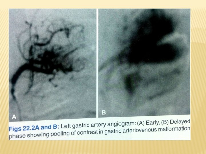
In mesentric studies flow rate of contrast � Coeliac -10 ml/sec � SMA-8 ml/sec � IMA 3 ml/sec -Imaging rapid during arterial phase (1 -2 frames/sec) -Reduced to 1 frame/2 -5 sec for capillary and venous phase -carried out for atleast 30 secs to look for extravasation, venous occlusion, vascular malformations -suspected bleeding vessel should be injected first -selective arteriograhy may reveal bleeding as low as 0. 5 -1 ml/min In apprx 70% of pts with gastric mucosal haemorrhage bleeding occurs frm left gastric artery hence essential that LGA be well opacified during selective angiography �
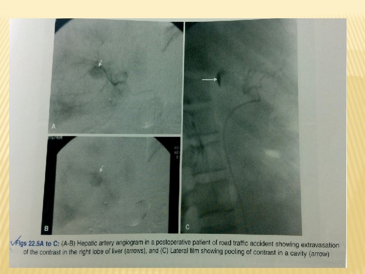
ANGIOGRAPHIC FINDINGS IN GIH � Extravasation of contrast : seen as puddling or staining that persists beyond capillary and venous phases � Delayed films may show opacified intestinal folds d/t luminal extravasation of contrast material � If lumen of GIT is filled with clotted blood “pseudovein ” appearance is seen as(as extravasated contrast flows through the clot to the most dependant part but unlike venous phase it persists much longer)
RADIONUCLIDE STUDIES � Minimal risk and invasiveness to pt � Used in pts where bleeding has stopped or is intermittent in nature � Not useful in massively bleeding patients � Commonly used are � Tc 99 m sulphur colloids: cleared frm blood pool by RE system
RADIONUCLIDE STUDIES � � � � Tc 99 m sulphur colloids: cleared frm blood pool by RE system Half life 2. 5 -3 min As tracer extravasate in bowel hence only active bleeding can be diagnosed Can detect blood loss of as low as 0. 1 ml/min Drawback is bleeding into hepatic or splenic flexure of colon and ugih will be obscured Tc 99 m labelled RBCs: RBCs in circulation extravasate during bleeding and can be detected Images can be obtained upto 2 hrs after initial injection hence useful in intermittent bleeding Tc 99 m pertechnate scanning: in cases of bleeding from meckels diverticulum , the tracer accumulates in ectopic gastric mucosa
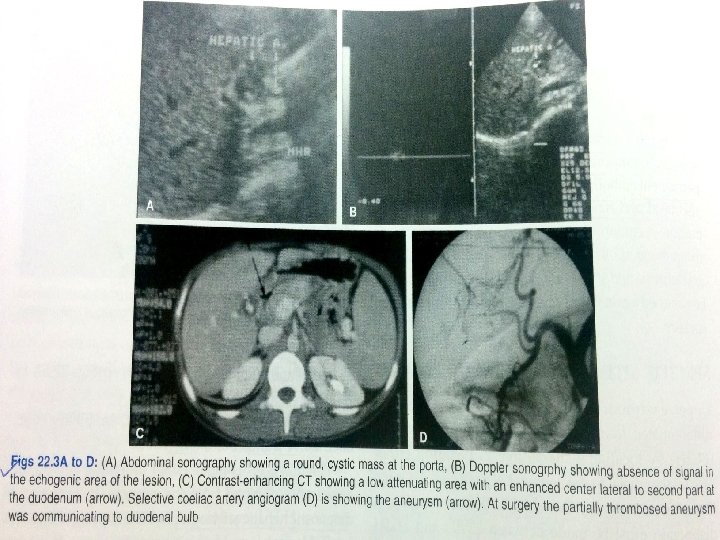
CT � Acute localization is possible on arterial phase of CT � Arterial phase ct demonstrates extravasated contrast in the bowel lumen greater than 90 HU. � In some cases it may detect parenchymal abnormalities such as hepatic artery aneurysms, arterioportal fistulae, tumours etc
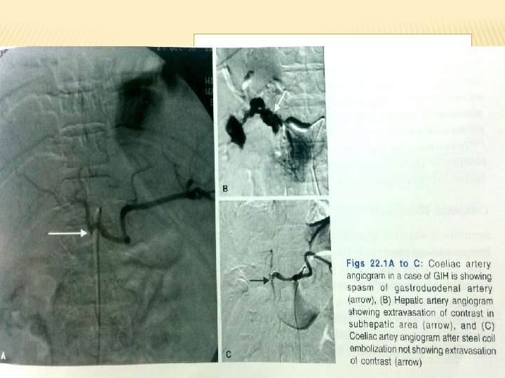
CONTROL OF GI HAEMORRHAGES � Endoscopic coagulation and mucosal injection of sclerosing agent can control bleeding in ulcers and variceal bleed � TRANSCATHETER EMBOLIZATION: � For lesion of stomach colon and small bowel Especially useful in t/t of bleeding pancreatic and hepatic artery pseudoaneurysm Advantages are Control is immediate Risk of visceral ischemia is minimum if embolization is superselective and collaterals are preserved. The most commonly used materials include gelatin sponge(a temporary occluding agent) or steel coils for permanent embolization. Polyvinyl alcohol particles have shown good results � � �
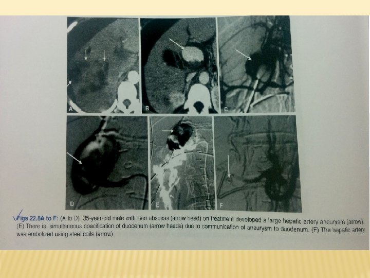
HAEMOBILIA � Presentation is billiary colic, obstructive jaundice and upper GI haemorrhage. � Diagnosis is made on endoscopy(by appearnce of blood at ampulla of vater � By diagnostic hepatic arteriography � Can be treated by angioembolization or surgical ligation of hepatic artery
TUMOURS � They bleed spontaneously but blood loss is characteristically chronic and intermittent. � On angiography some tumors like leiomyoma and leiomyosarcoma, adenoma and kaposi’s sarcoma are hypervascular and shows intense capillary stain
PANCREATITIS AND UGIH � Pseudocyst and pseudoaneurysm are the complications d/t extension of pancreatic inflammation to nearby splenic artery � Other complications are haemorrhage within cyst , gi haemorrhage d/t fistula formation, rupture of cyst into peritoneum, haemorrhage into deudenum via pancreatic duct. � Superior mesentric angiography is the investigation of choice for diagnosis and selective arterial embolization may control haemorrhage.
LOWER GI HAEMORRHAGE � COLONIC DIVERTICULA � most common in left colon � Bleeding is abrupt massive and often painless. � Bleeding demonstrated by direct endoscopic visualization or by filling the bleeding diverticulum with contrast medium at the time of angiography.
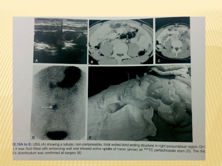
MECKEL’S DIVERTICULUM Congenital anomaly � Cause of acute hemorrhage in children and young adults � Acid secretion from gastric mucosa results in peptic ulceration most often in ileal mucosa and lead to gi bleeding � Tc 99 m pertechnate radionuclide scanning detect bleeding diverticula � � Angiography shows vessel supplying area of abnormality and increased contrast medium accumulation in distal ileum
ANGIODYSPLASIA � It is a mucosal telangiectasia often multiple, involve cecum and ascending colon � Represent dilated preexisting vascular structures � On angiography it is seen as vascular tuft appearing in arterial phase -> early opacification of draining veins -> delayed emptying of a dilated tortuous intramural vein.
TUMOURS � They bleed spontaneously but blood loss is characteristically chronic and intermittent. � On angiography some tumors like leiomyoma and leiomyosarcoma, adenoma and kaposi’s sarcoma are hypervascular and shows intense capillary stain
INTERVENTION RADIOLOGY IN PORTAL HYPERTENSION
PORTAL HYPERTENSION � � 1. 2. 3. 4. 5. 6. � 1. 2. Primary goal in treating portal hypertension is reducing the portal venous pressure and thus treating its complications like variceal, refractory ascites, hydrothorax and hypersplenism INTERVENTIONS THAT REDUCE PORTAL PRESSURE Transjugular intrahepatic portosystemic shunts. (TIPS) Revision of occluded surgical and radiological portosystemic shunts Embolization of arterioportal fistulas Recanalization of occluded portal vein Partial splenic embolization Recanalization of hepatic venous outflow INTERVENTIONS TO PALLIATE SYMPTOMS OF PORTAL HYPERTENSION Percutaneous transhepatic variceal embolization Balloon occluded retrograde transvenous obliteration of gastric varices
TIPS � Refers to creation of portosystyemic shunt by transjugular insertion of expandable metal stent between hepatic and portal vein under image guidance � Purpose is to decompress portal venous system and prevent its complication including rebleeding from varices � Normally procedure is performrd under conscious sedation , GA is used in case of haemodynamically unstable patient.
INDICATIONS OF TIPS � Acute variceal bleeding unresponsive to medical treatment � Sec. prevention of variceal bleeding � Refractory ascites � Portal hypertensive gastropathy � Refractory hepatic hydrothorax � Budd chiari syndrome � Veno occlusive disease
� ADVANTAGE: does not need general anaesthesia, postinterventional intensive care, parentral nutrition and immobilization. � Control rebleeding in patients with poor liver function. � DISADVANTAGE: � Like surgery risk of hepatic encephalopathy, � liver failure and � procedural complications. �
CONTRAINDICATIONS OF TIPS ABSOLUTE: � Primary prevention of variceal bleed � Congestive heart failure � Multiple hepatic cyst � Uncontrolled systemic infections and sepsis � Unrelieved billiary obstruction � Severe pulmonary hypertension RELATIVE: � Hepatoma especially with central obsrtuction of all veins � Portal vein thrombosis � Severe coagulopathy � Thrombocytopenia of less than 20000/cm 3 � Moderate pulmonary hypertension
STEPS OF TIPS 1. � � � 2. � � Cannulation of hepatic vein through transjugular approach which is performed via right transjugular route as it provides straight path to hepatic vein opening It is important to decide which hepatic vein to use which is done by using USG or fluroscopy Right hepatic vein is dorsal and cephalad to anterior superior branch of right portal vein After cannulation of hepatic vein, hepatic venography is performed to ensure that vein is large enough to provide outflow tract of sufficient diameter of 8 -10 mm Second step is passage of long needle from hepatic vein through liver parenchyma into the portal vein(the right pv branch typically runs at the level of 11 th rib about 0. 5 -1. 5 vertebral width from the lateral border of spine Subsequently portal vein pressure is measured and base line pressure gradient is measured A marking pigtail catheter is advanced into main portal vein for venography for determining suitability of portal vein puncture. 3. Third step involves balloon angioplasty with dilatation of parenchymal tract created by needle 4. Last step is stent deployment to ensure patency of tract The ends of stent should project for short distance into hepatic and portal vein to prevent displacement �
VIATORR PTFE covered stents are preferred nowadays � Stents of variable sizes are available with chances of shunt stenosis decreasing and encephalopathy increasing with larger size of shunt. � Successful outcome Is determined by decreasing portosystemic gradient to less than 12 mm of hg and resolution of complication of portal hypertension � At the end of procedure portal venogram is performed to document shunt patency and nonvisualization of gastro esophageal varices. �
COMPLICATIONS OF TIPS � � Thrombosis , occlusion, stenosis, transcapsular puncture, intraperitoneal bleed, hepatic infarction, fistulae, hemobilia, sepsis, haemolysis, encephalo pathy. Recurrence of symptoms should be managed by shunt venography and interventions. use of PTFE(polytetrafluoroethylene) film minimizes transmural permeation of bile and mucin thus prevent hyperplastic tissue ingrowth Another complication is hepatic encephalopathy for which most patients respond to medical conservative therapy or constraining stents and constraining grafts have been used to reduce shunt diameter
REVISION OF OCCLUDED SURGICAL STENTS: � � Shunt stenosis is most common cause of rebleeding Percutaneous transluminal angioplasty is alternative method of therapy for shunt stenosis ARTERIPORTAL FISTULAS: � Radiological intervention is therapy of first choice � Between portal vein and systemic arteris occur intraabdominally � Are not readily accesible � Presents with f/o portal hypertension , cardiac failure, intestinal ischemia � May be congenital; may be multiple sec to hereditary telangiectatic diseases, av malformation and aneurysms � May be acquired; sec to blunt or penetrating abdominal trauma or iatrogenic � Hepatic artery is the most common artery involved followed by splenic artery � Arteriography confirms by definitve demonstration of fistula feeding vessel, location and size of shunt, portal vein flow and collaterals � Selective and superselective catheterization is done and various embolizing materials like coils and wires , isobutyl-2 -acrylate gelfoam and balloons have been used to occlude fistula
� � � � RECANALIZATION OF OCCLUDED PORTAL VEIN AND ITS BRANCHES: 5 -10% of portal hypertension cases are d/t extrahepatic portal vein obstruction and ¼ th of them are d/t neoplasms Portal venous stents may be placed in all these carcinomas to resolve blockage of portal vein and alleviate the related symptoms BUDD CHIARI SYNDROME: Percutaneous radiological management is becoming procedure of choice Thrombolysis is the first line therapy and locoregional thrombolysis by selective catheterization as well as mechanical and hydrodynamic techniques are used for removal of obstructing thrombus. If thrombolysis is unsuccessful then TIPS may be done
PARTIAL SPLENIC VEIN EMBOLOZATION � � � � Hypersplenism is a common complication of portal hypertension and can be treated with splenectomy, splenic artery ligation and splenic artery embolization. In partial splenic embolization supply is occluded more peripherally in the splenic parenchyma Splenic artery is selectively cannulated through transfemoral route using 4 F catheter Tip should lie distal to last pancreatic artery. Gelfoam is then injected to induce splenic infarction and induce fibrosis in 45 -70% splenic parenchyma Embolizing agent such as gelfoam or PVA particle are soaked in antibiotic solution penicillin and gentamycin Procedure treats hypersplenism in convenient and noninvasive way and also reduces portal flow resulting in decreased portal pressure.
PERCUTANEOUS TRANSHEPATIC VARICEAL EMBOLIZATION � � Used to control variceal bleeding after failure of sclerotherapy or in cases of recurrent bleeding after sclerotherapy. Portal vein thrombosis is the major complication of this procedure ANGIOGRAPHIC VASOPRESSIN INFUSION: � � Intravenous systemic vasopressin has been used to control bleeding and vasopressin is infused through SMA at the rate of 0. 1 mg /min for 24 hrs. This causes reduced mesentric blood flow with drop in portal pressure resulting in decompression of varices and subsequent thrombosis of bleeding site.
INTERVENTIONAL TREATMENT OF LIVER TUMOURS � 1. 2. 3. 4. 5. 6. 7. Interventional techniques have been developed employing angiographic and percutaneous route, these are: Chemical ablation with ethanol(PEI) or acetic acid(PAI) Radiofrequency ablation Transcatheter arterial chemoembolization Transcatheter arterial radionuclide therapy Right portal vein embolization Hepatic vein stenting
TRETMENT OF LIVER TUMOURS HCC Used in patients with extensive lesions or with concurrent cirrhosis or with intrahepatic mets who cant undergo surgical resection of HCC CHEMICAL ABLATION: � Direct percutaneous injection of ethanol or acetic acid into a liver tumour under USG or CT guidance � Percutaneous acetic acid/ethanol result in coagulative necrosis and fibrosis of tumour. � 95% ethanol is most commonly used and highly effective method this technique is particularly useful for lesion less than 3 cm in diameter � It is contraindicated in surafce lesions, tumours with ascites or coagulopathy
� � � � Alcohol is injected in multiple sessions under radiological guidance using long needle Alcohol injection is started from deepest to superficial portion of tumour avoiding direct injection into hepatic vein, Multiple sessions are required (ie once or twice a week) In PAI 50% of acetic acid is used, amount in ml required to achieve complete response is equal to three times the diameter of lesion in cms It is done on outdoor basis under aseptic condition and local anesthesia At a time 1 -8 ml at different site is injected by one or more injections Perfused area usually spreads within a radius of 2 -3 cms and is seen as a patch of hyperechogenecity on USG
RADIOFREQUENCY ABLATION � New technique for small HCC and hepatic mets � It induced necrosis of tumour by deposition of thermal energy around the tip of electrode inserted in the tumour � RFA is ideally suited for smaller lesions less than 3 cms as well as for multiple lesions. even tumours upto 5 cms can be ablated (chances of complete ablation diminish with increase in size of tumour)
TECHNIQUE OF RFA Radiofrequency probe is inserted into the lesion under USG or CT guidance � AC current is passed through the probe with energies ranging from 60100 watts for a period of 6 -12 mins, rapid change in polarity of electric current results in fast oscillation of intracellular molecules which causes friction and heat generation � Local temperature of more than 60 degrees is maintained for more than 5 minutes. � Complications: � less common � Include severe pain after procedure, haemorrhage, peritonitis, cholecystitis, colitis, vascular injury and thrombosis. � Folllow up: to asses degree of necrosis and completeness of ablation. -f/u CT shows low density nonenhancing area which corrospend to necrosis -tumour response rate to RFA is inversely proportional to size of tumour �
TRANSCATHETER ARTERIAL CHEMOEMBOLIZATION � � � � Is mainstay of t/t for unresectable HCC It involves intrarterial delivery of high concentration of chemotheurapetic agent emulsified in lipiodal(iodine containing lipid) combined with embolic agent directly to tumour under angiographic guidance Commonly used chemotherapeutic agents are adriamycin, epirubucin, mitomycin c, and cisplatin. It results in selective tumour necrosis d/t ischemia and is a/w preservation of most of the normal liver (as drug concentrates in maligant cells d/t tumour vacscularity and lack of lymphatics but is cleared from nonmalignant cells of liver) So it allows chemotherapeutic agent to remain in contact with tumour cells for prolonged period Large exophytic masses and surface lesions may get extrahepatic arterial supply which are chemoembolized to prevent tumour recurrence. f/u is taken with CT or MRI to look for reduction in size, amount of necrosis and lipiodal retention. As the procedure targets tumours specificallly while preserving normal liver it delays progression to liver failure.
TRANSCATHETER ARTERIAL EMBOLIZATION � In this tumour is embolized with intrarterial temporary or permanent embolizing material in single or multiple sessions. � It is less effective for large and peripherally located HCC d/t development of collaterals. � It can be done as an emergency procedure to reduce gastrointestinal haemorrhage from HCC which can be d/t varices or direct invasion of deudenum, tranverse colon or stomach
TRANSARTERIAL RADIONUCLIDE THERAPY: � � � � Internal radiotherapy by injection of isotopes iodine 131 lipiodal, yttrium 90 into hepatic artery have shown usefullness. Can be used in HCC with portal vein thrombosis. RIGHT PORTAL VEIN EMBOLIZATION: Imp preoperative t/t in pts who are undergoing extensive liver resection d/t hepatic maligancy. It reduces post op hepatic failure in major hepatectomy. Procedure is done percutaneousy using both ultrasound angiography guidance usually 3 wks before surgery, Technique is useful in cirrhotic liver with malignant tumour confined to segment of right lobe.
HEPATIC VEIN STENTING � HCC is most common in patients with membranous obstruction of vena cava � Pts are managed with hepatic vein stenting or IVC dilatation to relieve venous obstruction combined with transarterial chemoembolization.
BENIGN LIVER TUMOURS � Hepatic artery embolization using both temporary or permanent occludinng material can be used in the management of symptomatic haemangioma, haemangioma with high risk of bleeding and for preoperative embolization. � Preoparative embolization of focal nodular hyperplasia, hepatic adenoma or other vascular tumours may help in surgical removal of tumour.
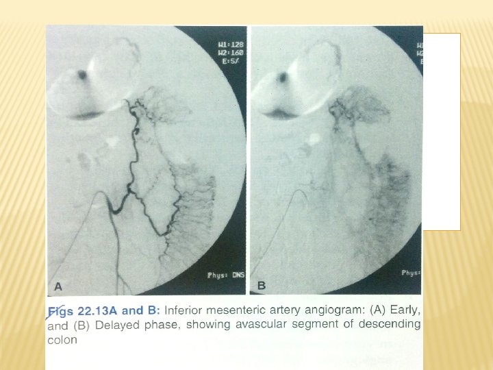
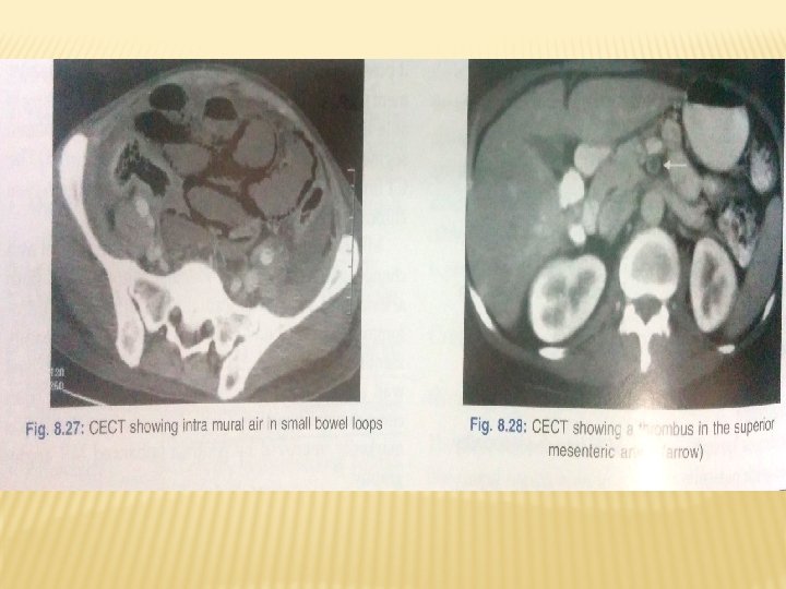
ISCHEMIC BOWEL DISEASE causes � Arterial occlusion(more common) Thromboembolic occlusion is the most common cause of acute mesentric ischemia � Venous occlusion � Low flow states � Types � Acute � chronic(it results from atherosclerosis of mesentric arteries)
INVESTIGATIONS � DUPLEX DOPPLER SONOGRAPHY: � It is preferred noninvasive modality for evaluation of stenosis of mesentric arteries. � SMA peak systolic velocity of 275 cm/sec or no flow signals are reliable indicators of 70% or greater stenosis. � CT � It detects ischemic changes in affected bowel loops and mesentry as well as determine cause of ischemia by allowing evaluation of mesentric vasculature
� � � � � MRI It is comparable to CT in detecting bowel changes and mesentric vascular abnormalities Intraluminal thrombus and collateral vessels can be readily identified It shows bowel wall thickening and increased in signal intensity from compropmised bowel on T 2 WI. DSA: Most accurate modality to evaluate degree of stenosis Allows diagnosis as well as interventional therapy. Percutaneous transluminal angioplasty with or without stent placement can be done. Invasive and time consuming.
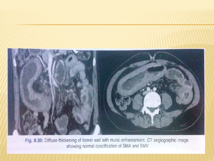
ACUTE ISCHEMIA � � � Most common CT finding is circumferential thickening of bowel wall which demonstrate low attenuating reflecting submucosal edema and inflammation or high attenuation d/t submucosal haemorrhage. may show halo appearance) It may demonstrate luminal dilatation and mesentric stranding After administartion of IV contrast material affected loops demonstrate decreased enhancement compared to normal bowel loops d/t compromised blood flow. In cases of infarction pneumatosis may be present which indicate damage. CT is more sensitive than plain radiography in detection of pneumatosis.
IN LOW FLOW STATES: � Mesentric arteries appear narrowed with limited opacification of branches d/t hypovolumia and spasm � CT shows diifuse thickening of bowel wall with or without mucosal enhancement and increased intraluminal fluid. � CHRONIC MESENTRIC ISCHEMIA: � Devlop collateral pathways � CT detects calcified plaques in aorta and mesentric arteries �
BARIUN STUDIES; � c/I In presence of suspected acute mesentric ischemia. � In clinically unsuspected patients barium findings are: -bowel dilatation -thumb printing -thickened folds -submucosal edema -ulceration � There is uniform thickening of bowel folds which produce symmetric spike like configuration simulating “stack of coins” or “picket fence” appearance. � In chronic cases there may be formation of strictures with smooth outlines. �
THANK YOU!!