Dr Sharon D Rasquinha Assistant Professor FALLOPIAN TUBE

Dr. Sharon D. Rasquinha Assistant Professor
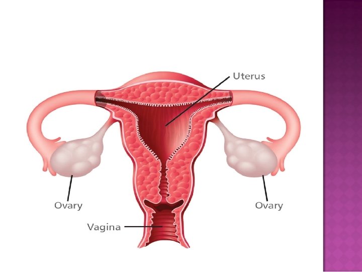

FALLOPIAN TUBE : � Also known as oviducts � Transport the ova from ovary to uterus each month. � In case of fertilization, tubes transport the fertilized egg to uterus for implantation.

Anatomy : � Uterine appendages located bilaterally at superior portion of uterine cavity. � They are present at the cornua. � 10 cm in length 1 cm in diameter and situated within mesosalpinix

Parts : � Isthmus � Ampulla � Infundibulum � Fimbriae

Blood supply : � Branches of uterine and ovarian arteries Nerve supply : � Both sympathetic and para sympathetic Lymphatic drainage : � Iliac and lateral aortic nodes

Microscopic : � 3 layers – �Mucosa �Muscularis �Serosa Cell type within mucosa - columnar ciliated epithelial cells (25%) • Secretory cells (60%) • Peg cells (10%) •

� Egg or nut � Analogous to tests in male � Gonads and endocrine glands

Structure : � Whitish in colour � Located along lateral wall of uterus in a region ovarian fossa � Fossa is beneath external iliac artery and in front of ureter and internal iliac artery � 4 cm x 3 cm x 2 cm in size

Ligaments : � Paired ovaries are within pelvic cavity. � Attached to uterus via cord – ovarian ligament

Extremities : � The end to which fallopian tube attaches – tubal extremity and ovaries connected to it infundibulopelvic ligament. � The other extrimity – uterine extrimity attached to uterus – ovarian ligament

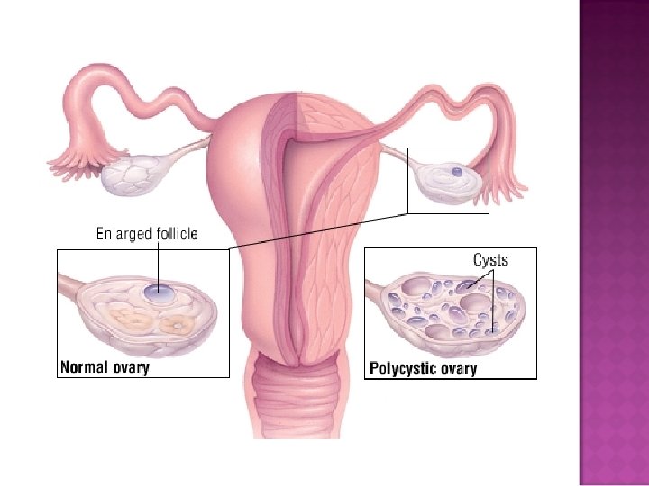
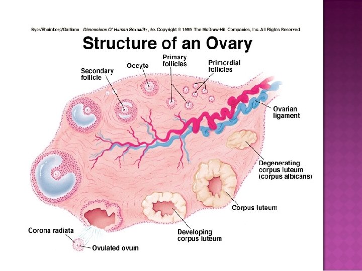
Histology : � Germinal epithelium � Ovarian cortex : Follicles and there is stroma in between them � Follicles : �Cumulus oophorus �Membrana granulosa �Corona radiata �Zona pellucida �Primary oocyte � Inner most layer – ovarian medulla
 � Endocrine – �Estrogen, testorterone and progesterone")
Functions : � Egg cells (oocyte) � Endocrine – �Estrogen, testorterone and progesterone
- Slides: 19