Dr Nazish Waheed By the end of the

Dr Nazish Waheed

By the end of the session my first year students will be able to: • Describe the histological features of bone tissue • Identify the bone tissue as it appear under microscope • Enlist the cells forming the bone tissue • Correlate its clinical features

�Extracellular matrix surrounding widely separated cells �Matrix 25% water 25% collagen fibers mainly type 1 50% crystallized mineral salts �The most abundant mineral salt is calcium phosphate

�A process called calcification is initiated by bone-building cells called osteoblasts �Mineral salts are deposited and crystalize in the framework formed by the collagen fibers of the extracellular matrix �Bone’s fibers flexibility depends on collagen

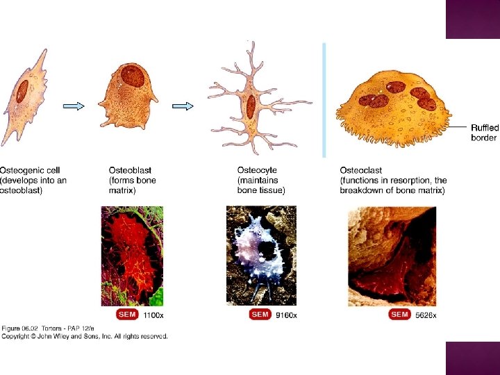
� Four types of cells are present in bone tissue � Osteogenic cells �Undergo cell division; the resulting cells develop into osteoblasts � Osteoblasts �Bone-building cells �Synthesize extracellular matrix of bone tissue � Osteocytes �Mature bone cells �Exchange nutrients and wastes with the blood
 �Regulate")
� Osteoclasts �Release enzymes that digest the mineral components of bone matrix (resorption) �Regulate blood calcium level �Parathyroid hormone increase activity �Calcitonin decrease activity �Howships lacunae

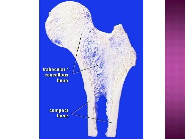
�Bone may be categorized as: �Compact �Spongy
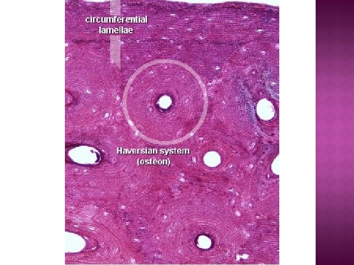

�Compact �Resists Bone the stresses produced by weight and movement �Components of compact bone arranged into repeating structural units called osteons or Haversian systems �Osteons consist of a central (Haversian) canal with concentrically arranged lamellae, lacunae, osteocytes, and canaliculi

�Osteon �Central canals run longitudinally through bone �Around the central canals are concentric lamellae (8 -15) Rings of calcified matrix (like the rings of a tree trunk) �Between the lamellae are small spaces called lacunae which contain osteocytes �Radiating in all directions from the lacunae are tiny canaliculi filled with extracellular fluid

� Osteon �Canaliculi connect lacunae, forming a system of interconnected canals Providing routes for nutrients and oxygen to reach the osteocytes �The organization of osteons changes in response to the physical demands placed on the skeleton

� penetrates the bone more or less perpendicular to its surface � establish connections of the Haversian canals with the inner and outer surfaces of the bone
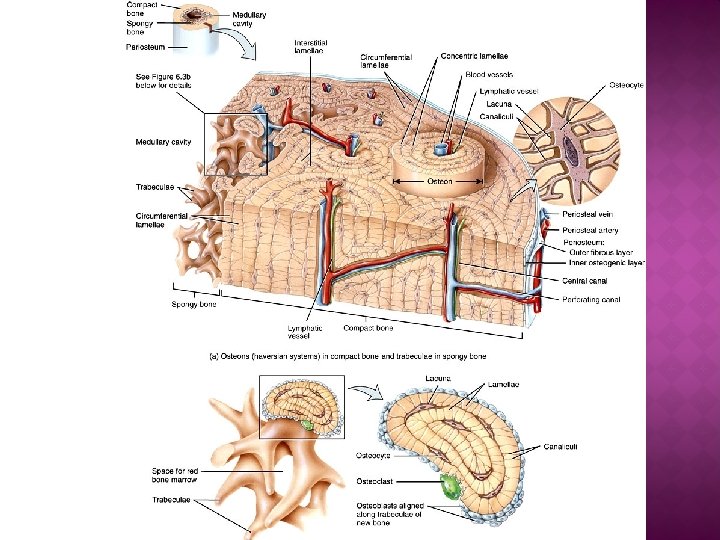

�Spongy Bone �Lacks osteons �Lamellae arranged in a lattice of thin columns called trabeculae Spaces between the trabeculae make bones lighter Trabeculae of spongy bone support and protect the red bone marrow Hemopoiesis (blood cell production) occurs in spongy bone
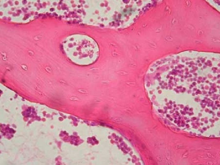

�Spongy Bone �Within each trabecula are lacunae that contain osteocytes �Osteocytes are nourished from the blood circulating through the trabeculae �Interior bone tissue is made up primarily of spongy bone �The trabeculae of spongy bone are oriented along lines of stress helps bones resist stresses without breaking

� The compact bone is also called the lamellar bone � The osteoclast is the bone forming cell � Lamella are present in concentric layers of spongy bone

� Laiq hussain � De fiores � Janqueira
- Slides: 20