DR Nazir KANAAN DIBETIC FOOT Definition q Infection

DR. Nazir KANAAN DIBETIC FOOT

Definition: q Infection, ulceration or destruction of deep tissues associated with neurological abnormalities & various degrees of peripheral vascular diseases in the lower limb ( based on WHO definition)

Diabetes Statistics � According to the American Diabetes Association, there are 18. 2 million people in the United States 6. 3% of the population who have diabetes. � While an estimated 13 million have been diagnosed there are 5. 2 million or 1/3 who have not yet been diagnosed. � About 50% of patients with diabetic foot infections who have foot amputations die within five years

�More than 30 % of patients attending diabetic clinics have evidence of peripheral neuropathy or vascular disease � about 40 % of non-trauma- related amputations in British hospitals are for complications of diabetes � Diabetic foot problems are a major health concern and are a common cause of hospitalization.

Patients with DM are 20 X More Likely to be Hospitalized for Non-traumatic Limb Amputation Public Health Agency of Canada (August 2011); using 2008/09 data from the Canadian Chronic Disease Surveillance System (Public Health Agency of

q 85% of diabetic related foot amputation are preceded by foot ulcer q 4 out of 5 ulcer in diabetics are precipitated by trauma q 4% -10% is the prevalence of foot ulcer in diabetics

Diabetic Foot �Most foot problems that people with diabetes face arise from two serious complications of the disease: nerve damage and poor circulation. One of the more critical foot problems these complications can cause is Charcot arthropathy, which can deform the shape of the foot and lead to disability.
 a predisposition to peripheral vascular disease; (2) damage")
Factors affecting the foot are: (1) a predisposition to peripheral vascular disease; (2) damage to peripheral nerves; (3) reduced resistance to infection; (4) osteoporosis.

�disease of the nervous system. The three major forms in people with diabetes are peripheral neuropathy, autonomic neuropathy, and mononeuropathy. The most common form is peripheral neuropathy, which affects mainly the legs and feet.

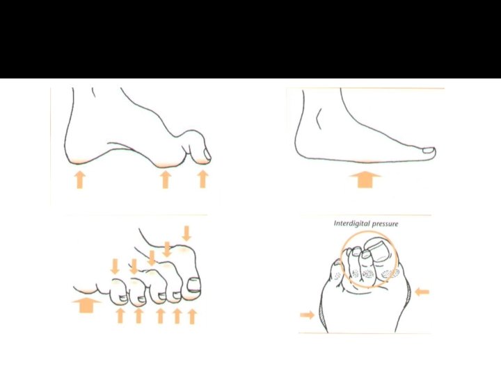
Peripheral neuropathy �Early on, patients are usually unaware of the abnormality but clinical tests will discover loss of vibration and joint position sense and diminished temperature discrimination in the feet. Symptoms, when they occur, are mainly due to sensory impairment: symmetrical numbness and paraesthesia, dryness and blistering of the skin, superficial burns and skin cracks or ulceration due to shoe scuffing or localized pressure. Motor loss usually manifests as claw toes with high arches and this, in turn, may predispose to plantar ulceration.

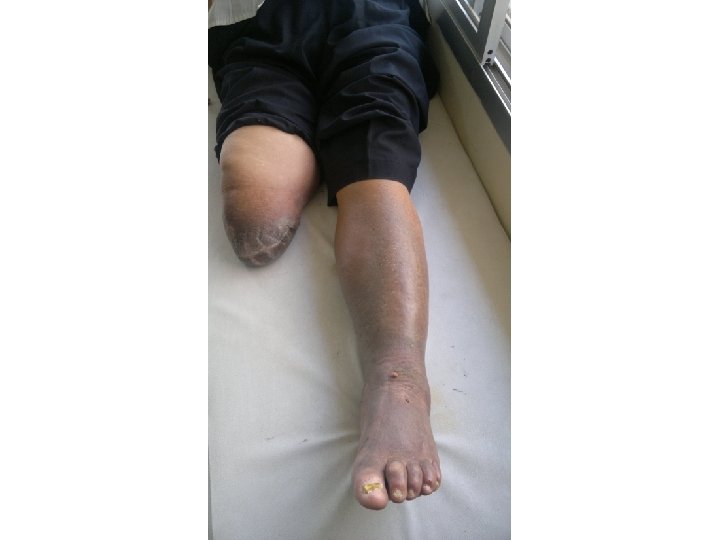
� Neuropathy may be mild with minimal somatic sensory changes and no autonomic nervous system changes. In its severest form, total anesthesia from the midtibia distally (somatic loss) and complete absence of sweating (autonomic loss) result in a dry, scaly, swollen, clumsy limb that the patient dissociates from the rest of the body because of the loss of sensory feedback

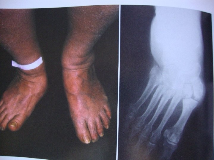
Neuropathic joint disease � ‘Charcot joints’ occur in less than 1% of diabetic patients, yet diabetes is the commonest cause of a neuropathic joint. The mid-tarsal joints are the most commonly affected, followed by the MTP and ankle joints. There is usually a provocative incident, such as a twisting injury or a fracture, following which the joint collapses relatively pain- lessly. X-rays show marked and fairly rapid destruction of the articular surfaces. These changes are easily mistaken for infection but the simultaneous involvement of several small joints and the lack of systemic signs point to a neuropathic disorder. Joint aspiration and micro biological investigation will also help to exclude infection.

�Vibratory and position senses are lost early, and the patient does not know where in space the foot is located at a given time. This is especially catastrophic if the patient also cannot see the foot.

� In late cases there may be severe deformity and loss of function. A rocker-bottom deformity from collapse of the midfoot is diagnostic.
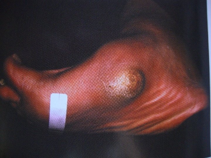

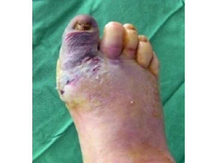
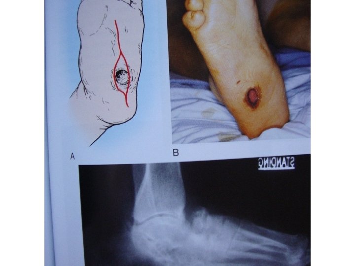
Peripheral vascular disease � Atherosclerosis affects mainly the medium-sized vessels below the knee. The patient may complain of claudication or ischaemic changes and � ulceration in the foot. The skin feels smooth and cold, the nails show trophic changes and the pulses are weak or absent. Doppler studies should corroborate the clinical findings. Superficial ulceration occurs on the toes, deep ulceration typically under the heel; unlike neuropathic ulcers, these are painful and tender. Digital vessel occlusion may cause dry gangrene of one or more toes; proximal vascular occlusion is less common but more serious, sometimes resulting in extensive wet gangrene.

Osteoporosis �There is a generalized loss of bone density in diabetes. In the foot the changes may be severe enough to result in insufficiency fractures around the ankle or in the metatarsals.

� There is a generalized loss of bone density in diabetes. In the foot the changes may be severe enough to result in insufficiency fractures around the ankle or in the metatarsals.

Infection Diabetes � if not controlled, is known to have a deleterious effect on white cell function. This, combined with local ischaemia, insensitivity to skin injury and localized pressure due to deformity, makes sepsis an ever-recurring hazard.

�Typically, diabetic foot infection are polymicrobial. The most common culprits are staph or strep but most infections have about 4 to 6 other bugs present. In recent years, staph strains have evolved to be more resistant to antibiotics. �MRSA, (methicillin-resistant Staphylococcus aureus) has become increasingly common, both in hospital strains and in community-acquired cases (CA-MRSA).

Diabetic foot infections typically take one of the following forms: �Cellulitis �Deep-skin and soft-tissue infections �Acute osteomyelitis �Chronic osteomyelitis

Cellulitis �Tender, erythematous, nonraised skin lesions are present, sometimes with lymphangitis �Lymphangitis suggests group A streptococcal infection �Bullae are typical of Staphylococcus aureus infection, but occasionally occur with group A streptococci � No ulcer or wound exudate is present.

Deep-skin and soft-tissue infections �The patient may be acutely ill, with painful induration of the soft tissues in the extremity �Wound discharge is usually not present �In mixed infections that may involve anaerobes, crepitation may be noted over the afflicted area �Extreme pain and tenderness may indicate compartment syndrome or clostridial infection (ie, gas gangrene) �The tissues are not tense, and bullae may be present �Discharge, if present, is often foul

Acute osteomyelitis �Unless peripheral neuropathy is present, the patient has pain at the site of the involved bone �Usually, fever and regional adenopathy are absent

Chronic osteomyelitis �The patient's temperature is usually less than 38. 5 ° �Discharge is commonly foul �No lymphangitis is observed �Pain may or may not be present, depending on the degree of peripheral neuropathy �Deep, penetrating ulcers and deep sinus tracts (diagnostic of chronic osteomyelitis) are usually located between the toes or on the plantar surface of the foot �The medial malleoli, shins, or heels are not usually sites of involvement.

Foot Ulcers �Foot ulcers affect one in ten diabetics during their lifetime �Wound healing is also impaired from affected collagen synthesis.

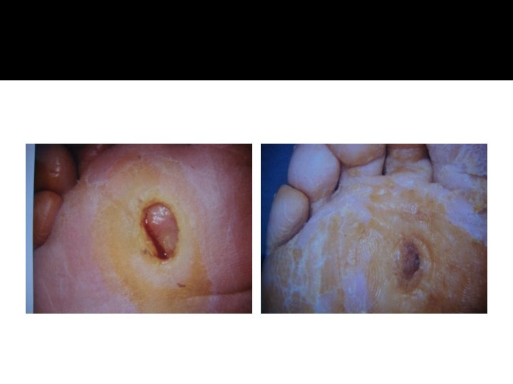
� Diabetic ulcers are most common in the forefoot beneath one of the metatarsal heads or the interphalangeal joint of the hallux.

STAGES OF ULCER DEVELOPMENT

STAGES OF ULCER DEVELOPMENT

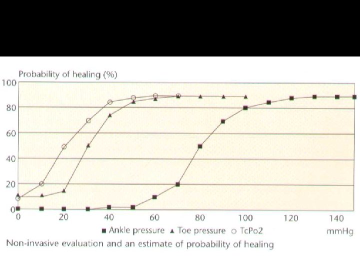
�The ulcerated foot with no palpable pedal pulses needs at least Doppler pressures.

Classification of Diabetic Ulcers � The ischemic index is the ratio of ankle to brachial pressure.

� Direct pressures in the toe probably are the most helpful.

Wagner classification �Grade 0—skin intact, but bony deformities produce a “foot at risk” �Grade 1—localized, superficial ulcer �Grade 2—deep ulcer to tendon, bone, ligament, or joint �Grade 3—deep abscess, osteomyelitis �Grade 4—gangrene of toes or forefoot �Grade 5—gangrene of entire foot.

Eichenholz classification staging system that is useful in Charcot arthropathy: �Stage 0—unilateral edema, erythema, and associated warmth; no break in skin integrity; radiographs negative or show local osteoporosis.

�Stage I—unilateral edema, erythema, and unilateral warmth; radiographs show osseous destruction, joint dislocation or subluxation �Stage II—decreased local edema, erythema, and associated warmth; radiographs show coalescence of small fracture fragments and absorption of fine bone debris �Stage III—no or minimal edema, erythema, or increased warmth; radiographs show consolidation and remodeling of fracture fragments.

Risk Factors for Diabetic Foot Ulcers �poorly fitted or poor quality shoes �poor hygiene (not washing regularly or thoroughly) �improper trimming of toenails �alcohol consumption �eye disease from diabetes �heart disease �kidney disease �obesity �tobacco use (inhibits blood circulation).

Diagnosis �Examination for early signs of neuropathy should include the use of Semmes–Weinstein hairs (for test- ing skin sensibility) and a biothesiometer (for testing vibration sense). Peripheral vascular examination is enhanced by using a Doppler ultrasound probe. Frequently, multiple bacterial types are isolated (anaerobes make a regular appearance). X-ray examination may reveal periosteal reactions, osteoporosis, cortical defects near the articular margins and osteolysis – often collectively described as ‘diabetic osteopathy’.

Cellulitis �The WBC and ESR are slightly or moderately elevated, but these elevations are not diagnostic �Blood culture results are usually negative; if positive, they usually indicate the presence of group A or group B streptococci �Cultures of skin via aspiration or biopsy are generally unrewarding; aspiration of a sample from the leading edge of the erythematous border has a low yield (likely < 5%) but may be used if the likely organism must be identified on initial presentation.

Skin and soft-tissue infections � The WBC and ESR are mildly or moderately elevated � If bullae are present, Gram stain and culture results from aspirated exudate from a bullous lesion may help identify the pathogen � Blood culture results may be positive � In suspected deep soft-tissue infection, plain radiography, CT, or MRI to evaluate for compartment syndrome or for gas or a foreign body ; excessive gas signifies a mixed aerobic-anaerobic infection, in contrast to gas gangrene (clostridial myonecrosis) � Cultures of samples aspirated from deep-skin and soft-tissue infections may be used to identify the organism.

Acute osteomyelitis � The WBC usually reveals leukocytosis, and the ESR is moderately or highly elevated. � Blood culture results are usually negative; when positive, the findings most frequently indicate the presence of S aureus � For affected long bones, plain radiographic findings generally become abnormal after 10 -14 days; softtissue swelling and periosteal elevation are the earliest signs � Bone scans are preferred to gallium or indium scans; bone-scan findings are positive within 24 hours � Bone biopsy is not necessary.

Chronic osteomyelitis � The WBC is often within the reference range; the ESR is usually very highly elevated and; the platelet count is also often elevated � Blood culture results are usually negative � Plain radiographic findings are invariably abnormal � Bone scans are usually unnecessary unless diagnostic confusion exists with another disorder (eg, bone tumor); an MRI scan would also be helpful in such a situation � Bone biopsy is the preferred way to identify the causative pathogen � Important pathogens include Bacteroides fragilis, E coli, Proteus mirabilis, and Klebsiella pneumoniae; Pseudomonas aeruginosa is usually not the causative organism.

Assessment and treatment �The diabetic patient presenting with a foot wound should be assessed at three levels- the patient as a whole, the affected limb and foot and the infected wound �There may be an obvious large wound or ulcer associated with erythema and pyrexia.

Management �Neuropathy, angiopathy, retinopathy, and nephropathy, alone or in combination and in varying degrees of severity, may influence the treatment of the diabetic foot

Management �The orthopaedic surgeon will usually be one member of a multidisciplinary team comprising a physician (or endocrinologist), surgeon, chiropodist and orthotist.

� There are treatment options for the wide range of diabetic foot problems. The most effective treatment, however, is prevention. � The best way of preventing complications is to insist on regular attendance at a diabetic clinic, full compliance with medication, examination for early signs of vascular or neurological abnormality, advice on foot care and footwear and a high level of skin hygiene � For people with diabetes, careful, daily inspection of the feet is essential to overall health and the prevention of damaging foot problems.

Preventing Diabetic Foot Problems � According to the American Podiatric Medical Association, 14 to 24% of Americans with diabetic foot ulcers have amputations. Preventive care is crucial. Chances of diabetes complications remain low when blood sugar is stable. Prevention diabetic foot problems by: � washing feet every day � keeping toenails adequately trimmed, but not too short � keeping feet dry and moisturized � changing socks frequently � seeing a podiatrist for corn and callus removal � wearing proper-fitting shoes

Management � Treatment of diabetic foot infections varies by type, as follows: � Cellulitis – Most responsive to antibiotics � Deep skin and soft-tissue infections – Usually curable, but additional debridement is usually indicated � Acute osteomyelitis – Infecting microorganisms and the likelihood of successful treatment with antimicrobial therapy are essentially the same as in patients without diabetes � Chronic osteomyelitis – Surgical debridement is essential, in addition to antibiotics; amputation may be necessary

ANTIBIOTIC THERAPY �Antibiotics should be used if an abscess is drained, if cellulitis is present, or in conjunction with surgical débridement of an area of osteomyelitis or pyarthrosis �Aerobic and anaerobic cultures should be obtained before initiation of antibiotic treatment �A broad-spectrum anti- biotic is begun, covering gram-positive and gram-negative organisms until sensitivities return and appropriate antibiotic choices are made

Medications � Antiplatelets, or anti-clotting. �health conditions that might increase risk of infections by these harmful bacteria, including HIV and liver problems.

Treating Diabetic Foot Ulcers � off-loading �wearing certain items to protect feet: �diabetic shoes �casts �foot braces �compression wraps �shoe inserts to prevent corns and calluses

�In addition to off-loading the ulcer, treatment with hyper- baric oxygen may help to heal an ischemic ulcer.

Nonsurgical Treatment � Casting. The early stages of Charcot are usually treated with a cast or cast boot to protect the foot and ankle. The use of a cast is very effective in reducing the swelling and protecting the bones � Casting requires that the patient not put weight on the foot until the bones begin to heal. Crutches, a knee-walker device, or a wheelchair are usually necessary. Healing can sometimes take 3 months or more. The cast will usually be changed every week or two to make sure that it continues to "fit" the leg as the swelling goes down.

� Custom-made shoes with total contact insoles must follow the successful healing of these ulcers to avoid recurrence.

Orthotics �Minimize the risk of a pressure sore. �An accommodative orthotic made from a soft material ( plastizote ). �The orthotics should not be hard, as this will increase the risk of a pressure ulcer. �The orthotic can be transferred from shoe to shoe and should be used at all times when standing or walking

�Many topical treatments are available for foot ulcers, including: �dressings containing silver or silver sulphadiazine cream �polyhexamethylene biguanide (PHMB) gel or solutions �iodine (either povidone or cadexomer) �medical grade honey in ointment or gel form

Surgical Treatment �Surgery may be recommended if the foot deformity puts the patient at a high risk for ulcers, or if protective shoewear is not effective. Unstable fractures and dislocations also require surgery to heal.

�New techniques for wound debridement include low frequency ultrasound therapy, hydrosurgery, monofilament polyester fibre pad and plasma-mediated bipolar radiofrequency ablation. �Skin grafting when no infection is present may be required.

�The diabetic foot infection classification system, along with a vascular assessment, would help determine which patients should be hospitalized, which may require special imaging procedures or surgical interventions including amputation.

�Ischaemic changes need the attention of a vascular surgeon who can advise on ways of improving the local blood supply. Arteriography may show that bypass surgery is feasible.

� Dry gangrene of the toe can be allowed to demarcate before local amputation; severe occlusive disease with wet gangrene may call for immediate amputation

Revascularisation �As diabetes is chronic and progressive, it makes sense to have a conservative surgical approach that include surgical revascularization. A successful surgical bypass of larger vessel disease may enable more conservative treatment of the diabetic foot. Revascularisation is, however, considered inappropriate in bedridden patients, in a functionally useless limb, in patients with life threatening sepsis, extensive muscle necrosis and where it is technically impossible. Primary amputation is better in these cases
 and luminal stenting or arterial reconstruction to improve blood")
�A percutaneous transluminal angioplasty (PTA) and luminal stenting or arterial reconstruction to improve blood flow would aid healing. Because in most cases ischaemia is secondary to larger vessel artherosclerosis rather than to 'small vessel disease. Thus lower extremity artherosclerosis can be amenable to angioplasty or vascular bypass

�The indications for a PTA in diabetic peripheral arterial disease are classically for disabling claudication and critical limb ischaemia, Patients with non-critical ischaemia (ankle/brachial pressure index (ABPI- 0. 4– 0. 9) can in some cases be successfully treated without a vascular procedure.

�Many centres have reported successful use of both aggressive endovascular interventions and distal bypass procedures for more severe vascular disease of the foot. The shortterm effects are satisfactory with healing of the foot ulcers and thus diminishing the risk of amputation.

�Interposition vein grafts to pedal arteries have preserved at least a portion of the foot suitable for weight bearing function to some degree �salvage of the foot or a functional part of the foot

�Mild deformity with tightness at the heel. In some cases, the deformity is mild and associated with tightness at the back of the heel. Ulcers in the front of the foot that do not respond to a period of casting and protective shoewear, may be treated by Achilles tendon lengthening. Surgically lengthening the tendon decreases the pressure on the midfoot and front of the foot. This allows the ulcer to heal and reduces the chance that it will return

�Bony prominence on the bottom of the foot. A more severe deformity is the appearance of a very large bony bump on the bottom of the foot. If this cannot be addressed with shoe modification, it requires surgery. The type of surgery depends on the stability of the bones and joints in the foot.

�Stable deformity. Surgery involves a simple removal of the prominent bone by shaving it off.

�Unstable deformity. When the bones are too loose at the sight of the prominence, a simple removal of the bump will not be effective. The loose bones will simply move and a new prominence will develop. In this situation, fusion and repositioning of the bones is needed.

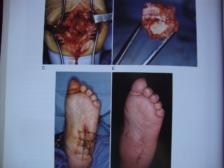
Unstable Charcot of the hindfoot. � A complex realignment and fusion was performed to prevent the patient from developing a prominence and ulceration �

�This operation is extremely difficult to perform and carries a higher risk of wound complications, infections, and amputation, compared to routine foot and ankle fracture surgery. �After this type of operation, there is typically a period of no weight on the foot for at least 3 months

�Fractures that occur in the softer bone of diabetics are typically more complex. Operations to fix them generally involve more hardware (plates and screws) than would normally be required in people without diabetes. The screws and plates may even be placed across normal joints to provide added stability.

�Ankle deformity. Charcot of the ankle is difficult to treat simply with a brace or shoe and commonly requires surgical fusion of both the ankle and the subtalar joint to hold the foot straight. Given the amount of destruction of the bone and the poor quality of the soft tissue, the risk of non-union and the risk of infection are very high. Amputation may be required, either as the first operation or to salvage a fusion that has not healed or has became infected

�If the subluxation at the tarsometatarsal joint is seen early, before extensive softtissue and bony disruption have occurred, closed reduction and pinning with several months of non–weight bearing cast immobilization may stabilize the midfootforefoot complex

�Limited arthrodesis with autogenous bone graft, rigid fixation, and prolonged non– weight bearing cast immobilization may be indicated, especially if only the first and second metatarsocuneiform articulations are arthrodesed.

�Tibiotalocalcaneal arthrodesis may be performed, and if fibrous nonunion occurs, it may be enough to provide stability and make bracing of the foot and ankle possible

�Heroic attempts are being made to preserve any part of a foot, even an insensitive, deformed one

�If a patient can have the medial two rays or lateral three rays (a general rule), the pedorthist-orthotist should be able to make a shoe and brace for the foot �Transmetatarsal amputation and even Chopart amputation , combined with appropriate tenotomies and a brace-shoe, allow some degree of function

�Partial foot, Lisfranc, Chopart, and Syme amputations �Prolonged convalescence, physical and emotional trauma from multiple surgical procedures, and the financial burden placed on a patient should influence care of this most complex fiscal and physical malady.

Amputation �Tissue death and infection can eventually become so painful and life threatening that amputation becomes necessary if all other measures such as antibiotics and debridement fail to work. �Damaged tissue will be removed during surgery with as much healthy tissue preserved as possible. The patient may require many days in the hospital and it can take up to eight weeks for the wound to completely heal.
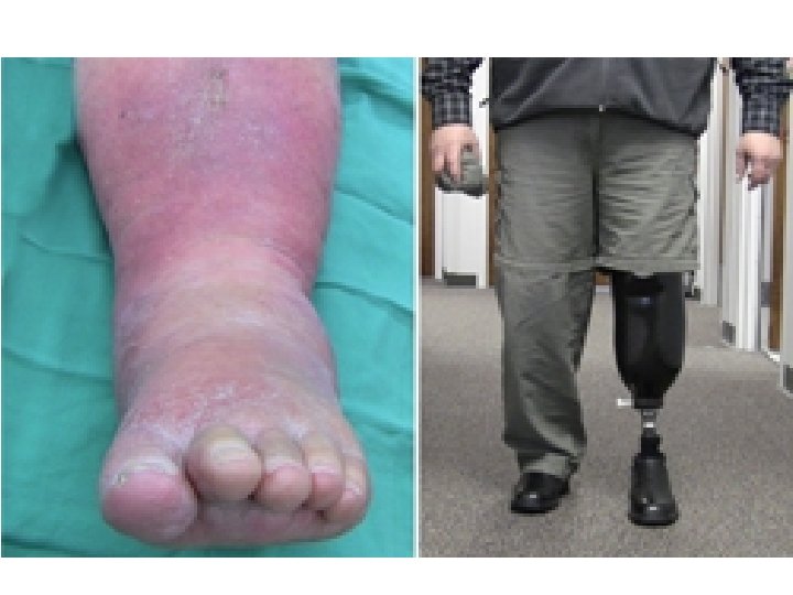

Rehabilitation �After surgery, the patient will be helped by a rehabilitation team to learn to deal with the physical and emotional challenges. A prosthesis may be needed and the team will help the patient adjust. The team will also be able to help with assistive devices, home adaptations, and learning to accomplish normal daily activities. Some amputees experience pain or discomfort in the missing limb. This is called phantom pain. Their team can help them learn how to deal with this problem.

Thank you
- Slides: 93