Dr Mazen Abou Chaaban Consultant Pediatrics and Pediatric

Dr. Mazen Abou Chaaban Consultant Pediatrics and Pediatric Nephrology Medical Director

Approach and Radiological Diagnosis

• Renovascular hypertension is an uncommon but important cause of hypertension in children. • It constitutes not more than 1% of children who do have hypertension in childhood. • The delay in the diagnosis is due to infrequent blood pressure measurement in children and high values are generally dismissed as inaccurate.

• Children with Renovascular disease may have other abnormalities of blood vessels like aorta, cerebral, intestinal or iliac. • The prevalence is much higher in patients with acute, severe or refractory hypertension resistant to treatment at young age.

Causes of RVH in Children • Fibromuscular dysplasia ØFocal ØMultifocal • Mid-aortic syndrome with RAS? • Syndromic RVH ØNF 1 ØWilliam syndrome ØAlagille syndrome ØOther (TSC, moya-moya? ) • Vasculitis ØTakayasu’s disease ØPolyarteritis nodoa ØKawasaki disease ØOther vasculitides • Extrinsic compression Ø tumors (neublasoma, Wilms tumor, paraganglioma, etc. ) • Other ØTrauma ØPost-surgical complications Øradiation

Renovascular Hypertension Pathophysiology • Elevation of blood pressure caused by RAAS (unilateral renal artery stenosis) and/or RAAS and sodium retention (bilateral renal artery stenosis). • Maintenance of elevated blood pressure with new threshold for natriuresis (bilateral RAS). • Kidney damage: ØIschemic kidney – ischemic nephropathy (decreased volume, tubular atrophy, tubulointerstitial fibrosis) ØHypertensive nephropathy (arteriolar and glomelular injury) in the other healthy kidney. • Hypertensive crisis

Whom to suspect with RVH to Investigate: • The clinical findings suggest a secondary hypertension rather than primary. Ø Severe and resistant hypertension Ø Acute rise in blood pressure over a previously stable value Ø Young onset hypertension with a negative family history Ø Unexplained an acute elevation in serum creatinine of at least 30% after administr. of ACE inhibitor or ARB Ø Recurrence episode of flash pulmonary edema ØUnilateral small kidney (cannot be explained by another reason) Ø Systolic or diastolic abdominal bruit

• The patient does not appear to have another cause of secondary hypertension Ø Primary kidney disease Ø Primary aldosteronism Ø Pheochromocytoma Ø Cushing’s syndrome Ø Sleep apnea syndrome Ø Coarctation of aorta Ø Hypothyroidism Ø Primary hyperparathyroidism • An intervention is planned if a significant stenotic lesion is found.

Because of the potential harm from invasive procedures, patients who have a high likelihood of benefitting from the procedure should be selected • Patients who experienced short duration (weeks or months) of blood pressure elevation prior to the diagnosis of renovascular disease. • Failure of optimal medical therapy to control the blood pressure. • Intolerance to optimal medical therapy (rise in serum creatinine after initiation of renin- angiotensin system inhibitor).
 • Suspected fibromuscular disease")
• Progressive deterioration of renal function (bilateral renovascular disease) • Suspected fibromuscular disease in a young patient (to avoid need of life-long antihypertensive therapy). • Recurrent flash pulmonary edema and/or refractory heart failure.

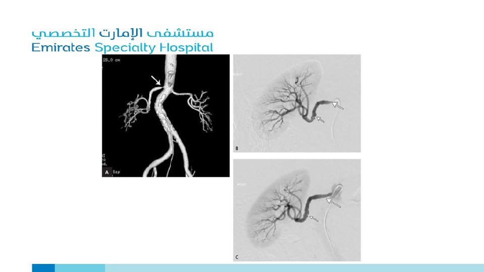
DIAGNOSTIC PROCEDURE OPTIONS Renal Artery Stenosis? • Renal Arteriography Ø Gold standard of diagnosis (invasive procedure) • Non-Invasive alternatives are: Ø Duplex Doppler ultrasonography Ø Computed tomographic angiography (CTA) Ø Spiral CT scan with CT angiography Ø Magnetic resonance angiography (MRA)

The choice of the appropriate procedure depends upon: ØLocal availability ØClinical expertise of each technique ØPresence or absence of renal impairment v. Radio contrast media and gadolinium are potentially harmful in patients with stage 4 -5 chronic kidney disease.

Duplex Doppler Ultrasonography ADVANTAGE • Provides functional assessment of the renal arteries and some anatomic information. • Can detect both unilateral and bilateral disease. • Inexpensive and suitable for serial measurements to determine disease progression. • Can be used to detect recurrent stenosis in patients previously treated with angioplasty or surgery.

Stenotic lesions can be detected by comparing the systolic flow velocity in the renal artery to that in the aorta ØVelocity of the flow increases as an artery narrows ØDiastolic velocity, maybe increased distal to a stenotic lesion

Duplex Doppler Ultrasonography DISADVANTAGE • It is time consuming (taking up to 2 hours to perform). • It is technically difficult (particularly in large patients). • Operator dependent. • A positive test is more informative that a negative test.

Duplex Doppler Ultrasonography with Resistive Index RESISTIVE INDEX • It is the peak systolic velocity minus end diastolic velocity divided by peak systolic velocity. • High resistive index is associated with a greater degree of intrinsic renal damage (likely a reversible small vessel injury) and, therefore, a diminished predicted benefit from revascularization. • In the hands of experienced operator improves the ability to predict outcome after revascularization.

Spiral CT Scan with CT Angiography • Rapid development in CT imaging with improved tissue reconstruction, multi-detector slice acquisition, high resolution with less contrast exposure continues to improve the value of CT Angiography as a diagnostic tool for renovascular disease. • Highly accurate non-invasive diagnostic test for imaging renovascular disease due to atherosclerosis but less so for fibromuscular disease.

• It combines the diagnostic accuracy of arteriography with the low risk of intravenous digital subtraction angiography. • Patients with fibromuscular dysplasia sensitivity and specificity of CTA were 64% and 92% respectively since fibromuscular disease usually involves distal arterial segments that are more difficult to image.

Magnetic Resonance Angiography ØUsed of breath-hold MRA with paramagnetic contrast material as gadopentetate dimeglumine substantially improves the ability to visualize the majority of accessory arteries in a better way. ØPhase contrast MRA produces result similar to digital subtraction angiography (Ann Intern Med 2004; 141: 730). ØIn patients with fibromuscular dysplasia sensitivity of only 22 -28% has been noted with MR and CT angiography.

Other Diagnostic Testing PLASMA RENIN ACTIVITY • Peripheral venous PRA is elevated in only 50 -80% of patients with renovascular hypertension. Ø Suppress by high dietary sodium intake Ø Bilateral renal artery disease Ø Volume expansion related to intrinsic kidney disease Ø Various antihypertensive drugs

• It has limited value for the diagnosis of renovascular hypertension. • The predicted value of PRA can be increased by measuring the rise in PRA one hour after the administration of 25 -50 mg captopril, patient with renal arteriosclerosis have an exaggerated increase in the PRA as compared with normal individuals (removal of the normal suppressive effect of high angiotensin II on renin secretion in stenotic kidney.

CAPTOPRIL RENOGRAM • DPTA and/or MAG 3 are being used as radionuclides in renography. • MAG 3 is more reliable in patients with renal insufficiency. • The predictive value can be increased by administration of oral captopril 25 -50 mg one hour before Isotope was injected. The efficacy of this test is based upon the expected ACE inhibitor-induced decline in glomerular filtration rate in stenotic kidney accompanied by an increased in GFR in the contralateral kidney due to removal of angiotensin II mediated vasoconstriction. • The sensitivity and specificity of the AC inhibitor scan varies substantially in different studies and have limited inter-observant agreement.

RENAL VEIN RENIN MEAUREMENT • Renin secretion should be increased in stenotic kidney by approx. ≥ 1. 5 x the value to contralateral kidney. • The large discrepancy in renin secretion between the two kidneys is present in less than 10% of normal individuals. • The accuracy of this measurement may be enhanced by the prior administration of AC inhibitor and/or diuretics which will increase renin secretion on the affected side. • Many false negative and occasional false positive result may be encountered.

• Renal vein renin have a limited role in estimating physiologic significance of a stenotic lesion.

• There is no sufficiently accurate, non-invasive radiologic or serologic diagnostic test, if negative will completely exclude the presence of renal artery stenosis. • Each tests is associated with strengths and weaknesses.

Patients without renal insufficiency • Duplex Doppler US, CT Angiography and MR Angiography all provide minimally invasive options for diagnostic evaluation prior to intra-arterial angiography. • In addition to diagnosing renal artery stenosis, these tests can identify anatomic abnormalities of kidneys, aortic disease and whether the stenosis is unilateral or bilateral.

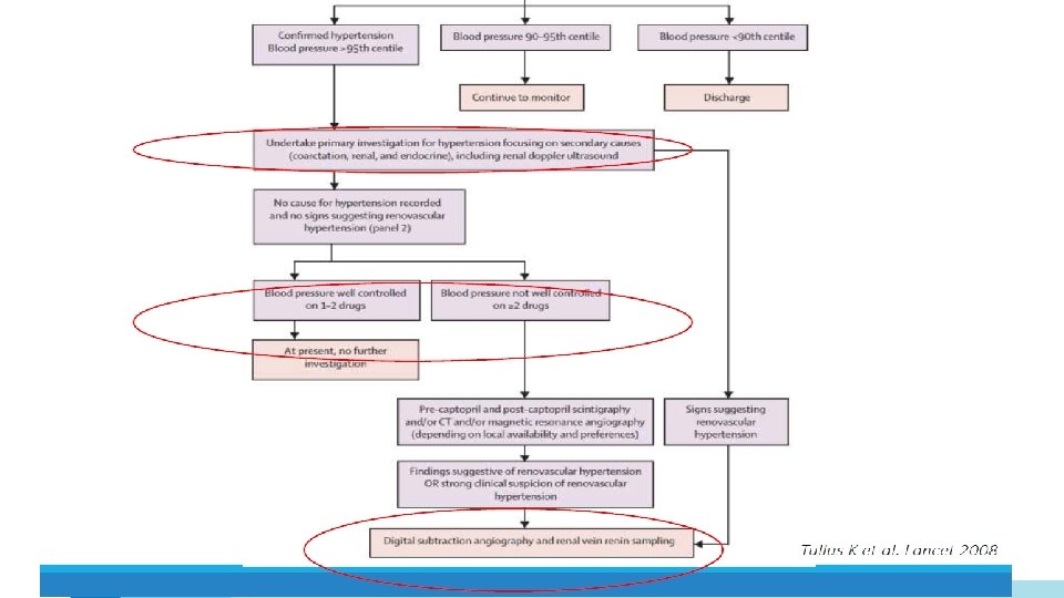
• Digital subtraction angiography should be performed when noninvasive testing is inconclusive. • If the Arteriogram is positive, endovascular revascularization often with primary renal artery stenting can be performed during the same procedure.

Patients with renal insufficiency ØEvaluation for Renovascular disease should be considered in Patients with Øchronic ischemic renal disease ØA progressive decline in renal function Ø Recurrent episode of flash pulmonary edema Ø Uncontrolled hypertension • The optimal approach in such patients is uncertain although some will have recovery of kidney function or slowing of renal function deterioration after successful revascularization.

Evaluation of Renovascular disease in these patients can be with: Ø Duplex Doppler ultrasonography, if technical expertise is available, is the preferred initial non-invasive diagnostic test. ØIf technical expertise is not available, Spiral CT scan with CT angiography in patient with GFR <30 ml/min. despite the risk of radiocontrast nephropathy. Ø MRA with gadolinium should be avoided if possible in patients with GFR <30 ml/min because of the risk of nephrogenic systemic fibrosis which is sometimes very severe.
 and atheroembolism are expected")
• Contrast induced kidney injury, nephrogenic systemic fibrosis (gadolinium) and atheroembolism are expected risks to such category of patients.

Conclusions • RVH is a leading cause of secondary Hypertensionin pediatric age group • Pharmacotherapy does not cure but may lower BP • RTA &/or Surgery may cure in 50% of case • RVH is not only disease of renal arteries.
- Slides: 36