
Dr Kumar Satish Ravi M B B S
Dr. Kumar Satish Ravi M. B. B. S. , M. D. (JIPMER), MAMS
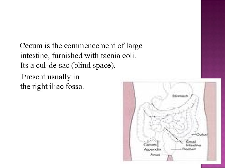
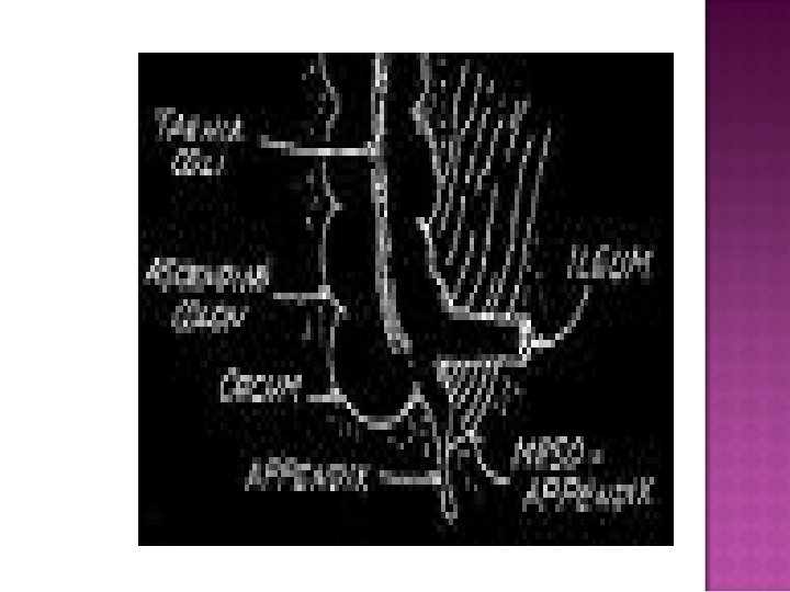
Cecum is the commencement of large intestine, furnished with taenia coli. Its a cul-de-sac (blind space). Present usually in the right iliac fossa.
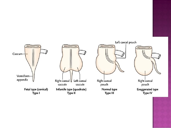
Size: Length-6 cm, Breadth-7. 5 cm Shape: Asymmetric. According to the shape, the cecum is divided into Infantile type Ø Foetal type Ø Adult type Ø Exaggerated type q Relations: Ø Peritoneal- Covered by peritoneum on all side
Relations Anteriorly: 1. Coils of small intestine. 2. Sometimes part of the greater omentum. 3. And the anterior abdominal wall in the right iliac region. Ø
Posteriorly: 1. The psoas and the iliacus muscles. 2. The femoral nerve. 3. The lateral cutaneous nerve of the thigh. 4. The appendix is commonly found behind the cecum. Ø Medially: The appendix arises from the cecum on its medial side. Ø
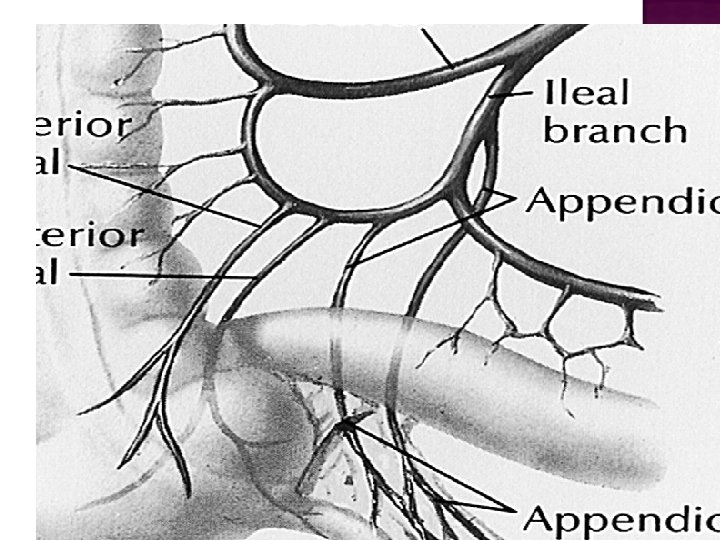
Blood Supply Arteries Anterior and posterior cecal arteries from the ileocolic artery, a branch of the superior mesenteric artery. Veins The veins correspond to the arteries and drain into the superior mesenteric vein.
Lymph Drainage The lymph vessels pass through several mesenteric nodes and finally reach the superior mesenteric nodes. Nerve Supply Branches from the sympathetic(T 11 -L 1) and parasympathetic (vagus) nerves form the superior mesenteric plexus.
q Interior of the cecum It has two orifices. Ø Ileo-ceacal Orifice Ø Appendicular Orifice
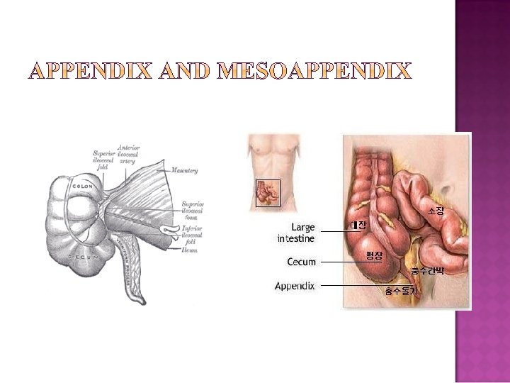
A narrow worm-like tubular diverticulum which arises from the postero-medial wall of the ceacum. Suspended by a peritoneal fold called mesoappendix. The body of appendix is kinked on itself, hence called vermiform appendix. Devoid of taenia coli, sacculations and appendices epiploicae. Large aggregations of lymphatic tissue. Abdominal Tonsil
Average length- 9 cm Presenting parts: Ø Base-attached to the posteromedial wall of ceacum. Ø Body-narrow, tubular and contains a canal which opens into ceacum. Ø Tip-least vascular and is directed in various positions.
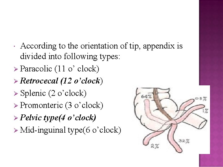
According to the orientation of tip, appendix is divided into following types: Ø Paracolic (11 o’ clock) Ø Retrocecal (12 o’clock) Ø Splenic (2 o’clock) Ø Promonteric (3 o’clock) Ø Pelvic type(4 o’clock) Ø Mid-inguinal type(6 o’clock)
Mesoappendix: Triangular peritoneal fol which invests the entire appendix Derived from the posterior layer of mesentery of the ileum. Appendicular vessels pass with in the free margin of the mesoappendix.
Blood Supply Arteries The APPENDICULAR artery- branch of the ILEO COLIC artery. Veins The appendicular vein drains into ILEO COLIC vein. Lymph Drainage The lymph vessels drain into one or two nodes lying in the mesoappendix and then eventually into the superior mesenteric nodes.
Nerve Supply The appendix is supplied by the sympathetic and parasympathetic (vagus) nerves from the superior mesenteric plexus. Afferent nerve fibers concerned with the conduction of visceral pain from the appendix accompany the sympathetic nerves and enter the spinal cord at the level of the 10 th thoracic segment.
APPENDICITIS Inflamation of appendix is called appendicitis. It is usually manifested by pain, temperature and vomitting – Murphy’s Syndrome. The pain is often felt first in umbilical region, then settles in the right iliac area due to local peritonitis.
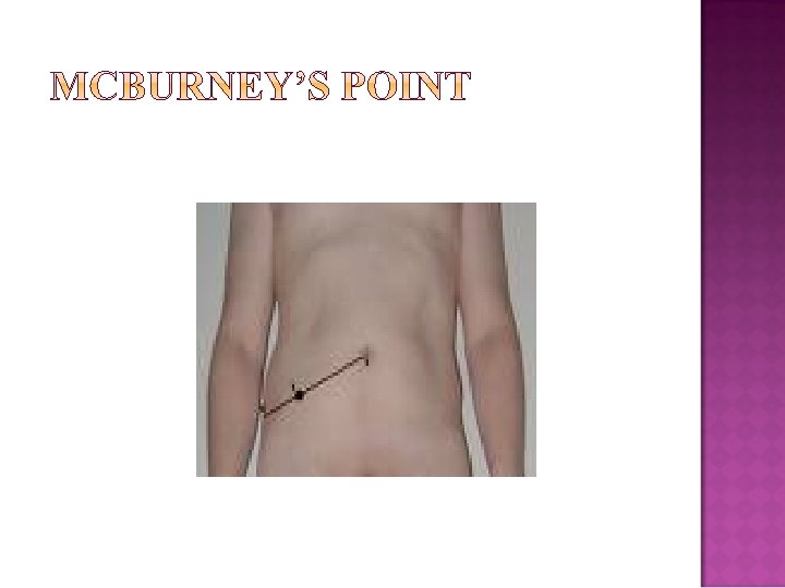
It is associated with tenderness and regidity at Mc. Burney’s Point-represented by a point at the junction of medial 2/3 rd & lateral 1/3 rd of a line which extends from umbilicus to Rt. Anterior superior iliac spine.
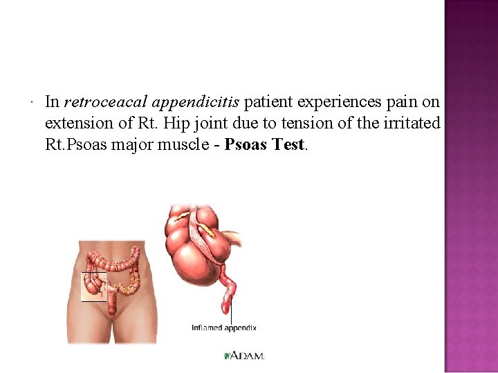
In retroceacal appendicitis patient experiences pain on extension of Rt. Hip joint due to tension of the irritated Rt. Psoas major muscle - Psoas Test.
Anatomical factors which cause appendicitis areØ Faecolith in the blind lumen of appendix. Ø Supplied by an end artery. Ø Presence of hiatus muscularis in the muscular coat. Ø Presence of numerous lymphatic follicles in the sub mucous coat.
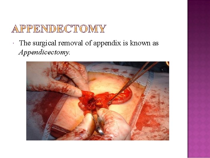
The surgical removal of appendix is known as Appendicectomy.
If the ceacum adheres to the inferior surface of the liver when it returns to the abdomen, it wil be drawn superiorly as the liver dimnishes in size. As a result ceacum & appendix remains in the fetal position. Its more common in males & occur in about 6% of fetuses. It may create a problem in the diagnosis of appendicitis & appendectomy.
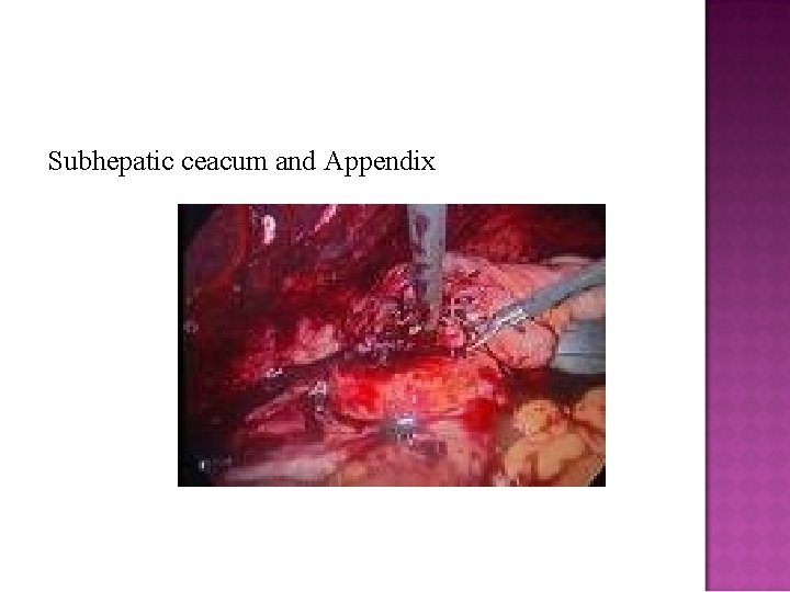
Subhepatic ceacum and Appendix
It results from incomplete fixation of the ascending colon. In approximately 10% people ceacum has an abnormal freedom & in unusual cases it may herniate in to Rt. inguinal canal. This condition is clinically significant as it can lead to Volvulus (twisting)of ceacum.
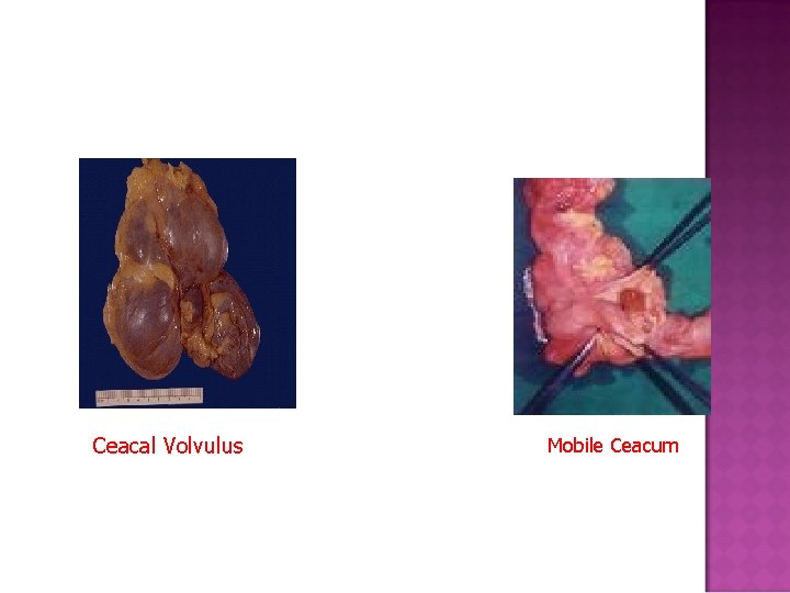
Ceacal Volvulus Mobile Ceacum
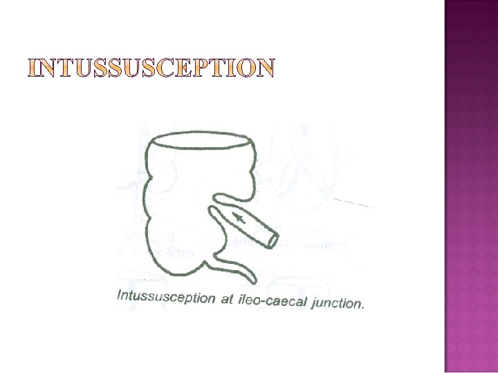
Sometimes the terminal part of the ileum is telescopically invaginated into ceacum and ascending colon at the ileo-ceacal junction and produces intestinal obstruction. This phenomenon is called intussusception.
Caecum acts as a guide in the operation of intestinal obstruction: Ø If the ceacum is distended, obstruction occur in the large gut. Ø If the ceacum is empty, the obstruction is of small gut.

- Slides: 32