
Dr John Trantalis ORTHOPAEDIC EMERGENCIES CARE OF THE

- Slides: 99
Dr John Trantalis ORTHOPAEDIC EMERGENCIES CARE OF THE INJURED LIMB
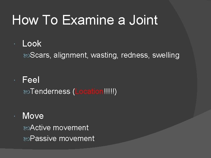
How To Examine a Joint Look Scars, alignment, wasting, redness, swelling Feel Tenderness (Location!!!!!) Move Active movement Passive movement
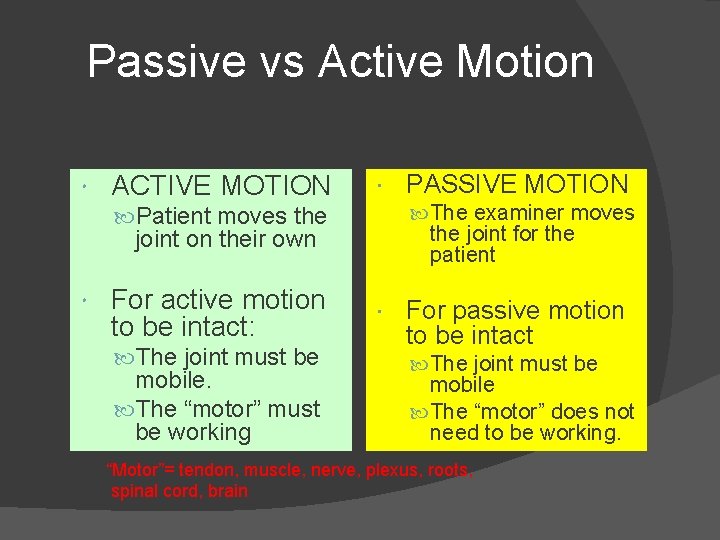
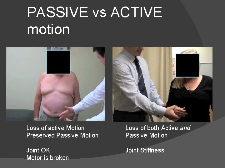
Passive vs Active Motion ACTIVE MOTION The examiner moves Patient moves the joint for the patient joint on their own For active motion to be intact: The joint must be mobile. The “motor” must be working PASSIVE MOTION For passive motion to be intact The joint must be mobile The “motor” does not need to be working. “Motor”= tendon, muscle, nerve, plexus, roots, spinal cord, brain
PASSIVE vs ACTIVE motion Loss of active Motion Preserved Passive Motion Loss of both Active and Passive Motion Joint OK Motor is broken Joint Stiffness
A White, Pulseless Limb after an Injury
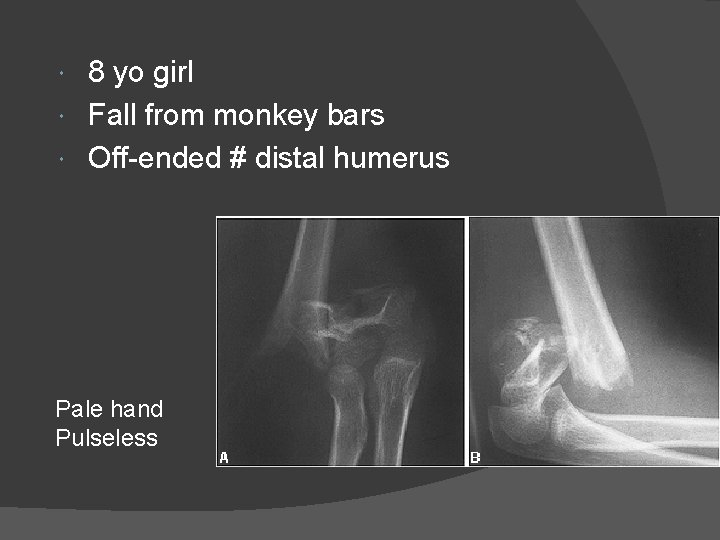
8 yo girl Fall from monkey bars Off-ended # distal humerus Pale hand Pulseless
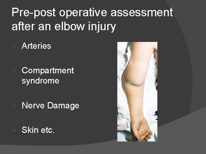
Pre-post operative assessment after an elbow injury Arteries Compartment syndrome Nerve Damage Skin etc.
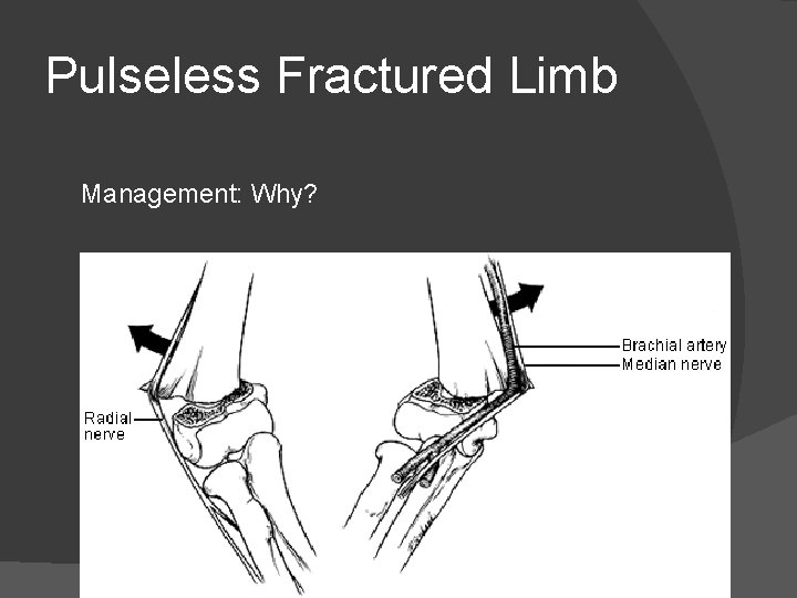
Pulseless Fractured Limb Management: Why?
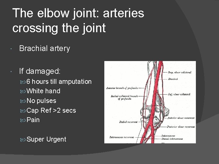
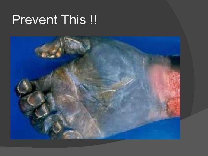
The elbow joint: arteries crossing the joint Brachial artery If damaged: 6 hours till amputation White hand No pulses Cap Ref >2 secs Pain Super Urgent
Prevent This !!
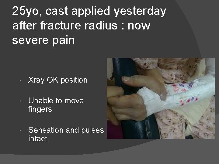
25 yo, cast applied yesterday after fracture radius : now severe pain Xray OK position Unable to move fingers Sensation and pulses intact
Diagnosis and Management?
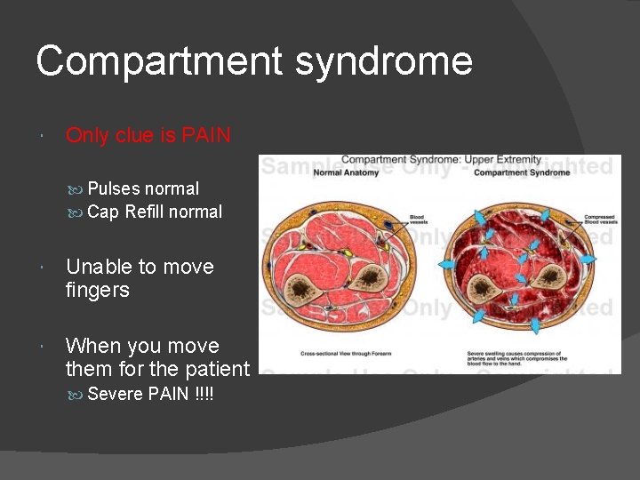
Compartment syndrome Only clue is PAIN Pulses normal Cap Refill normal Unable to move fingers When you move them for the patient Severe PAIN !!!!
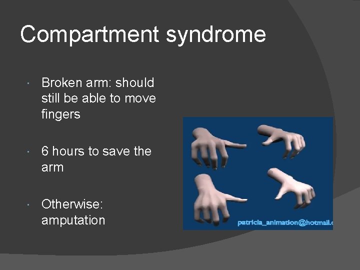
Compartment syndrome Broken arm: should still be able to move fingers 6 hours to save the arm Otherwise: amputation
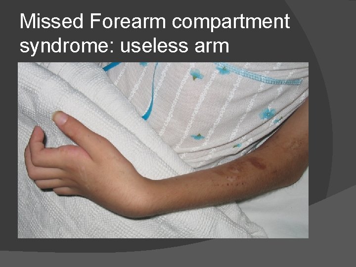
Missed Forearm compartment syndrome: useless arm
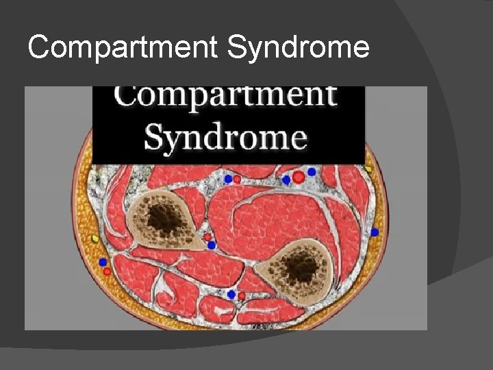
Compartment Syndrome
Why are the Pulses normal and the Fingers Pink? Ischaemia to muscles Capillaries 5 mm. Hg- shut down with small rise in compartment pressue Radial Artery Pressure of 120/80 mm. Hg. Therefore it stays open and hand stays pink
Therefore…. Only need one thing to diagnose compartment syndrome…. . PAIN
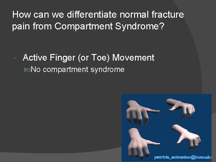
How can we differentiate normal fracture pain from Compartment Syndrome? Active Finger (or Toe) Movement No compartment syndrome
What to do if you suspect Compartment Syndrome…. CALL FOR HELP!!!!!! Speak to the orthopaedic team urgently Do not leave messages You must speak to somebody urgently Then… ○ Remove all encircling bandages… A tight bandage or plaster can cause compartment syndrome But it can also occur without anything wrapped around the limb… skin & fascia
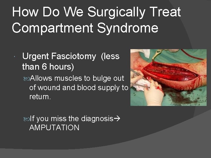
How Do We Surgically Treat Compartment Syndrome Urgent Fasciotomy (less than 6 hours) Allows muscles to bulge out of wound and blood supply to return. If you miss the diagnosis AMPUTATION
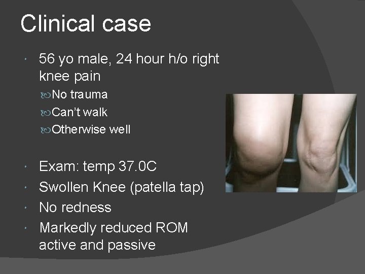
Clinical case 56 yo male, 24 hour h/o right knee pain No trauma Can’t walk Otherwise well Exam: temp 37. 0 C Swollen Knee (patella tap) No redness Markedly reduced ROM active and passive
Provisional Diagnosis? Septic Arthritis Differential Diagnosis? Gout Pseudogout Haemarthosis
Key Clinical Sign for Septic Arthritis in any Joint Decreased active and passive motion The joint is very inflamed and painful. Patient’s muscles spasm when movement is attempted.
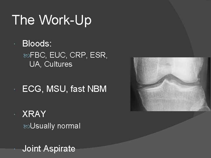
The Work-Up Bloods: FBC, EUC, CRP, ESR, UA, Cultures ECG, MSU, fast NBM XRAY Usually normal Joint Aspirate
Inflammatory Markers CRP C Reactive Protein Very Sensitive for inflammation or infection Indicative of what was happening in the body 1 day ago ESR Erythrocyte Sedimentation Rate Indicative of what was happening in the body 3 days ago.
Joint Aspirate Before any antibiotics are given. Never through red skin (can introduce skin infection into the joint) Send off for MCS, crystals, cell count.
Septic Arthritis: Treatment Joint Washout (arthroscopic) Removes the enzymes from white cells which otherwise destroy the articular cartilage IV antibiotics Empirical: cover Staph Aureus
Fractured Hips in The Elderly
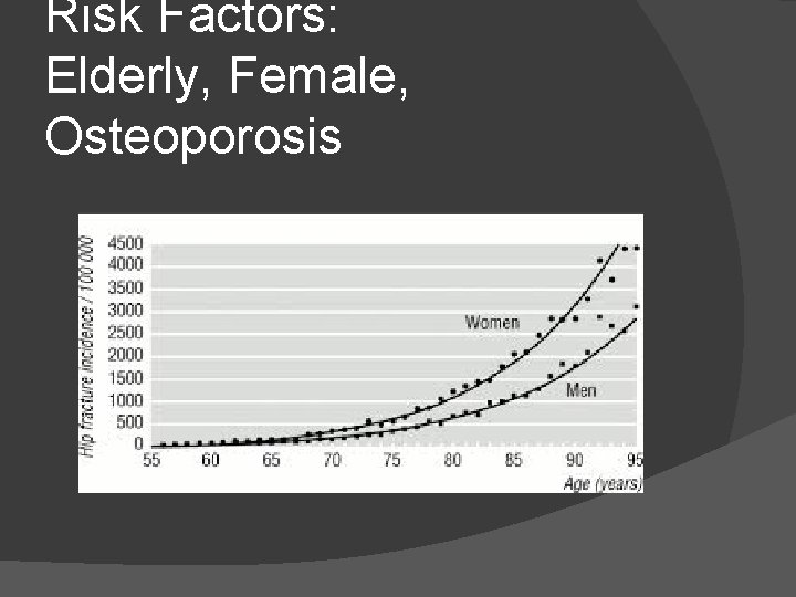
Risk Factors: Elderly, Female, Osteoporosis
One Year Mortality Rate for a Fractured NOF 30% Within 1 year, 30% or patients who sustain a fractured NOF will pass away. Due to comorbidities usually
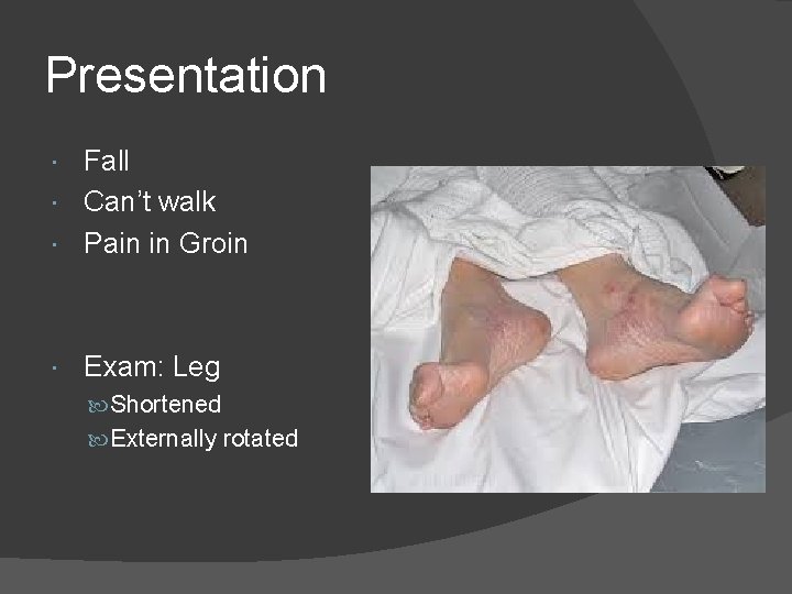
Presentation Fall Can’t walk Pain in Groin Exam: Leg Shortened Externally rotated
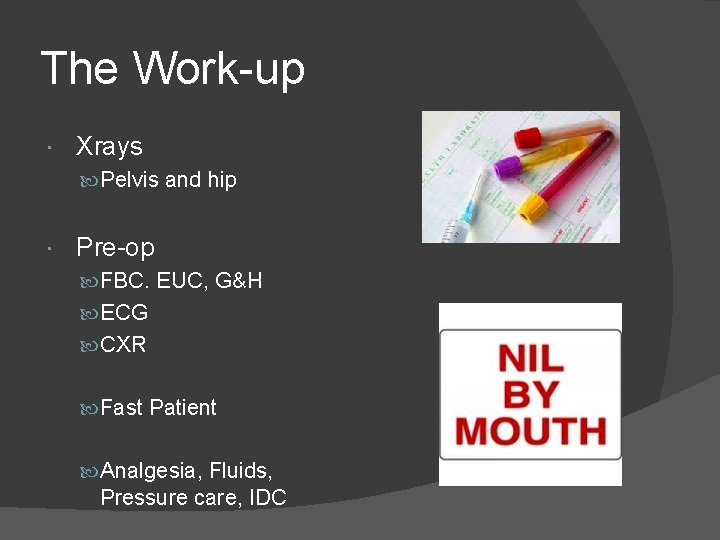
The Work-up Xrays Pelvis and hip Pre-op FBC. EUC, G&H ECG CXR Fast Patient Analgesia, Fluids, Pressure care, IDC
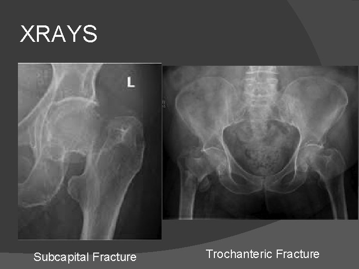
XRAYS Subcapital Fracture Trochanteric Fracture
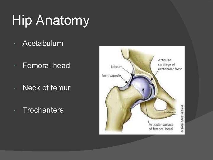
Hip Anatomy Acetabulum Femoral head Neck of femur Trochanters
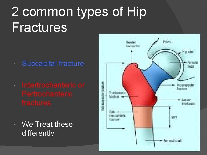
2 common types of Hip Fractures Subcapital fracture Intertrochanteric or Pertrochanteric fractures We Treat these differently
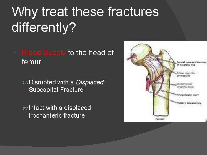
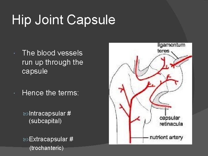
Why treat these fractures differently? Blood Supply to the head of femur Disrupted with a Displaced Subcapital Fracture Intact with a displaced trochanteric fracture
Hip Joint Capsule The blood vessels run up through the capsule Hence the terms: Intracapsular # (subcapital) Extracapsular # (trochanteric)
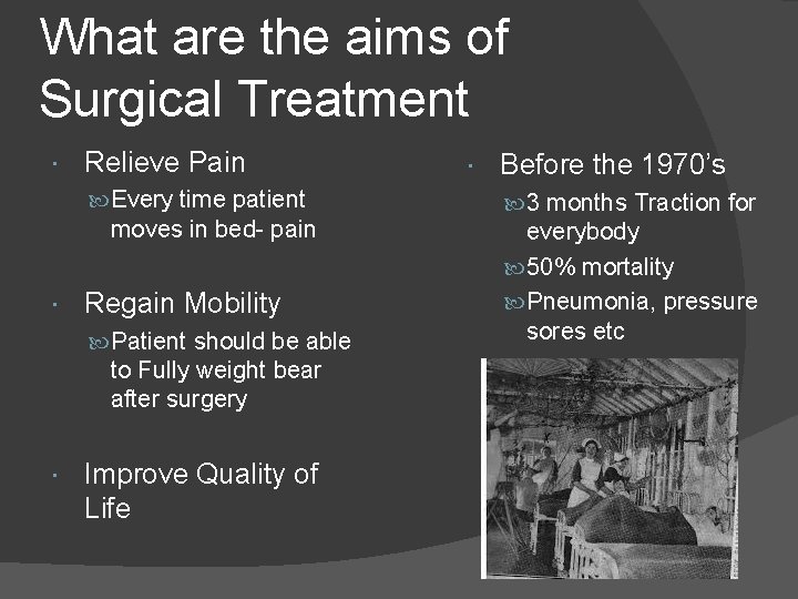
What are the aims of Surgical Treatment Relieve Pain Every time patient moves in bed- pain Regain Mobility Patient should be able to Fully weight bear after surgery Improve Quality of Life Before the 1970’s 3 months Traction for everybody 50% mortality Pneumonia, pressure sores etc
The Surgery Relieves Pain Patient with # NOF in bed…The fracture ends grind and cause pain with every movement Even with very ill patients, we still try to complete their surgery asap to relieve their pain and improve their quality of life (nursing etc) The faster the patient gets to surgery the less chance of pneumonia / pressure sores developing.
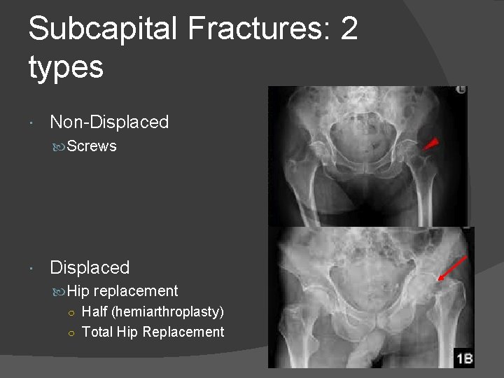
Subcapital Fractures: 2 types Non-Displaced Screws Displaced Hip replacement ○ Half (hemiarthroplasty) ○ Total Hip Replacement
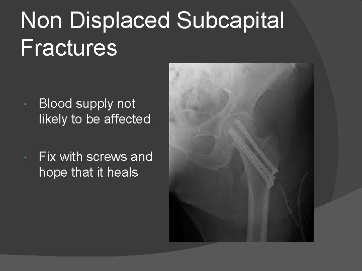
Non Displaced Subcapital Fractures Blood supply not likely to be affected Fix with screws and hope that it heals
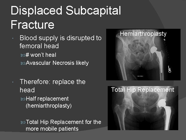
Displaced Subcapital Fracture Blood supply is disrupted to femoral head Hemiarthroplasty # won’t heal Avascular Necrosis likely Therefore: replace the head Half replacement (hemiarthroplasty) Total Hip Replacement for the more mobile patients Total Hip Replacement
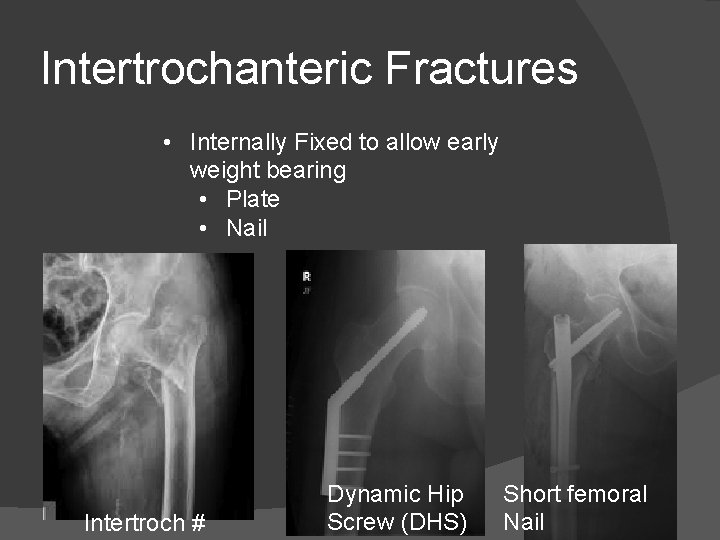
Intertrochanteric Fractures • Internally Fixed to allow early weight bearing • Plate • Nail Intertroch # Dynamic Hip Screw (DHS) Short femoral Nail
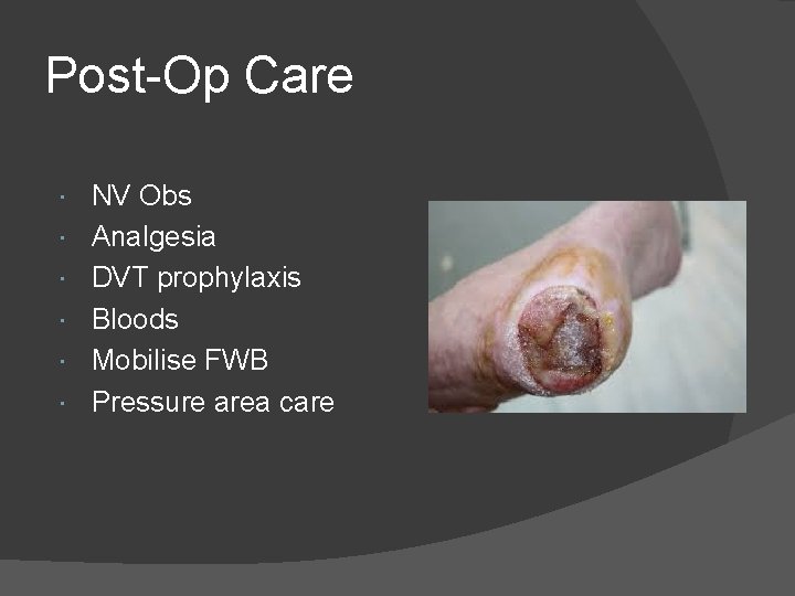
Post-Op Care NV Obs Analgesia DVT prophylaxis Bloods Mobilise FWB Pressure area care
THE INJURED LIMB Dr John Trantalis Orthopaedic Surgeon
Dislocated Joints Should all be reduced ASAP Pressure off NV structures Pain XRAY 2 views always CT if you are unsure Beware LOC ○ Trauma, Head injury Secondary survey You will detect decreased ROM ○ Seizures, electrocution
The Open Fracture
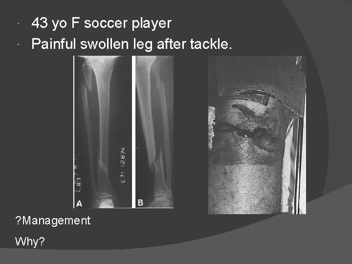
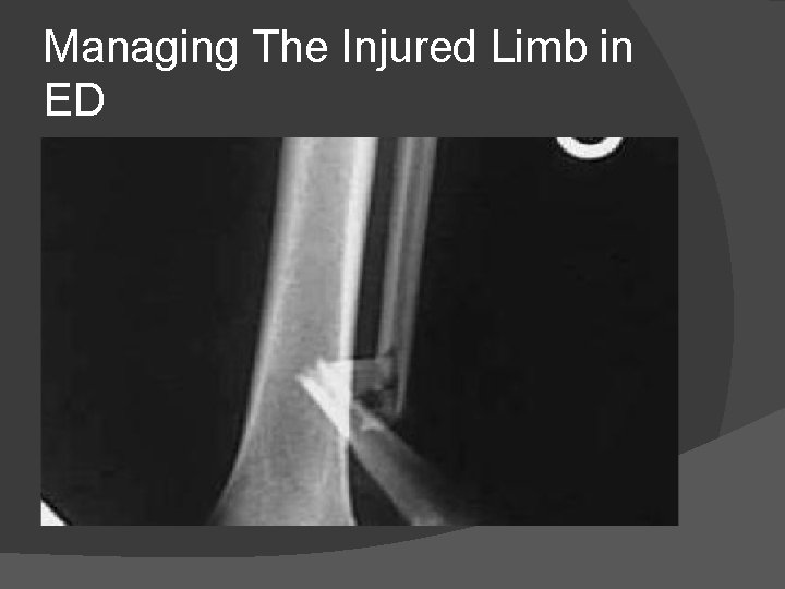
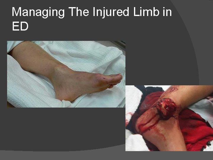
43 yo F soccer player Painful swollen leg after tackle. ? Management Why?
Managing The Injured Limb in ED
Managing The Injured Limb in ED
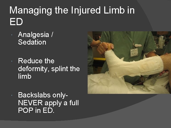
Managing the Injured Limb in ED Analgesia / Sedation Reduce the deformity, splint the limb Backslabs only. NEVER apply a full POP in ED.
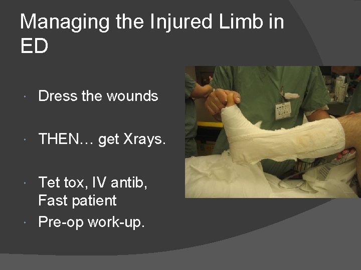
Managing the Injured Limb in ED Dress the wounds THEN… get Xrays. Tet tox, IV antib, Fast patient Pre-op work-up.
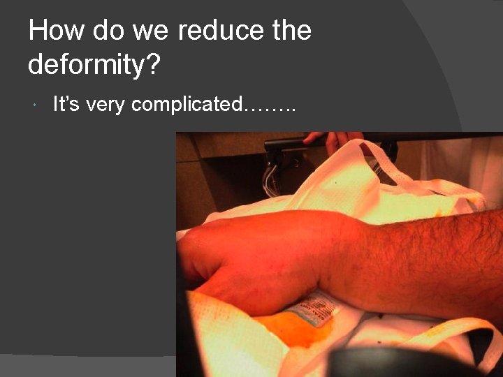
How do we reduce the deformity? It’s very complicated……. .
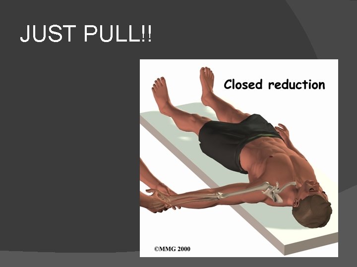
JUST PULL!!
How to describe a fracture
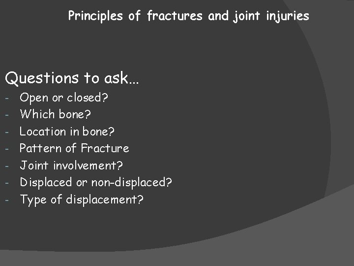
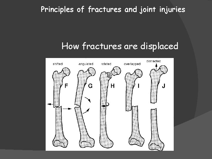
Principles of fractures and joint injuries Questions to ask… - Open or closed? Which bone? Location in bone? Pattern of Fracture Joint involvement? Displaced or non-displaced? Type of displacement?
Principles of fractures and joint injuries How fractures are displaced
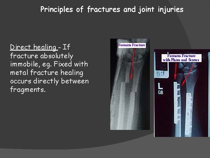
Principles of fractures and joint injuries Direct healing - If fracture absolutely immobile, eg. Fixed with metal fracture healing occurs directly between fragments.
Principles of fractures and joint injuries How Long Does It Take To for a Fracture to Heal? • Depends on…… • Patient Factors: Age, Comorbidities etc • Fracture Factors: which bone, type of fracture etc • Can take up to 6 months for a tibia versus 2 weeks for a phalanx. • Healing seen on XRAY always takes longer than clinical union
Clinical signs of fracture Union No tenderness, movement or crepitus at a fracture site.
The injured limb – Clinical features
Clinical Features If you remember nothing else about examining a limb… LOOK FEEL MOVE
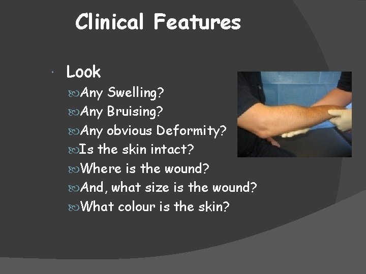
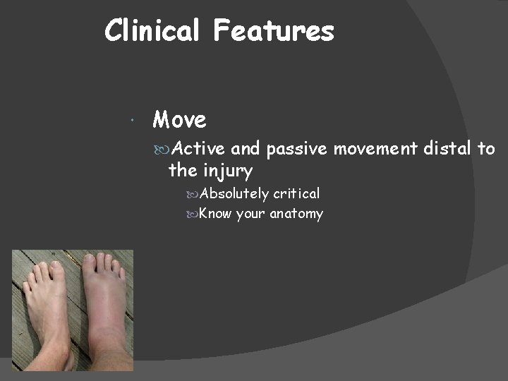
Clinical Features Look Any Swelling? Any Bruising? Any obvious Deformity? Is the skin intact? Where is the wound? And, what size is the wound? What colour is the skin?
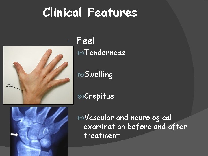
Clinical Features Feel Tenderness Swelling Crepitus Vascular and neurological examination before and after treatment
Clinical Features Move Active and passive movement distal to the injury Absolutely critical Know your anatomy
The injured limb Imaging
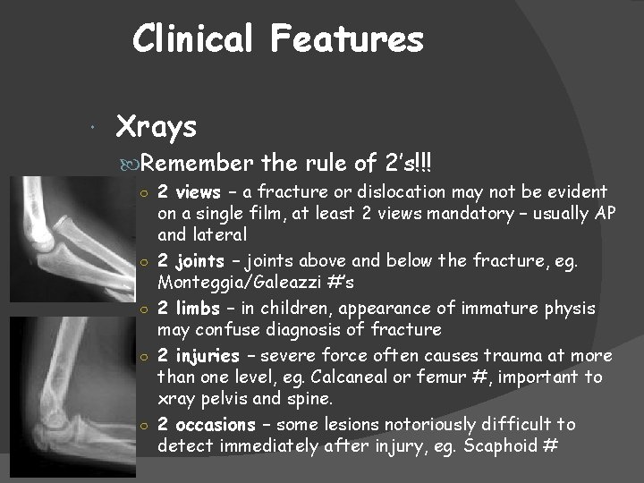
Clinical Features Xrays Remember the rule of 2’s!!! ○ 2 views – a fracture or dislocation may not be evident ○ ○ on a single film, at least 2 views mandatory – usually AP and lateral 2 joints – joints above and below the fracture, eg. Monteggia/Galeazzi #’s 2 limbs – in children, appearance of immature physis may confuse diagnosis of fracture 2 injuries – severe force often causes trauma at more than one level, eg. Calcaneal or femur #, important to xray pelvis and spine. 2 occasions – some lesions notoriously difficult to detect immediately after injury, eg. Scaphoid #
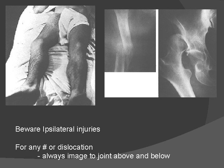
Beware Ipsilateral injuries For any # or dislocation - always image to joint above and below
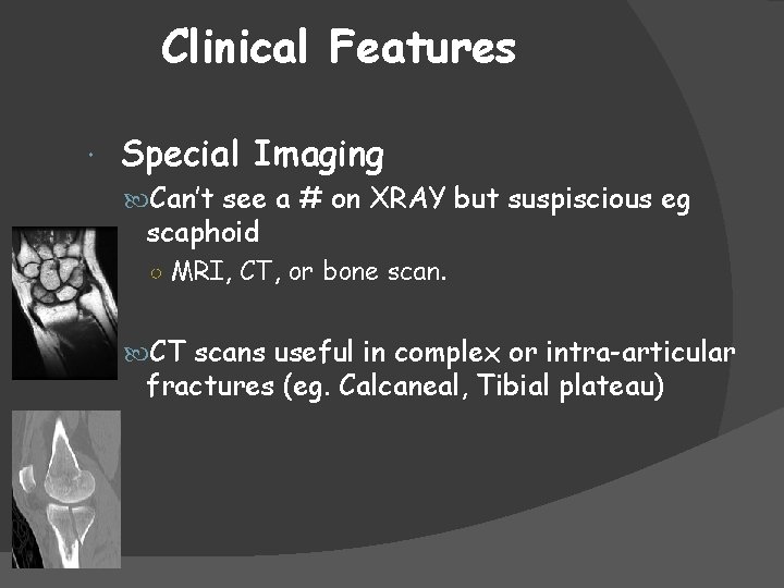
Clinical Features Special Imaging Can’t see a # on XRAY but suspiscious eg scaphoid ○ MRI, CT, or bone scan. CT scans useful in complex or intra-articular fractures (eg. Calcaneal, Tibial plateau)
The injured limb – Management principles
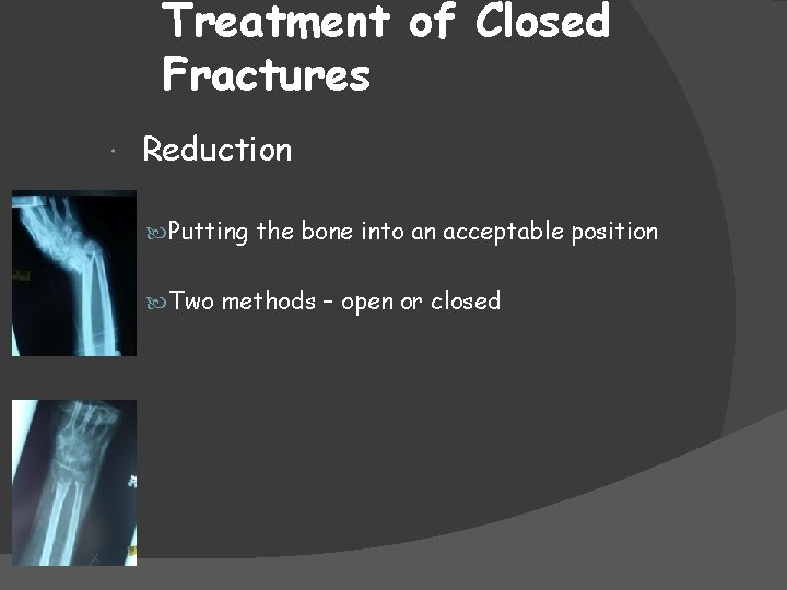
Treatment of Closed Fractures Reduction Putting the bone into an acceptable position Two methods – open or closed
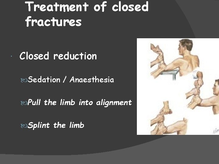
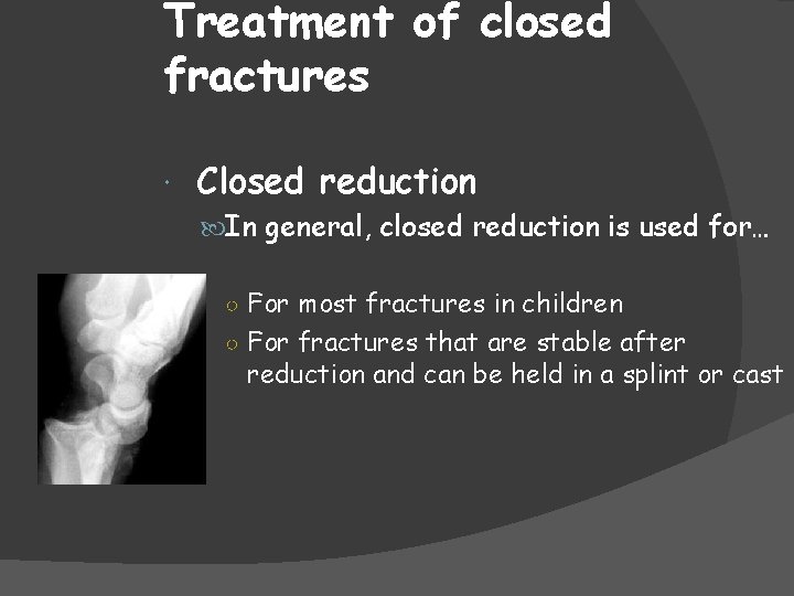
Treatment of closed fractures Closed reduction Sedation / Anaesthesia Pull the limb into alignment Splint the limb
Treatment of closed fractures Closed reduction In general, closed reduction is used for… ○ For most fractures in children ○ For fractures that are stable after reduction and can be held in a splint or cast
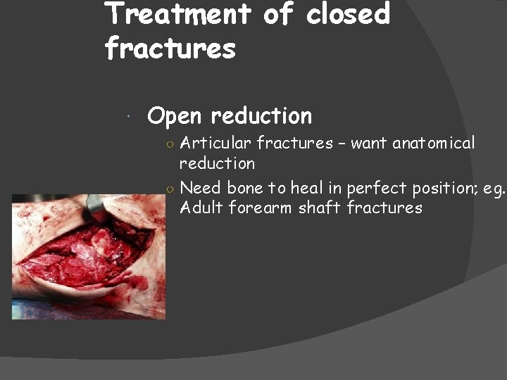
Treatment of closed fractures Open reduction ○ Articular fractures – want anatomical reduction ○ Need bone to heal in perfect position; eg. Adult forearm shaft fractures
Fracture Immobilisation Following reduction, the available methods of holding are… 1) cast splintage 2) Internal Fixation (plates, screws, nails) 3) external fixation 4) Traction
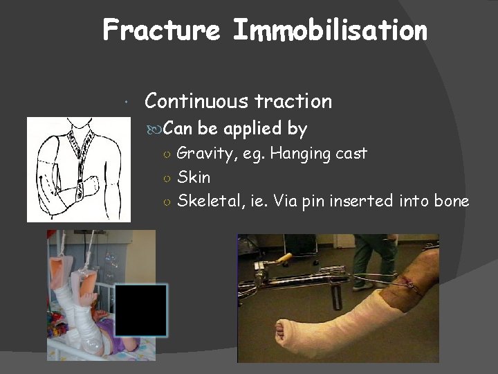
Fracture Immobilisation Continuous traction Can be applied by ○ Gravity, eg. Hanging cast ○ Skin ○ Skeletal, ie. Via pin inserted into bone
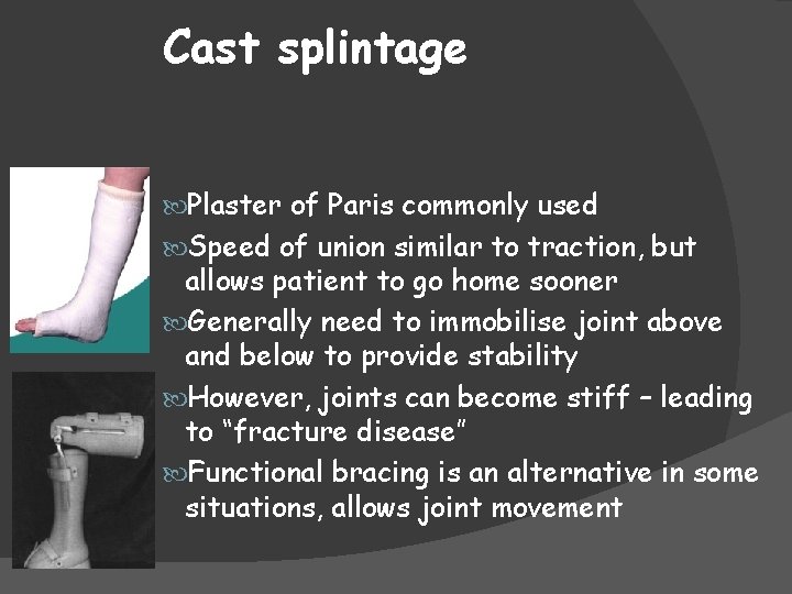
Cast splintage Plaster of Paris commonly used Speed of union similar to traction, but allows patient to go home sooner Generally need to immobilise joint above and below to provide stability However, joints can become stiff – leading to “fracture disease” Functional bracing is an alternative in some situations, allows joint movement
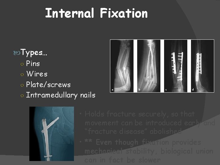
Internal Fixation Types… ○ Pins ○ Wires ○ Plate/screws ○ Intramedullary nails • Holds fracture securely, so that movement can be introduced early and “fracture disease” abolished • ** Even though fixation provides mechanical stability, biological union can in fact be slower
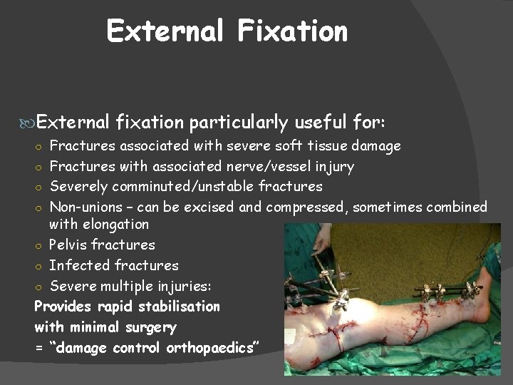
External Fixation External fixation particularly useful for: ○ Fractures associated with severe soft tissue damage ○ Fractures with associated nerve/vessel injury ○ Severely comminuted/unstable fractures ○ Non-unions – can be excised and compressed, sometimes combined with elongation ○ Pelvis fractures ○ Infected fractures ○ Severe multiple injuries: Provides rapid stabilisation with minimal surgery = “damage control orthopaedics”
Complications of fractures Early Complications, including: ○ Vascular injury ○ Nerve injury ○ Compartment syndrome ○ Infection ○ Fracture blisters (elevation of superficial layers of skin by oedema) Late Complications, including: ○ Delayed/Non-union ○ Malunion ○ Avascular necrosis ○ Growth disturbance ○ Stiffness, CRPS, post traumatic osteoarthritis, etc
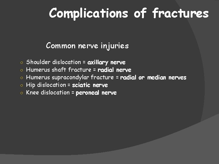
Complications of fractures Common nerve injuries ○ Shoulder dislocation = axillary nerve ○ Humerus shaft fracture = radial nerve ○ Humerus supracondylar fracture = radial or median nerves ○ Hip dislocation = sciatic nerve ○ Knee dislocation = peroneal nerve
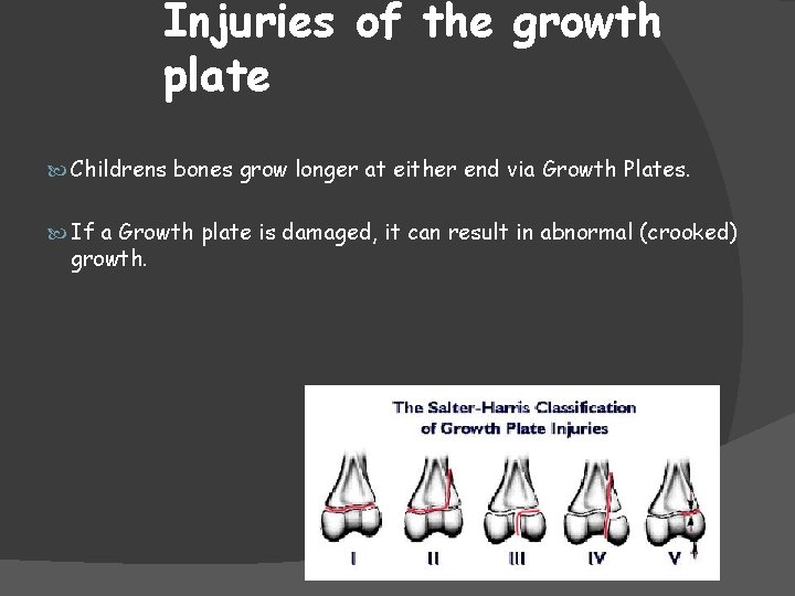
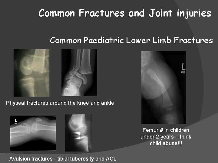
Injuries of the growth plate Childrens bones grow longer at either end via Growth Plates. If a Growth plate is damaged, it can result in abnormal (crooked) growth.
Complications of fractures Delayed Union and Non Union • Delayed union = prolonged time to fracture union • Non Union = failure of bone to unite Factors – multiple: Smoking increases risk 30%
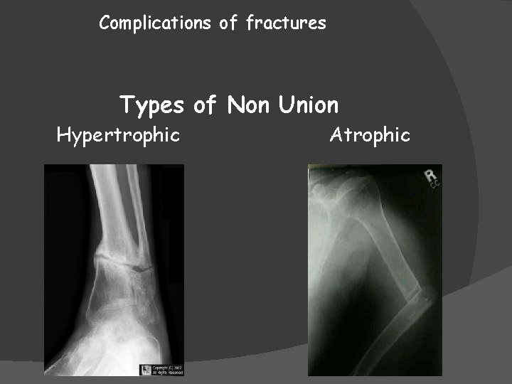
Complications of fractures Types of Non Union Hypertrophic Atrophic
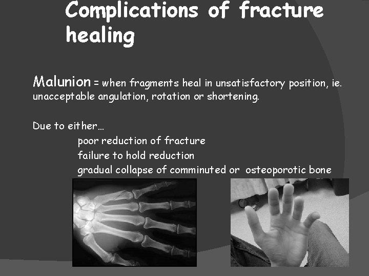
Complications of fracture healing Malunion = when fragments heal in unsatisfactory position, ie. unacceptable angulation, rotation or shortening. Due to either… poor reduction of fracture failure to hold reduction gradual collapse of comminuted or osteoporotic bone
Complications of fracture healing Avascular Necrosis (AVN) Certain fractures/injuries are notorious for their propensity to develop ischemia and subsequent bone necrosis… 1) Femoral head - #femoral neck (#NOF) or hip dislocation 2) Scaphoid – particularly with more proximal fractures, as blood supply is from distal to proximal 3) Talus – similar to scaphoid, blood supplies bone from distal to proximal, therefore body talus at risk AVN
Common Upper Limb Injuries
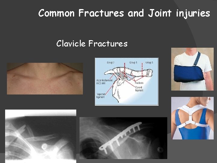
Common Fractures and Joint injuries Clavicle Fractures
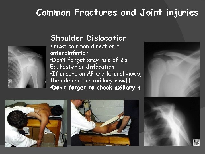
Common Fractures and Joint injuries Shoulder Dislocation • most common direction = anteroinferior • Don’t forget xray rule of 2’s Eg. Posterior dislocation • If unsure on AP and lateral views, then demand an axillary view!!! • Don’t forget to check axillary n.
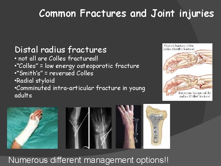
Common Fractures and Joint injuries Distal radius fractures • not all are Colles fractures!! • “Colles” = low energy osteoporotic fracture • “Smith’s” = reversed Colles • Radial styloid • Comminuted intra-articular fracture in young adults Numerous different management options!!
Common Lower Limb Injuries
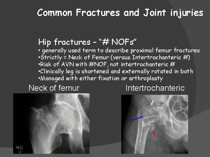
Common Fractures and Joint injuries Hip fractures – “# NOFs” • generally used term to describe proximal femur fractures • Strictly = Neck of Femur (versus Intertrochanteric #) • Risk of AVN with #NOF, not intertrochanteric # • Clinically leg is shortened and externally rotated in both • Managed with either fixation or arthroplasty Neck of femur Intertrochanteric
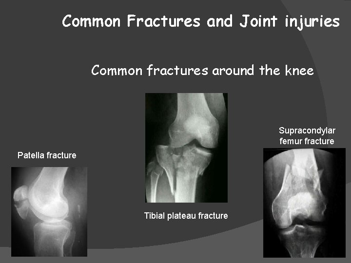
Common Fractures and Joint injuries Common fractures around the knee Supracondylar femur fracture Patella fracture Tibial plateau fracture
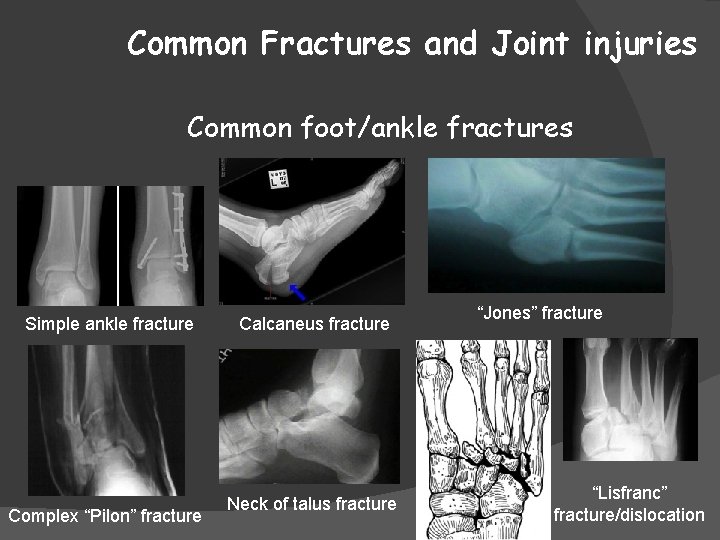
Common Fractures and Joint injuries Common foot/ankle fractures Simple ankle fracture Complex “Pilon” fracture Calcaneus fracture Neck of talus fracture “Jones” fracture “Lisfranc” fracture/dislocation
Common Paediatric Injuries
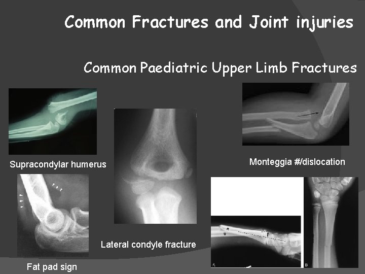
Common Fractures and Joint injuries Common Paediatric Upper Limb Fractures Supracondylar humerus Monteggia #/dislocation Galeazzi #/dislocation Lateral condyle fracture Fat pad sign
Common Fractures and Joint injuries Common Paediatric Lower Limb Fractures Physeal fractures around the knee and ankle Femur # in children under 2 years – think child abuse!!! Avulsion fractures - tibial tuberosity and ACL