Dr JANSHER SENIOR RESIDENT CARDIOLOGY CALICUT MEDICAL COLLEGE


 Most elective or non-emergency Cardioversions are performed : • To treat atrial")
 Cardioversion is used in emergency situations to correct a rapid abnormal rhythm")
 cardioversion: refers to")
")
; is a procedure whereby")

 guidelines: • Any patient with")


 is a device that")


- Slides: 40
Dr JANSHER SENIOR RESIDENT CARDIOLOGY CALICUT MEDICAL COLLEGE
7 Defibrillation History 1900 defibrillation discovery 1947 human defibrillation 1967 successful outside hospital defibrillation For VF or pulseless VT Defibrillation (shock success) termination of VF for at least 5 s following shock
9 Sternum Apex
8 Types 360 J 200 J http: //www. resuscitationcentral. com/
Monophasic Vs Biphasic • Defibrillators can deliver energy in various waveforms that are broadly characterized as monophasic or biphasic. • Monophasic defibrillation delivers a charge in only one direction. Biphasic defibrillation delivers a charge in one direction for half of the shock and in the electrically opposite direction for the second half. • Newer defibrillators deliver energy in biphasic waveforms. Biphasic waveform defibrillators deliver a more consistent magnitude of current. They tend to successfully terminate arrhythmias at lower energies than monophasic waveform defibrillators (Automated External defibrillator (AED)
10 Automated AED @ KLIA
Definition of Cardioversion is a synchronized administration of shock during the R waves or QRS complex of a cardiac cycle and restore a rapid heart beat back to normal.
Cardioversion (Elective) Most elective or non-emergency Cardioversions are performed : • To treat atrial fibrillation or atrial flutter to regain heart rhythm. • To treat disturbances originating in the upper Chambers (atria) of the heart.
Cardioversion (Emergency) Cardioversion is used in emergency situations to correct a rapid abnormal rhythm associated with faintness, low blood pressure, chest pain, difficulty breathing, or loss of consciousness.
Types Of Cardioversion can be "chemical" or "electrical". • Chemical (Pharmacological) cardioversion: refers to the use of antiarrhythmic medications to restore the heart's normal rhythm.
Pharmacological Cardioversion of Atrial Fibrillation< 7 days Duration Class of Recommendation (level of evidence) Drug Agents with proven efficacy Dofetilide I (A) Flecainide I (A) Ibutilide I (A) Propafenone I (A) Amiodarone IIa (A) Less effective or incompletely studied Disopyramide IIb (B) Procainamide IIb (B) Quinidine IIb (B) Should not be administered Digoxin III (A) Sotalol III (A) Modified from: ACC/AHA/ESC Practice Guidelines, Fuster et al. JACC 2006; 48: e 149 -246 9
Electric Cardioversion (Also known as " direct-current" or DC cardioversion); is a procedure whereby a synchronized electrical shock is delivered through the chest wall to the heart through special electrodes or paddles that are applied to the skin of the chest and back.
Basic principles • Transient delivery of electrical current causes a momentary depolarization of most cardiac cells allowing the sinus node to resume normal pacemaker activity. • In the presence of reentrant-induced arrhythmia, such as PSVT and VT, electrical cardioversion interrupts the selfperpetuating circuit and restores a sinus rhythm. • Electrical cardioversion is much less effective in treating arrhythmia caused by increased automaticity (eg, digitalisinduced tachycardia, catecholamine-induced arrhythmia) since the mechanism of the arrhythmia remains after the arrhythmia is terminated and therefore is likely to recur.
Indications • Based on advanced cardiac life support (ACLS) guidelines: • Any patient with narrow or wide QRS complex tachycardia (ventricular rate >150) who is unstable (eg, chest pain, pulmonary edema, lightheadedness, hypotension, signs of shock). • Supraventricular tachycardia due to reentry • Atrial fibrillation • Atrial flutter (types I and II) • Atrial tachycardia • Monomorphic VT with pulse
Contraindications • Presence of left atrial thrombus • Digitalis toxicity or hypokalemia • Sinus tachycardia caused by various clinical conditions and catecholamine-induced arrhythmia • Multifocal atrial tachycardia • VF (since the cardioverter may not sense a QRS wave and may therefore fail to deliver a shock)
Recommendations for Direct-current Cardioversion of Atrial Fibrillation • Class I 1. When a rapid ventricular response does not respond promptly to pharmacological measures for patients with AF with ongoing myocardial ischemia, symptomatic hypotension, angina or hear faliure, immediate R-wave synchronized direct-current cardioversion is recommended. (Level of Evidence: C) 2. AF involving preexcitation when very rapid tachycardia or hemodynamic instability occurs. (Level of Evidence: B) 3. Cardioversion is recommended in patients without hemodynamic instability when symptoms of AF are unacceptable to the patient. In case of early relapse of AF after cardioversion, repeated cardioversion attempts may be made following administration of antiarrhythmic medication. (Level of Evidence: C)
Recommendations for Direct-current Cardioversion of Atrial Fibrillation • Class IIa 1. Direct-current cardioversion can be useful to restore sinus rhythm as part of a long-term management strategy for patients with AF. (Level of Evidence: B) 2. Patient preference is a reasonable consideration in the selection of infrequently repeated cardioversions for the management of symptomatic or recurrent AF. (Level of Evidence: C) ACC/AHA/ESC Practice Guidelines, Fuster et al. JACC 2006; 48: e 149 -246
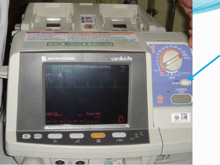
Equipment • Defibrillator with a synchronising button. • Emergency trolley with emergency drugs; ( lignocaine, atropine, and adrenaline ). • Oxygen mask, intubation equipment, airway. • Monitor and continuous recording facilities (BP, ECG, Sp. O 2). • Intravenous access • Suction device
Electrode Placement : Antero-apical position: one paddle is placed to the right of the sternum just below the clavicle. Another paddle is placed to the normal cardiac apex. Antero-posterior position: the anterior paddle placed over the apex, and the posterior paddle on the back in the left or right infrascapular region. Proper position of the paddles 22
Energy Requirements Atrial fibrillation energy requirements are as follows: • 200 Joules for monophasic devices • 120 -200 Joules for biphasic devices Atrial flutter energy requirements are as follows: • 100 Joules for monophasic devices • 50 -100 Joules for biphasic devices Ventricular tachycardia with pulse energy requirements are as follows: • 200 Joules for monophasic devices • 100 Joules for biphasic devices
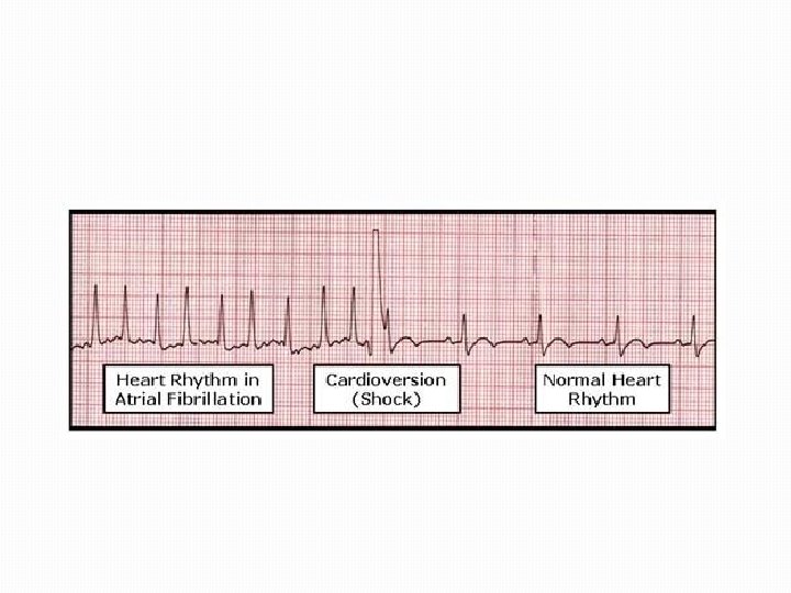
• ECG strip shows a atrial fibrillation terminated by a synchronized shock (synchronization marks [arrows] in the apex of the QRS complex) to normal sinus rhythm
Preparing for a Cardioversion Do not eat or drink for at least eight hours prior to the procedure. Blood thining medicines may be given with electrical cardioversion to prevent clots from moving to the heart Take your regularly scheduled medications the morning of the procedure unless your medical practitioner has told you otherwise. Stop digoxin 48 hours prior to the procedure Bring a list of all your medications with you.
Preparing for a Cardioversion Do not apply any lotions or ointments to chest or back as this may interfere with the adhesiveness of the shocking pads. Do not drive yourself home after receiving sedation anesthesia.
Defibrillation Is a medical technique used to counter the onset of ventricular fibrillation, a common cause of cardiac arrest, and pulseless ventricular tachycardia, In simple terms, the process uses an electric shock to stop the heart arrhythmias, in the hope that the heart will restart with rhythmic contractions.
Differences Between Cardioversion & Defibrillation One major difference between cardioversion and defibrillation with the timing of the delivery of electrical current. Another major difference concerns the circumstance defibrillation usually performed as an emergency treatment. Cardioversion is usually, but not always a planned procedure.
16 Defibrillation Cardioversion
17
Implantable Cardioversion Defibrillation An implantable cardioverter-defibrillator (often called an ICD) is a device that briefly passes an electric current through the heart. It is "implanted, " or put in your body surgically. It includes a pulse generator and one or more leads. The pulse generator constantly watches your heartbeat.
Procedure The most well-known type of electrode is the traditional metal paddle with an insulated handle. This type must be held in place on the patient's skin while a shock or a series of shocks is delivered.
Procedure Steps: • Place paddles so that they do not touch pts clothing or bed linens or not near direct oxygen supply. • Ensure monitor is attached to patient. • Do not charge the machine untill ready to shock.
Procedure • Exert 25 pound pressure on the paddle. • Ensure you and every body is free of the patient. Inspect skin for burns. • Record the delivered energy.
Treatment Shock may be delivered, but it is not regarded as the treatment of choice. Antiarrhythmic medications such as amiodarone, lidocaine or magnesium are given if ventricular dysrhythmia persists.
Special Considerations for Anticoagulation Prior to Cardioversion • For patients with AF > 48 hours of AF, or when duration is unknown, 3 weeks of anticoagulation are required prior to cardioversion. • Documentation of anticoagulation adequacy is important prior to cardioversion • Anticoagulation must be continued for at least 4 weeks post cardioversion • TEE can be used to assess LA for thrombus as alternative to 3 -week anticoagulation (however, anti-coagulation must continue for 4 weeks post cardioversion
Special Considerations • Cardioversion can be performed safely in pregnant women. The fetal heart rate should be monitored during the procedure using fetal monitoring techniques. • Cardioversion in patients with permanent pacemaker/ICD should be performed with extra care. Improper technique may damage the device, lead system, or myocardial tissue, resulting in device malfunction. The electrode paddle or patch should be at least 12 cm from the pulse generator and antero-posterior paddle position and lowest amount of energy be used.
Complications • • • Possible complications of cardioversion are uncommon but may include: Harmless arrhythmias, such as atrial, ventricular, and junctional premature beats. Serious complications include ventricular fibrillation (VF) resulting from high amounts of electrical energy, digitalis toxicity, severe heart disease, or improper synchronization of the shock with the R wave Thromboembolization is associated with cardioversion in 1 -3% of patients, especially in patients with atrial fibrillation who have not been anticoagulated prior to cardioversion. Bruising, burning or pain where the paddles were used. Allergic reactions from medicines used in pharmacologic cardioversion.
Complications • Myocardial necrosis can result from high-energy shocks. ST segment elevation can be seen immediately and usually lasts for 1 -2 minutes. ST segment elevation that lasts longer than 2 minutes usually indicates myocardial injury unrelated to the shock. • Pulmonary edema is a rare complication of cardioversion. It is probably due to transient left atrial standstill and left ventricular systolic dysfunction.