
DR IMRANA AZIZ Assistant Professor Surgical Department CYSTS



- Slides: 46
DR IMRANA AZIZ Assistant Professor Surgical Department
CYSTS & ULCERS
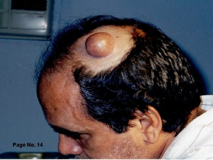
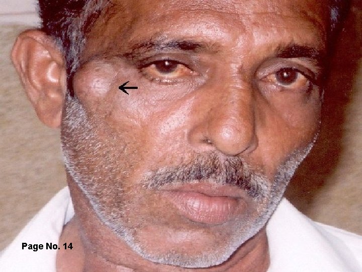
CYSTS • Cyst is a fluid filled sac bound by a wall. • Fluid is often clear, colorless or cholesterol crystals, or tooth paste like. • True Cyst: lined with epithelial or endothelial cells. • False Cyst: which are walled off fluid collection not by epithelium e. g. Pancreatic pseudocyst
Classification Ø Congenital Ø Acquired
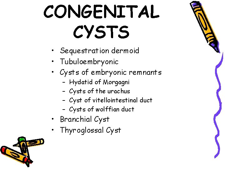
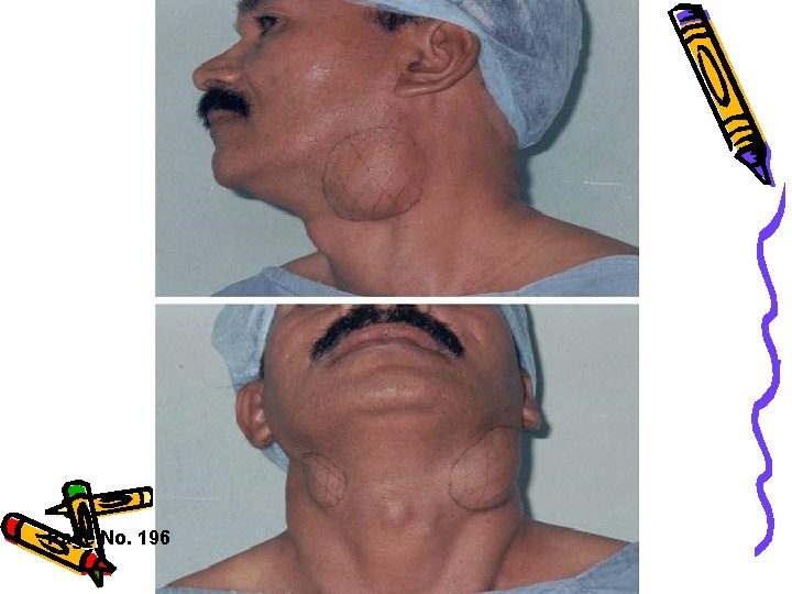
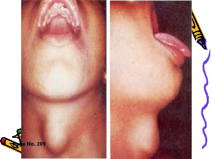
CONGENITAL CYSTS • Sequestration dermoid • Tubuloembryonic • Cysts of embryonic remnants – – Hydatid of Morgagni Cysts of the urachus Cyst of vitellointestinal duct Cysts of wolffian duct • Branchial Cyst • Thyroglossal Cyst
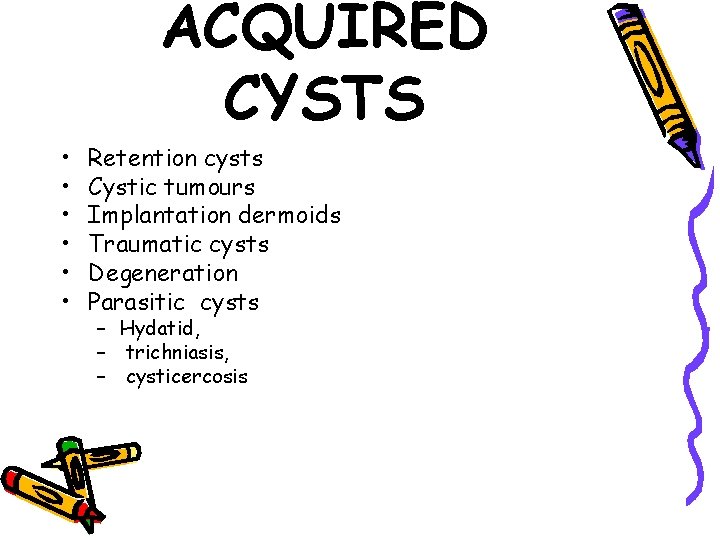
ACQUIRED CYSTS • • • Retention cysts Cystic tumours Implantation dermoids Traumatic cysts Degeneration Parasitic cysts – Hydatid, – trichniasis, – cysticercosis
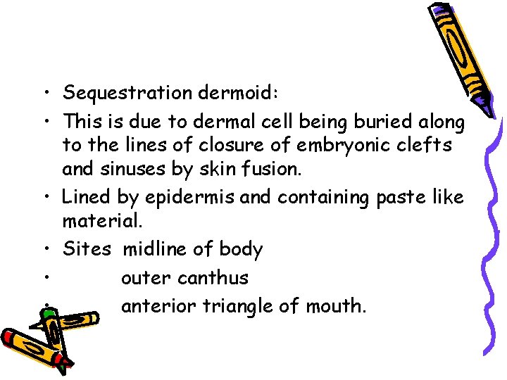
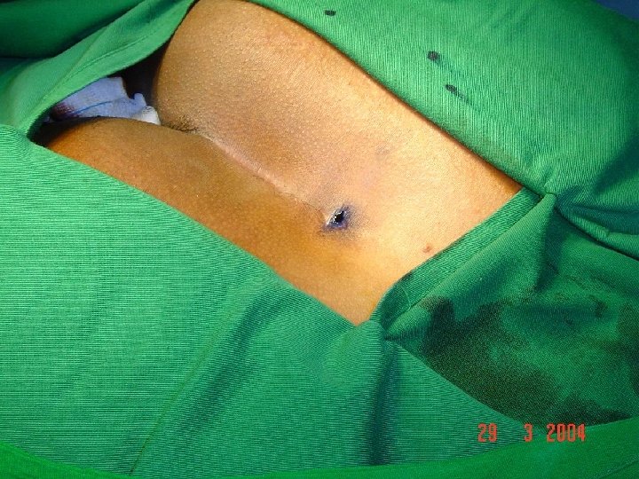
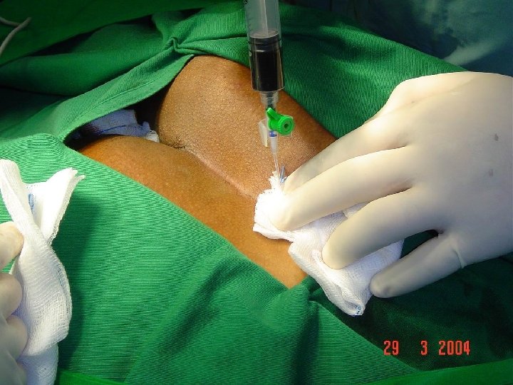
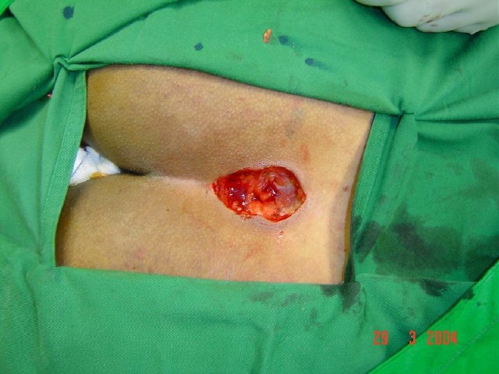
• Sequestration dermoid: • This is due to dermal cell being buried along to the lines of closure of embryonic clefts and sinuses by skin fusion. • Lined by epidermis and containing paste like material. • Sites midline of body • outer canthus • anterior triangle of mouth.
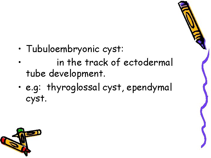
• Tubuloembryonic cyst: • in the track of ectodermal tube development. • e. g: thyroglossal cyst, ependymal cyst.
• Acquired cyst: • Retention cyst: • due to accumulation of secretion in gland behind an obstruction of a duct. • e. g: sebaceous cyst, Pseudu pancreatic cyst, parotid gland cyst.
• Distension cyst: • occur in thyroid from dilatation of acni. • cystic hygroma and lymphatic cyst.
Exudation Cyst • Exudative fluid accumulate in endothelium lined anatomical space. • e. g: Hydrocele, bursa.
Cystic tumor • e. g: cystic teratoma, cystadenoma. Implantation dermoid • Squamous epithelium driven beneath skin by penetrating wound.
Clinical Features • Varies according to the site & size • Pain → enlarging cysts, • Pain → Secondary to haemorrhage, infection, rupture, torsion • Acute abdominal emergency: torsion or rupture of ovarian cyst
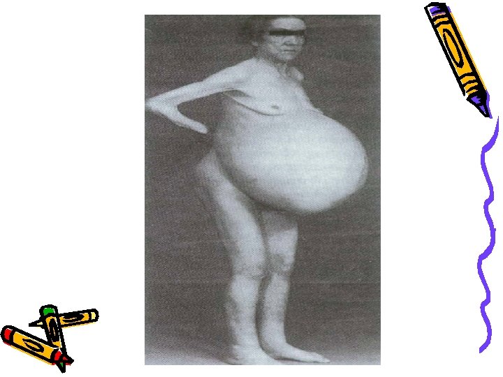
Clinical Features Compression symptoms resulting from compression of adjacent structures Haemorrhage in thyroglossal cyst → increase in size → compress the trachea Large ovarian cyst → abd: fullness & reduced appetite resulting from raises I/abd: pressure Obstruction to pelvic veins → varicose veins of lower limbs
COMPLICATIONS 1. 2. 3. 4. 5. 6. Infection Haemorrhage Torsion Obstruction Calcification Malignancy ( very rarely)
INVESTIGATIONS Signs: Fluctuant, transilluminant if containing clear fluid Diagnosis: obvious in cases of superficial cysts Deep seated intra-abdominal or thoracic cysts need U/sound, CT scan, MRI
Page No. 14
Page No. 14
Page No. 196
Page No. 209
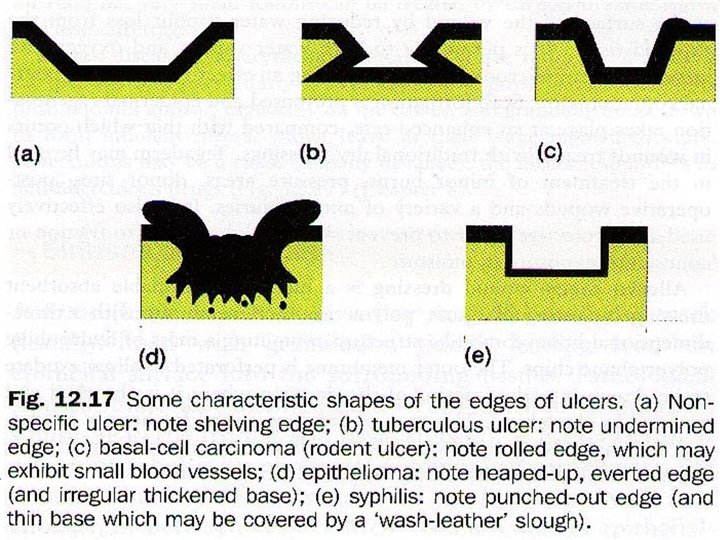
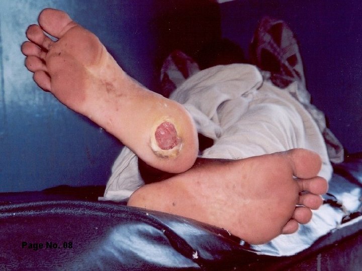
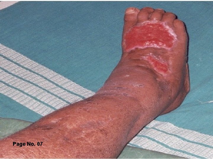
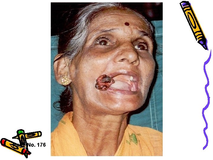
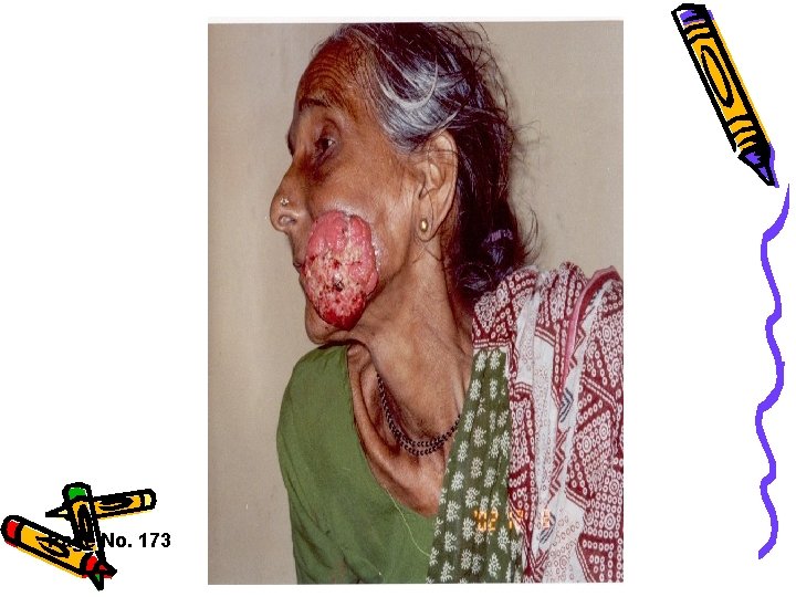
ULCER • An ulcer is a break in the continuity of an epithelial surface. • Characterized by progressive destruction of the surface epithelium and a granulating base which may clean, healthy or containing necrotic slough
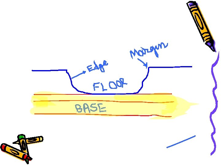
Clinical Examination • • • Size Shape Edge Floor Base Discharge • • • Surrounding area Lymph nodes Pain General exam: Pathological exam:
Marjolin’s ulcer • Malignant change occurring in any long standing benign ulcer irrespective of its cause. • Change usually occurs at the edge of a chronic ulcer
Management • • • Treatment of cause Accurate assessment of the ulcer Identify & correct the co - morbid factors Adequate drainage & desloughing Antiseptics and topical antibiotics Wound dressings – – – Hydrogel Alginates Lyofoam Tegaderm Alleyvn
Page No. 08
Page No. 07
Page No. 176
Page No. 173
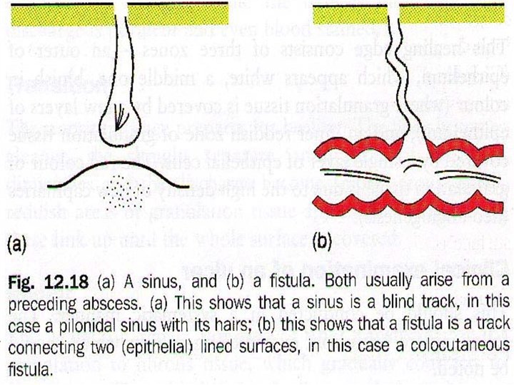
SINUSES • A sinus is a blind tract usually lined with granulation tissue that leads from an epithelial surface into the surrounding tissue. • e. g. pilonidal sinus
FISTULA • It is a communicating track between two epithelial surfaces, commonly between a hollow viscus and the skin ( external fistula) or between two hollow viscera ( internal fistula) • The track is lined with granulation tissue which is subsequently epithelialzed
CLASSIFICATION v Congenital v Acquired
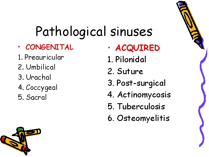
Pathological sinuses • CONGENITAL 1. Preauricular 2. Umbilical 3. Urachal 4. Coccygeal 5. Sacral • ACQUIRED 1. Pilonidal 2. Suture 3. Post-surgical 4. Actinomycosis 5. Tuberculosis 6. Osteomyelitis
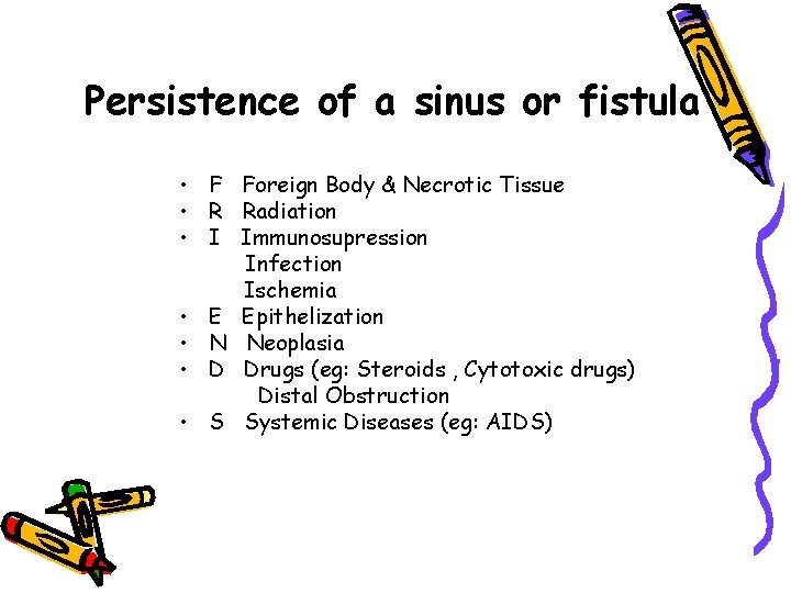
Persistence of a sinus or fistula • F Foreign Body & Necrotic Tissue • R Radiation • I Immunosupression Infection Ischemia • E Epithelization • N Neoplasia • D Drugs (eg: Steroids , Cytotoxic drugs) Distal Obstruction • S Systemic Diseases (eg: AIDS)
Clinical features • • Asymptomatic Recurrent or persistent discharge Pain Infection
Diagnosis • Assess the accurate direction, depth & presence of multiple tracts. • Microbiological examination of discharge ( gut organism, actinomycosis, tuberculosis) • Sinogram
Management of Sinus • • Complete excision of all sinus tract. Sinus is laid open or excised Biopsy of tissue is sent Removal of the cause