
Dr Haider Yousif The Microscopic Techniques Introduction to

- Slides: 34
Dr. Haider Yousif The Microscopic Techniques
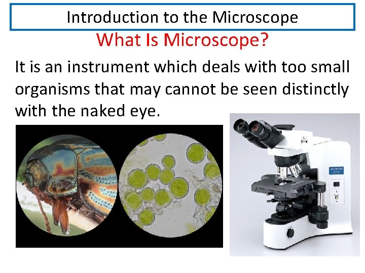
Introduction to the Microscope What Is Microscope? It is an instrument which deals with too small organisms that may cannot be seen distinctly with the naked eye.
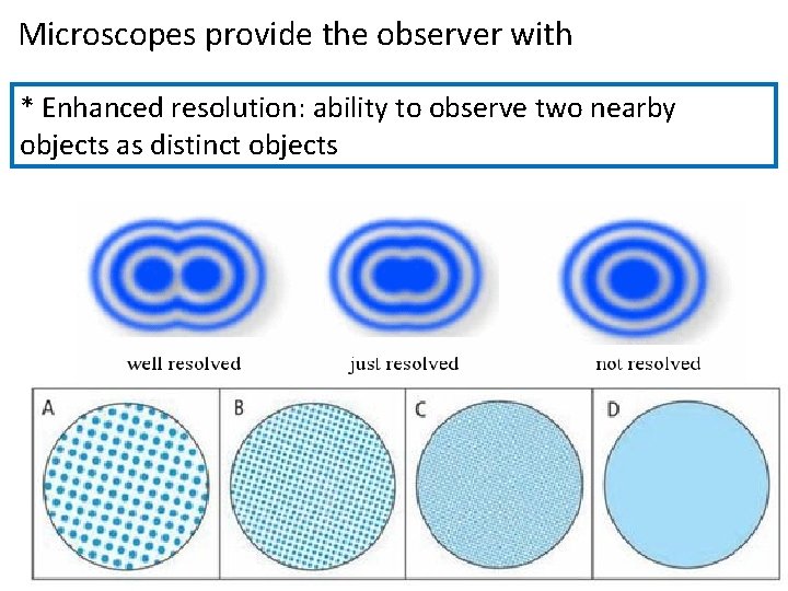
Microscopes provide the observer with * Enhanced resolution: ability to observe two nearby objects as distinct objects
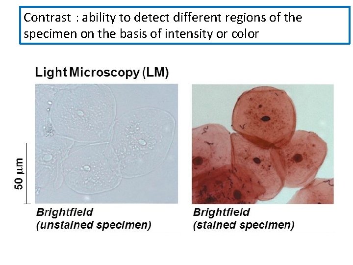
Contrast : ability to detect different regions of the specimen on the basis of intensity or color
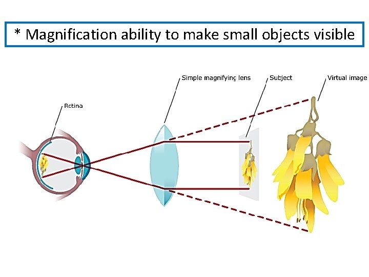
* Magnification ability to make small objects visible
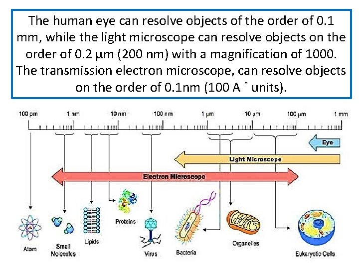
The human eye can resolve objects of the order of 0. 1 mm, while the light microscope can resolve objects on the order of 0. 2 μm (200 nm) with a magnification of 1000. The transmission electron microscope, can resolve objects on the order of 0. 1 nm (100 A ˚ units).
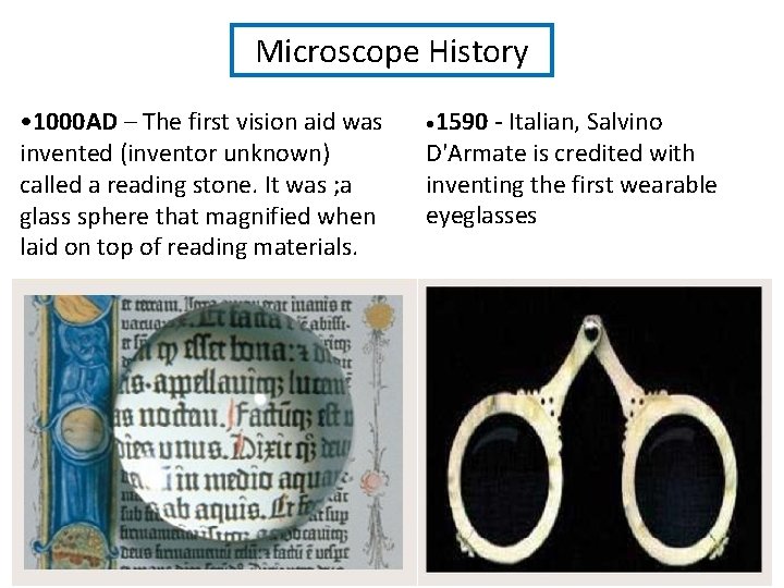
Microscope History • 1000 AD – The first vision aid was invented (inventor unknown) called a reading stone. It was ; a glass sphere that magnified when laid on top of reading materials. • 1590 - Italian, Salvino D'Armate is credited with inventing the first wearable eyeglasses
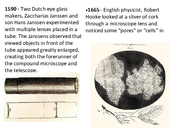
1590 - Two Dutch eye glass • 1665– English physicist, Robert makers, Zaccharias Janssen and Hooke looked at a sliver of cork son Hans Janssen experimented through a microscope lens and with multiple lenses placed in a noticed some "pores" or "cells" in tube. The Janssens observed that viewed objects in front of the tube appeared greatly enlarged, creating both the forerunner of the compound microscope and the telescope.
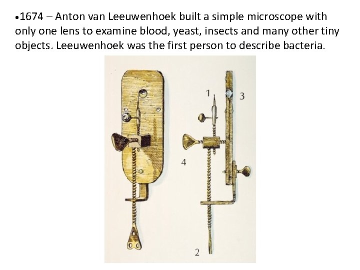
• 1674 – Anton van Leeuwenhoek built a simple microscope with only one lens to examine blood, yeast, insects and many other tiny objects. Leeuwenhoek was the first person to describe bacteria.
• 18 th century – Technical innovations improved microscopes, leading to microscopy becoming popular among scientists. Lenses combining two types of glass reduced the "chromatic effect" the disturbing halos resulting from differences in refraction of light. • 1830 – Joseph Jackson Lister: reduces spherical aberration or the "chromatic effect" by showing that several weak lenses used together at certain distances gave good magnification without blurring the image. This was the prototype for the compound microscope. • 1872 – Ernst Abbe wrote a mathematical formula called the "Abbe Sine Condition". His formula provided calculations that allowed for the maximum resolution in microscopes possible.
• 1903 – Richard Zsigmondy developed the ultra microscope that could study objects below the wavelength of light. He won the Nobel Prize in Chemistry in 1925. • 1932 – Frits Zernike invented the phase-contrast microscope that allowed for the study of colorless and transparent biological materials for which he won the Nobel Prize in Physics in 1953.
• 1931 – Ernst Ruska co-invented the electron microscope for which he won the Nobel Prize in Physics in 1986. An electron microscope depends on electrons rather than light to view an object, electrons are speeded up in a vacuum until their wavelength is extremely short, only one hundred thousandth that of white light. Electron microscopes make it possible to view objects as small as the diameter of an atom. • 1981 – Gerd Binnig and Heinrich Rohrer invented the scanning tunneling microscope that gives three-dimensional images of objects down to the atomic level. Binnig and Rohrer won the Nobel Prize in Physics in 1986.
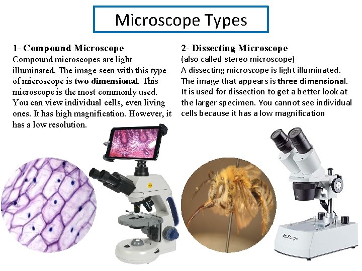
Microscope Types 1 - Compound Microscope 2 - Dissecting Microscope Compound microscopes are light illuminated. The image seen with this type of microscope is two dimensional. This microscope is the most commonly used. You can view individual cells, even living ones. It has high magnification. However, it has a low resolution. (also called stereo microscope) A dissecting microscope is light illuminated. The image that appears is three dimensional. It is used for dissection to get a better look at the larger specimen. You cannot see individual cells because it has a low magnification
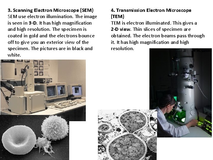
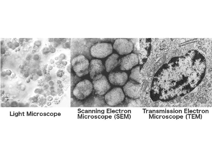
3. Scanning Electron Microscope (SEM) SEM use electron illumination. The image is seen in 3 -D. It has high magnification and high resolution. The specimen is coated in gold and the electrons bounce off to give you an exterior view of the specimen. The pictures are in black and white. 4. Transmission Electron Microscope (TEM) TEM is electron illuminated. This gives a 2 -D view. Thin slices of specimen are obtained. The electron beams pass through it. It has high magnification and high resolution.
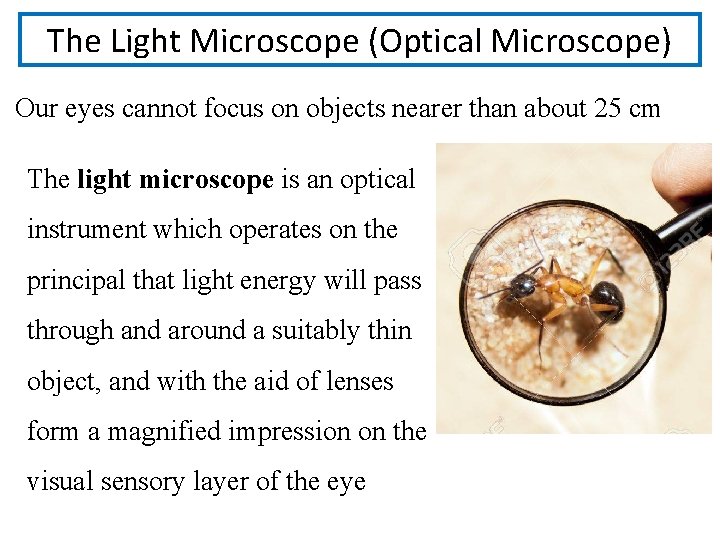
The Light Microscope (Optical Microscope) Our eyes cannot focus on objects nearer than about 25 cm The light microscope is an optical instrument which operates on the principal that light energy will pass through and around a suitably thin object, and with the aid of lenses form a magnified impression on the visual sensory layer of the eye
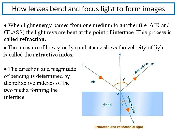
How lenses bend and focus light to form images When light energy passes from one medium to another (i. e. AIR and GLASS) the light rays are bent at the point of interface. This process is called refraction. . The measure of how greatly a substance slows the velocity of light is called the refractive index The direction and magnitude of bending is determined by the refractive indexes of the two media forming the interface
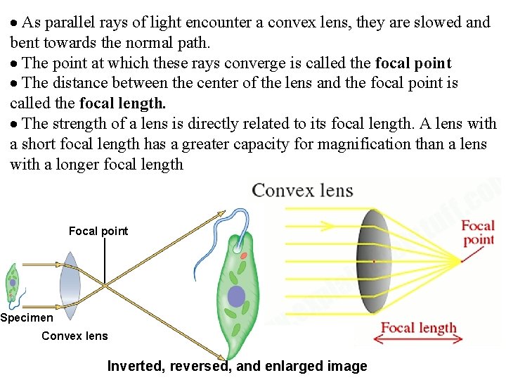
As parallel rays of light encounter a convex lens, they are slowed and bent towards the normal path. The point at which these rays converge is called the focal point The distance between the center of the lens and the focal point is called the focal length. The strength of a lens is directly related to its focal length. A lens with a short focal length has a greater capacity for magnification than a lens with a longer focal length Focal point Specimen Convex lens Inverted, reversed, and enlarged image
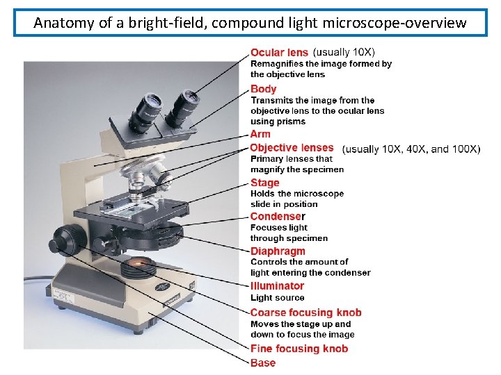
Anatomy of a bright-field, compound light microscope-overview
Microscope Care • Always carry with 2 hands • Never touch the lenses with your fingers. • Only use lens paper for cleaning • When you are finished with your "scope", rotate the nosepiece so that it's on the low power objective, roll the stage down to lowest level
Using the Microscope 1 - start with the lowest power objective lens first and while looking from the side, put the lens down as close to the specimen as possible without touching it. 2 - Look through the eyepiece lens and focus upward only until the image is sharp. If you can't get it in focus, repeat the process again. 3 - Once the image is sharp with the low power lens, you should be able to simply click in the next power lens and do minor adjustments with the focus knob. 4 - If your microscope has a fine focus adjustment, turning it a little bit should be all that's necessary. 5 - Continue with subsequent objective lenses and fine focus each time.
Using High Power 1 - Rotate to 40 x objective, locate desired portion of specimen in the center of the field. 2 - Refocus very carefully so that the specimen is focused as sharply as possible. (Do not alter focus for the Following steps) Partially rotate so that 40 x and 100 x objectives include the specimen. 3 - Place a small drop of oil on the slide in the center of the lighted area. (Take care not to drop on the stage. ). 4 - Rotate so that the 100 x oil immersion objective touches the oil and clicks into place. 5 - Focus only with fine focus. Hopefully, the specimen will come into focus easily. Do not change focus dramatically. Clean up: When you have finished for the day, wipe the 100 x oil immersion objective carefully with lens paper to remove all oil. Wipe oil from the slide thoroughly with a Kim wipe.
Types of Light Microscope There a variety of light microscopes mostly employed in Microbiology: 1. Bright-field 2. Dark-field 3. Phase-contrast 4. Fluorescence
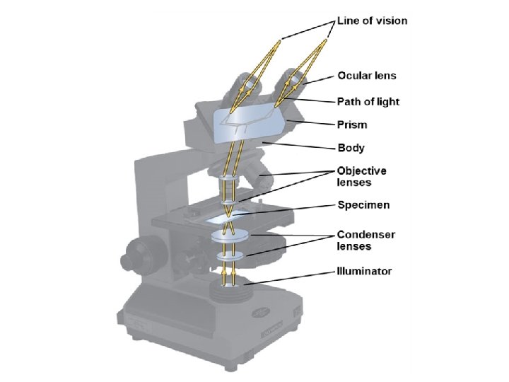
1 -The Bright-field Microscope Called the ordinary microscope because it forms a dark image against a brighter background. Image should remain in focus when objectives are changed. The objective lens forms an enlarged real image within the microscope and the eyepiece lens further magnifies this primarily image. Upon looking in the microscope, the enlarged specimen image (Virtual image) appears to lie just beyond the stage about 25 cm away. Total magnification is calculated by multiplying the objective and eyepiece magnification together.
Three factors determine the quality of an optical image: a. Magnification b. Resolution c. Contrast.
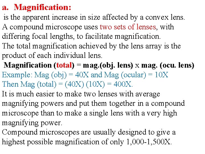
a. Magnification: is the apparent increase in size affected by a convex lens. A compound microscope uses two sets of lenses, with differing focal lengths, to facilitate magnification. The total magnification achieved by the lens array is the product of each individual lens. Magnification (total) = mag. (obj. lens) x mag. (ocu. lens) Example: Mag (obj) = 40 X and Mag (ocular) = 10 X Then Mag (total) = (40 X) (10 X) = 400 X. It is much easier to make two lenses with average magnifying powers and put them together in a compound microscope than to make a single lens with a very high magnifying power. Compound microscopes are usually designed to give a highest possible magnification of only 1, 000 -1, 500 X.
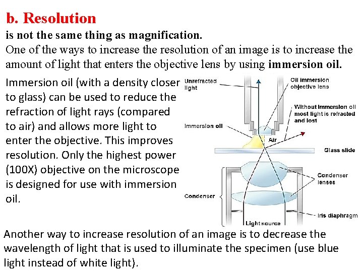
b. Resolution is not the same thing as magnification. One of the ways to increase the resolution of an image is to increase the amount of light that enters the objective lens by using immersion oil. Immersion oil (with a density closer to glass) can be used to reduce the refraction of light rays (compared to air) and allows more light to enter the objective. This improves resolution. Only the highest power (100 X) objective on the microscope is designed for use with immersion oil. Another way to increase resolution of an image is to decrease the wavelength of light that is used to illuminate the specimen (use blue light instead of white light).
c. Contrast Microbes are composed of water, nucleic acids, proteins, and lipids. Most appear colorless against a colorless background when observed using bright field microscopy. Therefore in order to see them, we must devise a way to increase the contrast. Direct staining of the microorganisms and Indirect (negative) staining of the background In order to stain a specimen, it must first be fixed to the slide and chemically altered. This results in the death of a specimen.
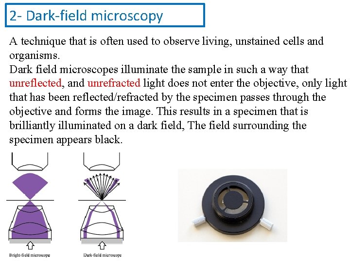
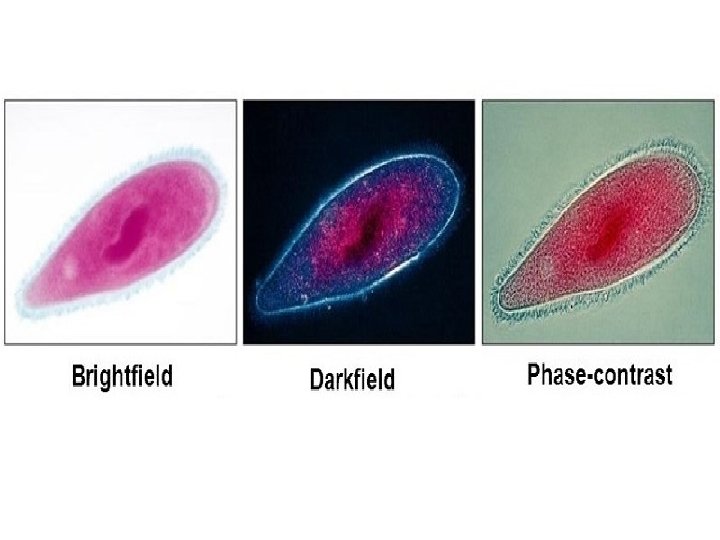
2 - Dark-field microscopy A technique that is often used to observe living, unstained cells and organisms. Dark field microscopes illuminate the sample in such a way that unreflected, and unrefracted light does not enter the objective, only light that has been reflected/refracted by the specimen passes through the objective and forms the image. This results in a specimen that is brilliantly illuminated on a dark field, The field surrounding the specimen appears black.
3 - Phase-contrast microscopes Convert slight differences in refractive index and density into variations in light intensity. This allow organelles of the living cell to become visible with fair contrast in them
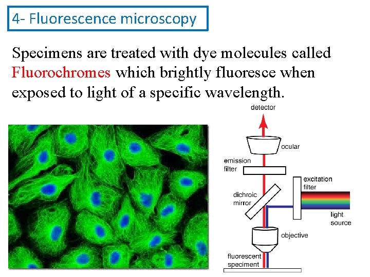
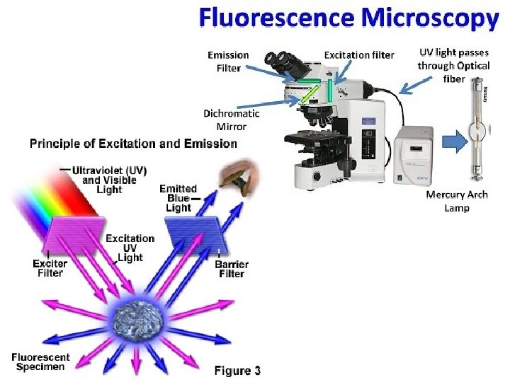
4 - Fluorescence microscopy Specimens are treated with dye molecules called Fluorochromes which brightly fluoresce when exposed to light of a specific wavelength.