Dr Ahmed Fathalla Ibrahim MITOSIS DIFFERENCE BETWEEN MITOSIS

Dr. Ahmed Fathalla Ibrahim

MITOSIS

DIFFERENCE BETWEEN MITOSIS & MEIOSIS

DEFINITION The process of formation of mature male & female gametes. • Spermatogenesis: sequence of events by which the primitive germ cells (spermatogonia) are transformed into mature sperms or spermatozoa • Oogenesis: sequence of events by which the primitive germ cells (oogonia) are transformed into mature oocytes

SPERMATOGENESIS • AIM: formation of sperms with haploid number of chromosomes • SITE: in the seminefrous tubules of testis • DURATION: takes about two months • OCCURRENCE: starts at puberty & continues throughout life

SPERMATOGENESIS

SPERMATOGENESIS STAGES: • PROLIFERATION: each spermatogonium divides by mitosis into 2 daughter spermatogonia (with diploid number of chromosomes: 44 + XY) • GROWTH: spermatogonium enlarges to form a primary spermatocyte (with diploid number) • MATURATION (BY MEIOSIS): 1. 1 st meiotic division: a reduction division by which a primary spermatocyte divides into two secondary spermatocytes (haploid number of chromosomes: 22 + X or 22 + Y) 2. 2 nd meiotic divison: a process of mitosis without a normal interphase (without DNA replication) through which a secondary spermatocyte divides into two spermatids (with haploid number of chromosomes) • SPERMIOGENESIS: a process by which a spermatid is transformed into a mature sperm (with haploid number)

SPERMIOGENESIS
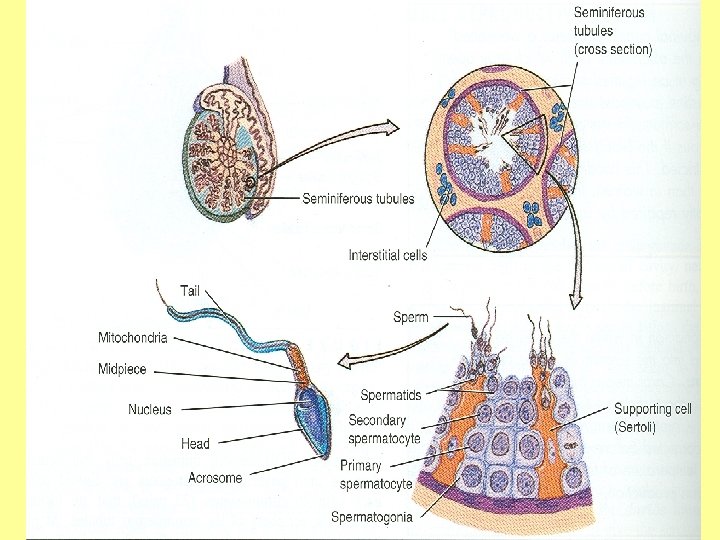

SPERM

SEMINAL FLUID • SOURCE: secretions from seminal vesicles, prostate gland & bulbourethral glands • VOLUME: 3 – 5 ml • SPERM COUNT: about 100 millions/ml • MOTILITY: about 3 mm/min, at least 70% of sperms should be motile • ABNORML FORMS: should not exceed 10%

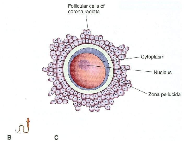
OOGENESIS • AIM: formation of secondary oocytes with haploid number of chromosomes • SITE: in the cortex of ovary • OCCURRENCE: starts during fetal life , becomes completed after puberty & continues till menopause

OOGENESIS

OOGENESIS STAGES: • DURING FETAL LIFE: 1. Proliferation: each oogonium divides by mitosis into 2 daughter oogonia (with diploid number of chromosomes: 44 + XX) 2. Growth: oogonium enlarges to form primary oocyte (with diploid number). Primary oocytes begin 1 st meiotic division & stop at prophase • DURING EACH OVARIAN CYCLE (AFTER PUBERTY): 1. 1 st meiotic division is completed: a reduction division by which a primary oocyte divides into one secondary oocyte (haploid number of chromosomes: 22 + X) & 1 st polar body (degenerates) 2. 2 nd meiotic divison begins: begins at ovulation, progresses only to metaphase and becomes arrested • AFTER FERTILIZATION (IN THE FALLOPIAN TUBE) 1. 2 nd meiotic division is completed: 2 ry oocyte divides into one mature ovum (haploid number) & 2 nd polar body (degenerates) N. B. : NO PRIMARY OOCYTES FORM AFTER BIRTH IN FEMALES

OVULATION
- Slides: 17