Dr Ahmed Fathalla Ibrahim ABDOMINAL VISCERA FOR EACH

Dr. Ahmed Fathalla Ibrahim

ABDOMINAL VISCERA FOR EACH PART YOU MUST KNOW: 1. SURFACE ANATOMY 2. RELATIONS 3. PERITONEAL COVERING 4. BLOOD SUPPLY 5. NERVE SUPPLY 6. LYMPHATIC DRAINAGE 7. SUPPORT (IN SOME PARTS)
")
SMALL INTESTINE • DUODENUM: fixed part • JEJUNUM & ILEUM: movable part (with mesentery)
 Beginning: duodenojejunal flexure")
JEJUNUM & ILEUM • • • Length: 6 meters (20 feet) Beginning: duodenojejunal flexure Termination: ileocecal junction Embryological origin: midgut Peritoneal fold: mesentery of small intestine Arterial supply: jejunal & ileal branches of superior mesenteric • Lymphatic drainage: superior mesenteric lymph nodes • Nerve supply: superior mesenteric plexus: sympathetic & parasympathetic (vagus)
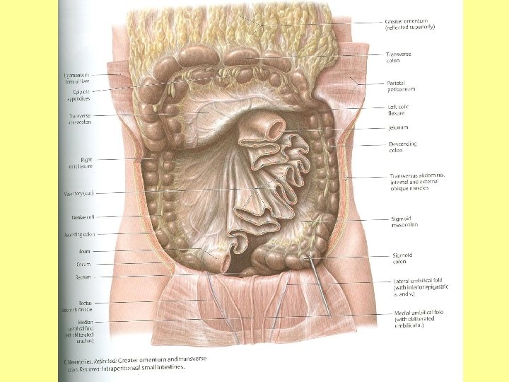
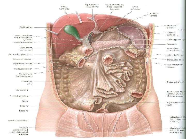
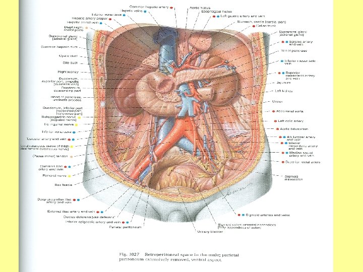
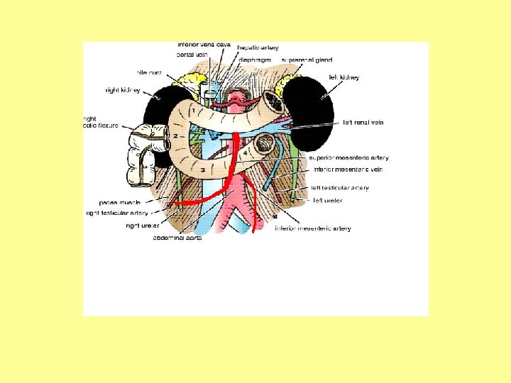

MESENTERY OF SMALL INTESTINE • • 1. 2. 3. 4. 5. Extent: from duodenojejunal flexure to ileocecal junction Formation: formed of 2 layers Free border (6 meters long): encloses jejunum & ileum Root (6 inches long, J-shaped): crosses superficial to the following structures: Third part of duodenum Abdominal aorta Inferior vena cava Right psoas major Right ureter

MESENTERY OF SMALL INTESTINE • • • 1. 2. 3. 4. 5. Shape: Fan-shaped with undulations Content of root: superior mesenteric vessels Contents (structures between its 2 layers): Jejunal vessels: form few arcades Ileal vessels: form many arcades Mesenteric lymph nodes Autonomic nerve fibers Mesenteric fat

MESENTERY OF SMALL INTESTINE
 Diameter: wider")
JEJUNUM 1. 2. 3. 4. 5. 6. 7. Length: shorter (proximal 2/5) Diameter: wider Wall: thicker (more numerous plicae circulares: circular folds of mucosa) Appearance: more red in color (more vascular) Vessels: less arcades, long terminal branches Mesenteric fat: small amount near intestinal border Aggregations of lymphoid tissue: few
 Diameter: narrower")
ILEUM 1. 2. 3. 4. 5. 6. 7. Length: longer (distal 3/5) Diameter: narrower Wall: thinner (less numerous plicae circulares: circular folds of mucosa) Appearance: light red in color (less vascular) Vessels: more arcades, short terminal branches Mesenteric fat: large amount near intestinal border Aggregations of lymphoid tissue: numerous (Peyer’s patches)

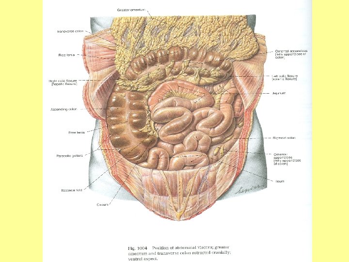
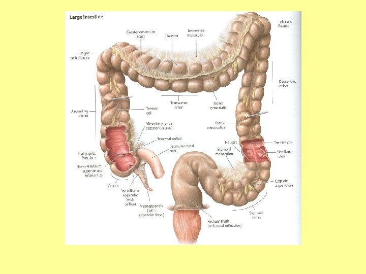
LARGE INTESTINE PARTS: 1. Cecum 2. Appendix 3. Ascending colon 4. Transverse colon 5. Descending colon 6. Sigmoid (pelvic) colon 7. Rectum 8. Anal canal 9. N. B. : Parts of large intestine in abdomen: from 1 to 5
: teniae")
LARGE INTESTINE CHARACTERISTICS: 1. Teniae coli: 3 longitudinal muscle bands 2. Sacculations (haustrations): teniae coli are shorter than large intestine 3. Appendices epiploicae: short peritoneal fold filled with fat • N. B. : characteristics are present in all large intestine EXCEPT: in rectum & anal canal

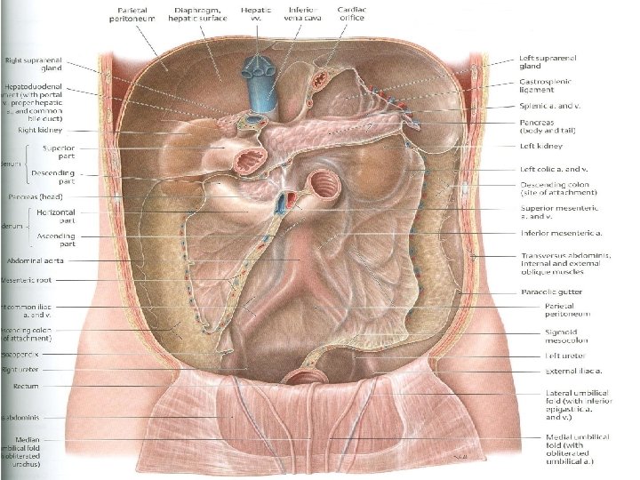
LARGE INTESTINE • Embryological origin: 1. From midgut: cecum, appendix, ascending colon, right 2/3 of transverse colon 2. From hindgut: left 1/3 of transverse colon, descending & sigmoid colon, rectum, upper half of anal canal • Peritoneal fold: 1. Appendix, transverse & sigmoid colon: have mesentery 2. Cecum: completely covered by peritoneum, but has no mesentery 3. Ascending & descending colon: covered anteriorly & on the sides 4. Rectum & anal canal: discussed later

LARGE INTESTINE • Arterial supply: 1. Midgut: colic branches of superior mesenteric 2. Hindgut: inferior mesenteric • Lymphatic drainage: 1. Midgut: superior mesenteric lymph nodes 2. Hindgut: inferior mesenteric lymph nodes • Nerve supply: 1. Superior mesenteric plexus: sympathetic & parasympathetic (vagus) 2. Inferior mesenteric plexus: sympathetic & parasympathetic (pelvic splanchnic nerves)

LARGE INTESTINE SURFACE ANATOMY

APPENDIX • Surface anatomy: the base of appendix is marked by Mc’Burney’s point: point A point at the junction of lateral 1/3 & medial 2/3 of a line traced from right anterior superior iliac spine to umbilicus • Opening: at posteromedial aspect of cecum, 1 inch below ileo-cecal junction
 1. Retrocecal: Retrocecal most common position")
APPENDIX • Positions: (from most to least common) 1. Retrocecal: Retrocecal most common position 2. Pelvic 3. Subcecal 4. Preilieal 5. Postileal

CECUM, ASCENDING & DESCENDING COLON • 1. 2. 3. ANTERIOR RELATIONS: Coils of small intestine Greater omentum Anterior abdominal wall

1 2 Iliac crest 5 3 4 6 1: Iliohypogastric nerve; 2: Ilioinguinal nerve; 3: lateral cutaneous nerve of thigh 4: Femoral nerve; 5: Genitofemoral nerve; 6: Obturator nerve P. M. = psoas major; Q. L. =quadratus lumborum; I. =iliacus; T. A. = transversus abdominis

1 2 5 3 6 4 1: Iliohypogastric nerve; 2: Ilioinguinal nerve; 3: lateral cutaneous nerve of thigh 4: Femoral nerve; 5: Genitofemoral nerve; 6: Obturator nerve P. M. = psoas major; Q. L. =quadratus lumborum; I. =iliacus; T. A. = transversus abdominis

CECUM, ASCENDING & DESCENDING COLON • POSTERIOR RELATIONS: • Cecum: psoas major, genitofemoral nerve, iliacus, lateral cutaneous nerve of thigh, femoral nerve • Ascending colon: iliacus, lateral cutaneous nerve of thigh, quadratus lumborum, ilioinguial nerve, iliohypogastric nerve, iliac crest, origin of transversus abdominis from lumbar fascia • Descending colon: relations of cecum + relations of ascending colon + left kidney

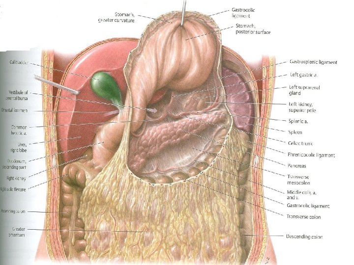
RELATIONS OF TRANSVERSE COLON

RELATIONS OF TRANSVERSE COLON • ANTERIOR: greater omentum, anterior abdominal wall • POSTERIOR: 2 nd part of duodenum, head of pancreas, coils of small intestine • SUPERIOR: liver, gall bladder, stomach • INFERIOR: coils of small intestine
- Slides: 31